Implications
Pain severely affects the welfare of horses and often remains underestimated. Measuring pain-related parameters in a reliable way can be challenging; however, an accurate determination and quantification of pain in horses is critical. Several physiological, endocrine and behavioural parameters have been investigated to identify pain conditions and severity in clinical studies. To date, no molecular indicators have been available to assess horse pain. Tackling the challenge of improving the welfare of horses affected by laminitis, the present study highlights the possible use of molecular biomarkers in assessing and quantifying pain in horses. Three candidate biomarkers have been identified in the serum of horses with acute laminitis. An integrated analysis between behavioural indicators (Horse Grimace Scale (HGS)) and molecular biomarkers could be used to assess and monitor painful conditions in horses.
Introduction
MicroRNAs (miRNAs) belong to a class of non-coding single-stranded RNA of 19 to 24 nucleotides with the ability to modulate gene expression post-transcriptionally. They are crucial modulators of cellular homoeostasis and their deregulation has been associated with a wide range of pathological conditions and with neuropathic, inflammatory, traumatic and cancer-associated pain in humans (Bali and Kuner, Reference Bali and Kuner2014; Zhang and Banerjee, Reference Zhang and Banerjee2015; Pang et al., Reference Pang, Tang and Zhang2016) and laboratory animals (Gong et al., Reference Gong, Lu, Huang, Ruan, Chen, Liang, Wang, Yue and Feng2015; Pang et al., Reference Pang, Tang and Zhang2016; Qureshi et al., Reference Qureshi, Tian, McDonald, Capasso, Douglas, Gao, Orlova, Barrett, Ajit and Sacan2016). The modulation of miRNA expression is an early event in pathogenic processes (Schwarzenbach et al., Reference Schwarzenbach, Nishida, Calin and Pantel2014). They act by guiding the RNA-induced silencing complex to partially complementary sequences in target messenger RNA (mRNA) to suppress gene expression by a combination of translation inhibition and mRNA decay (Lagos-Quintana et al., Reference Lagos-Quintana, Rauhut, Lendeckel and Tuschl2001). The level and composition of extracellular miRNAs in certain body fluids are tightly correlated to various human pathological conditions, such as cancer, diabetes, cardiovascular diseases and drug-induced organ damage (Ghai and Wang, Reference Ghai and Wang2016). A change in the expression pattern of miRNAs targeting key regulators of pain processing has been observed both in inflammatory and neuropathic pain (Zhao et al., Reference Zhao, Lee, Cendan, Shepherd, Baker, Asante, Bee, Bethry, Perkins, Nassar, Abrahamsen, Dickenson, Cobb, Merkenschlager and Wood2010; Genda et al., Reference Genda, Arai, Ishikawa, Tanaka, Okabe and Sakamoto2013). Although some investigations have already evaluated miRNAs as suitable biomarkers to assess stress associated with endurance exercise in horses (Mach et al., Reference Mach, Plancade, Pacholewska, Lecardonnel, Rivière, Moroldo, Vaiman, Morgenthaler, Beinat, Nevot, Robert and Barrey2016) and with road transportation in turkeys (Lecchi et al., Reference Lecchi, Marques, Redegalli, Meani, Vinco, Bronzo and Ceciliani2016), the diagnostic potential of circulating miRNAs deserves to be further explored in veterinary medicine in relation to stressful or painful conditions that might affect animal welfare.
Acute laminitis is a disease associated with ischaemia of digital dermal tissues whose exact aetiology is still not completely understood (Wylie et al., Reference Wylie, Collins, Verheyen and Newton2013). However, there are evidences that support a possible link between insulin resistance, obesity and acute laminitis (Geor, Reference Geor2008). Evidences suggest that the pain state associated with chronic equine laminitis have a neuropathic component (Jones et al., Reference Jones, Viñuela-Fernandez, Eager, Delaney, Anderson, Patel, Robertson, Allchorne, Sirinathsinghji, Milne, MacIntyre, Shaw, Waran, Mayhew and Fleetwood-Walker2007) indicating that both inflammatory and neuropathic pain should be considered in the management of this condition. In equine practice, acute laminitis provides a valuable pain model as it severely affects animal welfare causing severe, long-lasting debilitating pain in horses (Minero and Canali, Reference Minero and Canali2009; Dalla Costa et al., Reference Dalla Costa, Murray, Dai, Canali and Minero2014b). Since 2010, World Horse Welfare, an international horse charity, has launched a campaign about the importance of recognising the early signs of laminitis to protect horse welfare and minimise the associated pain. This disease has a worldwide estimated prevalence of 7% to 14% and is characterised by lameness, inability or reluctance to walk, frequent weight shifting, and abnormal weight distribution on hind feet (Collins et al., Reference Collins, Pollitt, Wylie and Matiasek2010; Wylie et al., Reference Wylie, Collins, Verheyen and Newton2013). Albeit challenging, accurate pain assessment is critical to recognise early signs associated with laminitis (e.g. strong digital pulse, shortening of the stride, abnormal foot lifting) (Wylie et al., Reference Wylie, Collins, Verheyen and Newton2013) and ensure appropriate treatment (de Grauw and van Loon, Reference de Grauw and van Loon2016). Among commonly used systems, the Composite Pain Scale (CPS), a multifactorial numerical rating scale, considers physiological response to treatment and behavioural data. The CPS was originally developed on an experimental model of acute orthopaedic pain in horses (Bussières et al., Reference Bussières, Jacques, Lainay, Beauchamp, Leblond, Cadoré, Desmaizières, Cuvelliez and Troncy2008) and then applied to monitor pain after surgery (e.g. castration, colic, orthopaedic and soft-tissue surgery) (Van Loon et al., Reference Van Loon, Back, Hellebrekers and René van Weeren2010). Recently, a facial expression-based pain coding system, the HGS was developed as a welfare indicator for pain assessment of horses undergoing surgical castration (Dalla Costa et al., Reference Dalla Costa, Minero, Lebelt, Stucke, Canali and Leach2014a) and acute laminitis (Dalla Costa et al., Reference Dalla Costa, Stucke, Dai, Minero, Leach and Lebelt2016). Horse Grimace Scale is a fast method based on behavioural observation. It does not cause disturbance to the horses in their boxes, does not require their walking and/or trotting as well as any palpation of their painful area. Horse Grimace Scale has potential to provide insights into the experience of pain in horses and in the assessment of horse welfare, however, the authors concluded that further validation studies were needed to apply the HGS in a clinical setting (Dalla Costa et al., Reference Dalla Costa, Minero, Lebelt, Stucke, Canali and Leach2014a, Reference Dalla Costa, Stucke, Dai, Minero, Leach and Lebelt2016 and Reference Dalla Costa, Bracci, Dai, Lebelt and Minero2017). Considering the diagnostic potential of miRNAs and the evidence of their identification in the equine genome, tissues and serum (Kim et al., Reference Kim, Lee, Ryu, Cui, Bhak and Kim2014; Pacholewska et al., Reference Pacholewska, Mach, Mata, Vaiman, Schibler, Barrey and Gerber2016), the aims of the present study were to (a) ascertain whether acute pain associated with laminitis may modulate the expression of circulating miRNAs; (b) investigate the potential use of differentially expressed (DE)-miRNAs as biomarkers to measure pain in horses; and (c) gather additional evidence on concurrent validity of HGS and CPS by investigating how they correlate with miRNAs.
Material and method
Ethics statement
The study design was approved by the Brandenburg State Veterinary Authority (V3-2347-A-42-1-2012) in compliance with German legislation on animal experiments. Individual horse owner’s consent was obtained for all horses participating in this study. Horses involved in this study were admitted for routine veterinary treatment of acute laminitis at the request of their owner on a voluntary basis. Control samples were obtained from healthy stallions referred to the clinic for surgical castration.
Animals and husbandry
Between 2012 and 2014, horses admitted to the Havelland Equine Clinic presenting acute laminitis with no prior treatment were included in the present study. Nine horses of different breed and gender, aged between 4 and 17 years (mean=9.4±5.0) were recruited. As a control group, nine healthy stallions of mixed age (mean=2.4±1) referred to the clinic for routine castration were recruited. In order to be included in this study, control horses had to be deemed healthy by an equine veterinarian after physical examination and behavioural evaluation. All horses, after admission, were stabled in standard single boxes (4×3 m with an outside window) on wood shavings (German Horse Span Classic, German Horse Pellets, Wismar Germany), and in visual contact with conspecifics. They were fed hay twice a day (~3/100 kg BW per day) and water was provided ad libitum by automatic drinkers.
Each horse with laminitis underwent a routine treatment including: oral administration of phenylbutazone (initial 4.5 mg/kg BW every 12 h for 2 days and subsequent 2.5 mg/kg BW every 12 h; Phenylbutariem®; Ecuphar, Greifswald, Germany); subcutaneous injections of heparin (50 IU/kg BW every 12 h for 5 days; Heparin-Natrium-25000-ratiopharm®; Ratiopharm, Ulm, Germany); padded hoof bandages with frog support and elevated heels (3 to 4 cm); ice water applications every 2 h for the first 3 days of treatment and strictly restricted movement in an individual box with a deep and soft bedding of wood shavings.
Horses with acute laminitis were assessed, as described in the following sections, at admission before any treatment (‘pre-treatment’) and 7 days after the treatment, before being discharged (‘post-treatment’). Control horses (‘control’) were assessed once, after 2 days of acclimatisation to the clinic setting and before surgery. Assessments were carried out in the morning between 0800 and 1200 h. Horses were monitored routinely at least three times a day, or more frequently if needed, by an experienced vet or nurse by collecting data about heart rate, breath frequency, appetite, defecation, weight shifting and obvious signs of pain.
MicroRNAs extraction and real-time quantitative PCR (qPCR)
Blood was collected by jugular venipuncture into Monovette® tubes (Sarstedt Company, Nümbrecht, Germany) and serum was stored at −80°C until RNA extraction. Total RNA was extracted using miRNeasy Serum/Plasma Kit (Qiagen, Milan, Italy, catalog number 217184). Serum was thawed on ice and centrifuged at 3000×g for 5 min at 4°C. An aliquot of 200 μl per sample was transferred to a new tube and 1 ml of Qiazol (Qiagen, Milan, Italy) was added. The Caenorhabditis elegans miRNA cel-miR-39 (Qiagen, Milan, Italy, catalog number 219610) was used as synthetic spike-in control because of a lack of sequence homology to equine miRNAs. After incubation at room temperature for 5 min, 3.75 μl (25 fmol final concentration) of spike-in control was added and the samples were vortexed to ensure complete mixing. RNA extraction was then carried out according to manufacturer’s instruction. Reverse transcription was performed using the TaqMan MicroRNA Reverse Transcription Kit (Applied Biosystems; Thermo Fisher Scientific, Monza, Milan, Italy, catalog number 4366596) and using miRNA-specific stem-loop reverse transcriptase (RT) primers, according to manufacturer’s instructions. Reverse transcription reactions were performed in 15 μl volume reactions containing 1.5 µl 10× miRNA RT buffer, 1 µl MultiScribe reverse transcriptase (50 U/µl), (Thermo Fisher Scientific, Monza, Milan, Italy), 0.30 µl 100 mM deoxynucleotides mix, 0.19 µl RNase Inhibitor (20 U/µl), 6 μl of custom RT primer pool and 3.01 μl of nuclease-free water. The custom RT primer pool was prepared combining 10 µl of each individual 5× RT primer in a final volume of 1000 µl; the final concentration of each primer in the RT primer pool was 0.05× each; 3 μl serum RNA was added to each RT reaction. Every RT reaction mixture was incubated on ice for 5 min, 16°C for 30 min, 42°C for 30 min and then 85°C for 5 min.
The qPCR experiments were designed following the Minimum Information for Publication of Quantitative Real-Time PCR Experiments (MIQE) guidelines. Small RNA TaqMan assays were performed according to manufacturer’s instruction. MicroRNAs were selected according to previous publications where they were correlated to pain in humans and mice (Bali and Kuner, Reference Bali and Kuner2014; Elramah et al., Reference Elramah, Landry and Favereaux2014; Qureshi et al., Reference Qureshi, Tian, McDonald, Capasso, Douglas, Gao, Orlova, Barrett, Ajit and Sacan2016). The selected primer/probe assays (Life Technologies; Thermo Fisher Scientific, Monza, Milan, Italy) included cel-miR-39-3p (assay ID000200), hsa-miR-532-3p (assay ID002355), hsa-miR-219-5p (assay ID00522), mmu-miR-134-5p (assayID001186), mmu-miR-124a-3p (assay ID001182), hsa-miR-200b-3p (assay ID002251), hsa-miR-146a-5p (assay ID000468), hsa-miR-23b-3p (assay ID000400), hsa-miR-145-5p (assay ID002278) and hsa-miR-181a-5p (assay ID000480). Quantitative reactions were performed in duplicate in scaled-down (12 μl) reaction volumes using 6 μl TaqMAN 2×Universal Master Mix II (Applied Biosystems, catalog number 4440044; Thermo Fisher Scientific, Monza, Milan, Italy), 0.6 μl miRNA-specific TaqMan Assay 20× and 1 μl of the RT product per reaction on Eco Real-Time PCR detection System (Illumina, Milan, Italy). The standard cycling programme was 50°C for 2 min, 95°C for 10 min and 40 cycles of 95°C for 15 sec and 60°C for 60 sec. Data were normalised relative to the expression of cel-miR-39. MicroRNAs expression levels are presented in terms of fold change normalised to cel-miR-39 expression using the formula 2−ΔΔCq . To investigate the pain relevance, predicted targets of the significant DE-miRNAs were computationally retrieved from the TargetScan database (http://www.targetscan.org/vert_71/) and mRNA enrichment was performed using DAVID bioinformatics resources (https://david.ncifcrf.gov/). The list of target genes was employed in further analyses and biological pathways in the Kyoto Encyclopedia of Genes and Genomes (KEGG) were examined for enrichment (http://www.genome.jp/kegg/). As alterations in neuronal excitability are believed to contribute to the initiation and maintenance of neuropathic pain following peripheral injury, an enrichment of mRNA targets that encode for ion channels – known to contribute and modulate neuronal excitability – was performed. Based on knockout studies, the predicted mRNA targets have already been associated with hypersensitivity in various pain models (http://www.jbldesign.com/jmogil/enter.html) (Lacroix-Fralish et al., Reference Lacroix-Fralish, Ledoux and Mogil2007).
Behavioural recordings
An experienced veterinarian conducted the live evaluation of the CPS developed by Bussières et al. (Reference Bussières, Jacques, Lainay, Beauchamp, Leblond, Cadoré, Desmaizières, Cuvelliez and Troncy2008) with slight modifications, as previously reported (Dalla Costa et al., Reference Dalla Costa, Minero, Lebelt, Stucke, Canali and Leach2014a). Twenty-minute videos were simultaneously recorded using two video cameras (Panasonic, HDC-SD99, Osaka, Japan). In order to collect videos without influencing the horse’s behaviour, the video cameras were positioned on the opposite sides of the box on top of their grate section. Following the methods of Dalla Costa et al. (Reference Dalla Costa, Minero, Lebelt, Stucke, Canali and Leach2014a), where images (one or two per animal) per time point were included, still frames of the face of each horse were extracted from the videos whenever they directly faced the video camera. The selection was based on the quality of the picture. On a sample of 27 still images (nine for control horses, nine for pre-treatment and nine for post-treatment), randomly selected by a non-participating assistant, the HGS was assessed by one trained treatment and time point blind veterinarian (not involved in the CPS assessment). A detailed handout with the description of the six Facial Action Units (FAUs) was used as training material. Facial Action Units are independently scored on a 3-point scale, with 0 indicating that the assessor is confident that the action unit is not present, 1 indicating that the assessor is confident that the action unit is only moderately present and 2 indicating that the assessor is confident that the action unit is obviously present. The possible maximum HGS score was 12 (two for each FAUs).
Statistical analysis
Statistical analysis was performed using SPSS 23 (SPSS Inc., Chicago, IL, USA). Statistical significance was accepted at P⩽0.05. The CPS total score was calculated. The HGS total score for each image consisted of the sum of the scores across the six FAUs. Data were tested for normality and homogeneity of variance using the Kolmogorov–Smirnov and Levene test, respectively. As data were not normally distributed, non-parametric statistical tests were applied. Mann–Whitney test was used to assess differences in miRNAs concentrations (only for those miRNAs that had a significant differential expression in the blood of horses), CPS and HGS scores between control group and pre-treatment group, and control and post-treatment group. Match paired Wilcoxon test was run to compare miRNA concentrations (miR-23b-3p, miR-145-5p and miR-200b-3p), CPS and HGS scores pre- and post-treatment. Receiver operating characteristic (ROC) analysis was performed to determine the diagnostic accuracy of targets that statistically differed between pre-treatment and controls. The diagnostic values were calculated for those miRNAs (alone and in combination) that showed a significant differential expression in the blood of horses, namely miR-23b-3p, miR-145-5p and miR-200b-3p. The ROC analysis was carried out by plotting the true positive (sensitivity) v. the false positive (1−specificity). Linear regression was used to investigate any relationship between miR-23b-3p, miR-145-5p and miR-200b-3p and age of the horses. Spearman’s ρ test was performed to evaluate whether there was any correlation among the expression levels of the various miRNAs, HGS and CPS.
Results
Acute laminitis alters expression levels of miR-23b-3p, miR-145-5p and miR-200b-3p in blood serum
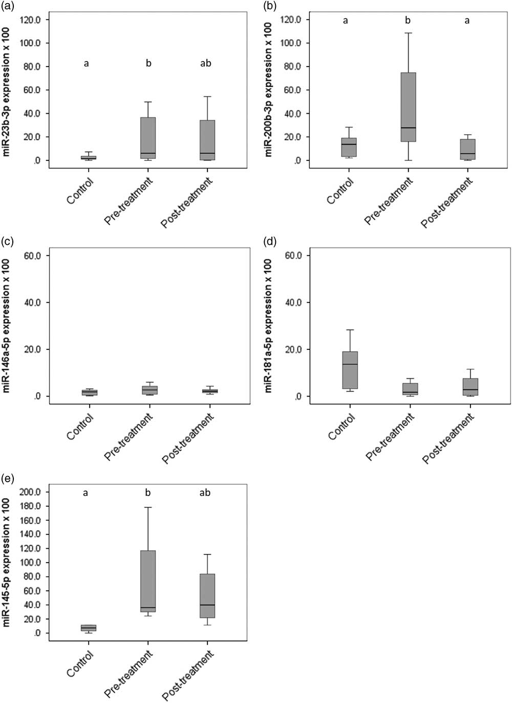
The levels of miRNAs were normalised to the levels of the artificial spike-in cel-miR-39, which was used as internal control. The selected target miRNAs were detected in all samples, with exception of the miRNAs has-miR-219-5p, has-miR-532-3p, mmu-miR-134-5p and mmu-miR-124a-3p. Statistical analyses was performed on detected five miRNAs, among which three circulating miRNAs, namely miR-23b-3p, miR-145-5p and miR-200b-3p, had a significant differential expression in the blood of horses. Figure 1 presents an overview of the results. In detail, the level of miR-23b-3p (fold change=14.6; P=0.029), miR-145-5p (fold change=4.4; P=0.015) and miR-200b-3p (fold change=3.4; P=0.023) was higher in pre-treatment compared with controls. Only miR-200b-3p significantly decreased from pre- to post-treatment (Match paired Wilcoxon, P=0.043). The levels of circulating miR-23b-3p, miR-145-5p and miR-200b-3p were not affected by the age of the horses (linear regression, P>0.05).
Box plots for circulating pain-related microRNAs levels: (a) miR-23b-3p, (b) miR-200b-3p, (c) miR-146a-5p, (d) miR-181a-5p and (e) miR-145-5p. a,b,abIndicate significant differences among the groups (control, pre-treatment and post-treatment) with a significance level at P<0.05. The black lines inside the boxes mark the medians. Whiskers indicate variability outside the upper and lower quartiles.

Diagnostic performance of miR-23b-3p, miR-145-5p and miR-200b-3p
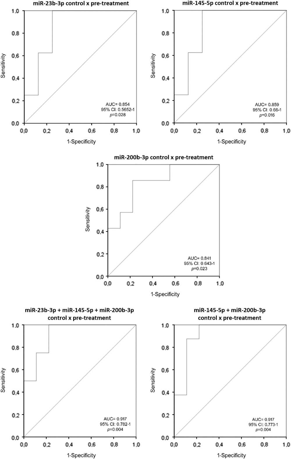
Diagnostic accuracy of miR-23b-3p, miR-145-5p and miR-200b-3p, as measured by the area under the curve (AUC), was good for all targets for the discrimination of pre-treatment from controls (Figure 2) and it was 0.854 (95% confidence interval (CI) 0.5652 to 1; P=0.028), 0.859 (95% CI 0.66 to 1; P=0.016) and 0.841 (95% CI 0.643 to 1; P=0.023), respectively.
Receiver–operator characteristics curve analysis of candidate pain-related microRNAs. AUC=area under the curve; CI=confidence interval.

Further statistical analysis was performed considering the average relative quantification values of the three pain-related miRNAs. The combination of miRNAs resulted in an improved discrimination of the pre-treatment. The created model included all three miRNAs (miR-23b-3p, miR-145-5p and miR-200b-3p) or two (miR-145-5p and miR-200b-3p). The AUC from ROC analysis increased to 0.917 (95% CI 0.782 to 1; P=0.004; sensitivity 100% and specificity 78%, cut-off 0.1936) and 0.917 (95% CI 0.773 to 1; P=0.004; sensitivity 87.5% and specificity 89%, cut-off 0.279), respectively.
Composite Pain Scale and Horse Grimace Scale and their correlation to microRNAs
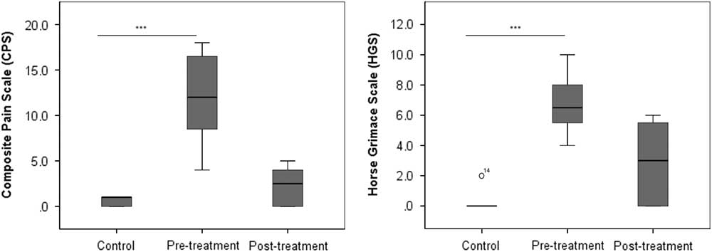
Both CPS and HGS scores were significantly lower in the control group (Mann–Whitney test, P<0.001) than in horses with acute laminitis (pre-treatment) (Figure 3). Control horses did not differ from post-treatment for both CPS and HGS scores (Mann–Whitney test, P=0.167 and P=0.059, respectively).
Composite Pain Scale (CPS) and Horse Grimace Scale (HGS) scores of the control, pre-treatment and post-treatment groups drawn in a box plot. Differences between the groups (control, pre-treatment and post-treatment) are indicated as follows: ***P<0.000. The black lines inside the boxes mark the medians. Whiskers indicate variability outside the upper and lower quartiles. Circles, labelled with the individual case numbers, represent outliers (1.5 to 3 times length of the box).

A positive correlation was observed among miR-145-5p, HGS (R 2=0.482; P=0.015) and CPS (R 2=0.575; P=0.003). A positive correlation was also found between the combination of miR-145-5p and miR-200b-3p with both HGS (R 2=0.559; P=0.004) and CPS (R 2=0.595; P=0.002).
MicroRNA target prediction and pathway enrichment
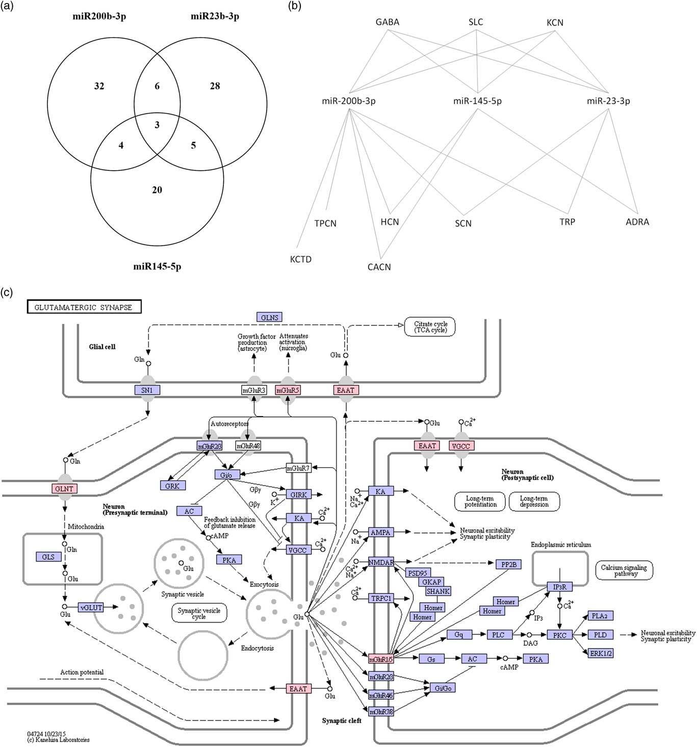
The predicted mRNA targets were 97, three of which were regulated by all three miRNAs and 15 by at least two miRNAs (Figure 4a). The target mRNA are grouped in ion channel families: solute carrier family transporters (SLC), potassium voltage-gated channel, voltage-gated sodium channel, γ-aminobutyric acid (GABA) A or B receptor, voltage-dependent calcium channel, adrenoceptor α, potassium channel (tetramerization domain), hyperpolarization-activated cyclic nucleotide-gated potassium channel, two pore segment channel, transient receptor potential cation channel (Figure 4b). The KEGG pathway analysis demonstrated that most of these genes code for proteins involved in the glutamatergic pathway (Figure 4c).
Predicted messenger RNA (mRNA) targets for differentially expressed microRNAs (DE-miRNAs). (a) Venn diagram showing the genes that are potential targets of DE-miRNAs. (b) TargetScan software was used to identify potential mRNA targets. The list was filtered by selecting those genes that encode for ion channels: solute carrier family transporters (SLC), potassium voltage-gated channel (KCN), voltage-gated sodium channel (SCN), γ-aminobutyric acid (GABA) receptor, voltage-dependent calcium channel (CACN), adrenoceptor α (ADRA), potassium channel (tetramerization domain) (KCTD), hyperpolarization-activated cyclic nucleotide-gated potassium channel (HCN), two pore segment channel (TPCN), transient receptor potential cation channel (TRP). (c) Glutamatergic pathway identified by KEGG pathway analysis using the mRNA targets. Proteins coded by mRNA targets genes are highlighted in red. Glutamine transporter (GLNT) and excitatory amino acid transporters (EAAT) belong to SLC; voltage-gated Ca2+ channels (VGCC) to CACN.

Discussion
The present study investigated the circulating levels of nine miRNAs and assessed their diagnostic value in serum of horses affected by acute laminitis. In particular, their incremental diagnostic value was investigated. We report five major findings. First, circulating levels of miR-23b-3p, miR-145-5p and miR-200b-3p were significantly higher in animals with acute laminitis than in controls. Second, diagnostic accuracy for acute laminitis was good (0.80<AUC<0.90) for miR-23b-3p, miR-145-5p and miR-200b-3p. Third, diagnostic accuracy of the combination of two or three DE-miRNAs was excellent (0.90<AUC<1). Fourth, the level of miR-200b-3p significantly decreased from pre- to post-treatment. Lastly, the correlation between miRNAs and other behavioural pain measures (HGS and CPS) was positive.
Accurate determination of pain is crucial, given that it is often difficult to measure pain-related parameters in a quantitative way. In horses affected by acute laminitis, the common challenging question for clinicians is whether their level of pain and suffering is acceptable to continue their treatment. An important development in understanding pain level and mechanisms is represented by miRNAs (Bali and Kuner, Reference Bali and Kuner2014). The use of miRNAs as diagnostic and prognostic biofluid-derived markers is advancing in various fields, including oncology and cardiology. The presence of an altered circulating miRNA profile has been demonstrated for several painful conditions, including neuropathic, inflammatory, traumatic and cancer-associated pain in humans and rodents (Andersen et al., Reference Andersen, Duroux and Gazerani2014; Qureshi et al., Reference Qureshi, Tian, McDonald, Capasso, Douglas, Gao, Orlova, Barrett, Ajit and Sacan2016). Recent studies reported that miRNome is involved in equine osteochondrosis physiopathology and quickly reacts to endurance exercise stress (Desjardin et al., Reference Desjardin, Vaiman, Mata, Legendre, Laubier, Kennedy, Laloe, Barrey, Jacques, Cribiu and Schibler2014; Mach et al., Reference Mach, Plancade, Pacholewska, Lecardonnel, Rivière, Moroldo, Vaiman, Morgenthaler, Beinat, Nevot, Robert and Barrey2016). Our results suggest that circulating levels of miR-23b-3p, miR-145-5p and miR-200b-3p were effective in accurately differentiating horses affected by acute laminitis from controls. To the best of our knowledge, the effect of phenylbutazone on circulating miRNAs has never been investigated, whereas contradictory results have been reported on heparin. Some authors indicated that heparin might have an impact on the circulating level of miRNA expression in patients undergoing cardiac catheterisation (Kaudewitz et al., Reference Kaudewitz, Lee, Willeit, McGregor, Markus, Kiechl, Zampetaki, Storey, Channon and Mayr2013), whereas others did not find any difference in patients with acute myocardial infarction (Wang et al., Reference Wang, Zhao, Liu, Zeng, Mao, Li, Zhang, Jiang, Zhou, Qian, Feng, Guan, Tang, Cheng and Chen2016). Our results showed that miR-200b-3p upregulated by acute laminitis was restored by drug administration, suggesting that molecular changes induced by drugs can be assessed from serum. Thus, increased understanding of miRNA expression pattern in the serum of pain-affected animals would improve both pre-treatment evaluation and monitoring of the disease course.
When facing intense and long-lasting painful diseases, such as laminitis, a combination of non-invasive pain measures and molecular biomarkers should be considered in the attempt of increasing the overall accuracy of the former. Previous studies adopted a similar approach to assess concurrent validity of new molecular pain indicators or to investigate the effectiveness of pain mitigation methods in laboratory animals (Tiwari et al., 2012; Amin et al., Reference Amin, Taheri and Hosseinzadeh2014) or in dairy cattle (Rialland et al., Reference Rialland, Otis, Courval, Mulon, Harvey, Bichot and Troncy2014). The authors generally reported that combining behavioural and molecular biomarkers meaningfully contributed in providing a global picture of the animal’s state. For the first time this paper addressed this question in horses: interestingly, positive correlations were found between miRNA expression levels and other non-invasive pain measures based on observation of horse demeanour (HGS and CPS). These findings suggest directions for a more accurate pain assessment that may help the equine clinician focus onto the peculiar issue of animal welfare in the course of laminitis. This implies, for example, acceptance or refusal of long-lasting pain and suffering, likely to range from weeks to months, even in the presence of medical treatment. Our results suggest that an integrated analysis of non-invasive pain measures (CPS, HGS) and molecular biomarkers may be useful for more reliable assessment and monitoring of painful conditions.
Pathway enrichment of the predicted targets of the DE-miRNAs was identified. Despite the differences in the lists of genes targeted by DE-miRNAs, the glutamatergic pathway was found to be enriched by all DE-miRNAs. Glutamate is the major excitatory neurotransmitter in the mammalian central nervous system and mediates many aspects of sensory function, including acute and chronic pain in both humans and animals (Brearley, Reference Brearley2000; Stephens, Reference Stephens2011). After release from presynaptic nerve terminals, glutamate is quickly removed from the synaptic cleft by a family of five glutamate transporters, the excitatory amino acid transporters (EAATs) (Fahlke et al., Reference Fahlke, Kortzak and Machtens2016). Our analysis demonstrated that EAATs might be regulated by DE-miRNAs. Excitatory amino acid transporters belong to solute carrier 1 family (SLC) and they can function both as transporters, by mediating the re-uptake of synaptic-released glutamate, and as anion-selective channels (Fahlke et al., Reference Fahlke, Kortzak and Machtens2016). As glutamate is the major excitatory neurotransmitter released in the first central synapse of the pain-transmitting afferent neurons, the modulation of transporters and ion channels activities, which regulate extracellular levels of glutamate, may be an important target for pain management strategies both in humans and in animals (Brearley, Reference Brearley2000; Stephens, Reference Stephens2011). An excessive accumulation of extracellular glutamate is implicated in the pathogenesis of trauma, ischaemia and neurodegenerative diseases in humans (Kim et al., Reference Kim, Lee, Kegelman, Su, Das, Dash, Dasgupta, Barral, Hedvat, Diaz, Reed, Stebbins, Pellecchia, Sarkar and Fisher2011). The modulation of EAATs by DE-miRNAs as identified in the present study might involve decrease in glutamate re-uptake leading to excitotoxicity and neuronal damage (Kim et al., Reference Kim, Lee, Kegelman, Su, Das, Dash, Dasgupta, Barral, Hedvat, Diaz, Reed, Stebbins, Pellecchia, Sarkar and Fisher2011). A previous study demonstrated that neuronal exosomal miR-124a regulates the expression of EAAT2 by modulation of translation process in mice (Morel et al., Reference Morel, Regan, Higashimori, Ng, Esau, Vidensky, Rothstein and Yang2013). Although miR-124a resulted not expressed in our model, the outcomes reported by Morel et al. (Reference Morel, Regan, Higashimori, Ng, Esau, Vidensky, Rothstein and Yang2013) demonstrated that miRNAs are involved in the regulation of EAATs and, consequently, in glutamate re-uptake. Augmentation of synaptic strength in nociceptive pathways represents a cellular model of pain amplification (Clark et al., Reference Clark, Gruber-Schoffnegger, Drdla-Schutting, Gerhold, Malcangio and Sandkuhler2015). Ding et al. (Reference Ding, Shen and Hu2017) demonstrated that miRNAs modulate the expression levels of n-methyl-D-aspartate (NMDA) receptor during chronic pain in rats. Our data may therefore suggest that, under chronic pain formation, the activation of NMDA receptors (mediated by miR-23b-3p, miR-145-5p and miR-200b-3p overexpression) may drive the facilitation of excitatory synaptic transmission in the dorsal horn, which contributes to maintaining allodynic and hyperalgesic pain in chronic pain states. Given that miR-200b-3p expression is restored to the control group level in animals affected by acute laminitis after treatment, the predicted targets of this miRNA was investigated using KEGG bioinformatics sources. Interestingly, the GABAergic pathway was found to be enriched. γ-Aminobutyric acid is the main inhibitory transmitter of the nervous system and GABA receptors play important roles in the dampening of neuropathic hyperexcitability by depolarising neurons, thus blocking the information transmission from dorsal root ganglia to the spinal cord (Gwak and Hulsebosch, Reference Gwak and Hulsebosch2011). Naik et al. (Reference Naik, Pathirathna and Jevtovic-Todorovic2008) demonstrated that GABA receptors are downregulated in injured neurons after spinal nerve injury in rats and hypothesised that they play an important role in the development of increased synaptic transmission in neuropathic pain. The influence of miRNAs on GABA receptors has been previously demonstrated, that is miR-33 regulates hippocampal extrasynaptic GABAA receptors in a mouse model of contextual fear conditioning (Jovasevic et al., Reference Jovasevic, Corcoran, Leaderbrand, Yamawaki, Guedea, Chen, Shepherd and Radulovic2015) and miR-181a downregulates GABA(Aα-1) receptor in adult spinal cords from rats with neonatal cystitis (Sengupta et al., Reference Sengupta, Pochiraju, Kannampalli, Bruckert, Addya, Yadav, Miranda, Shaker and Banerjee2013). We speculate that high levels of miR-200b-3p might downregulate the expression of GABA receptors during acute phase of laminitis, whereas the inhibition of miRNA expression after treatment might increase receptor density and thus sensitivity of GABA receptors.
The present study has some limitations to be pointed out. Its sample size is hampered because accessing cases of horses with acute laminitis having no prior treatment is difficult. However, conducting this initial study on a limited number of animals showed the advantage of testing the research design adequacy in view of future larger-scale studies. G-Power analysis suggests that clinical studies involving more than 20 horses per group are needed to confirm the diagnostic value of DE-miRNAs for acute laminitis. Moreover, future studies should elucidate whether DE-miRNAs could further discriminate between pain induced by laminitis and other acute or chronic pain conditions. In addition, exosome-mediated transfer of miRNAs may be involved in cell-to-cell communication (Valadi et al., Reference Valadi, Ekström, Bossios, Sjöstrand, Lee and Lötvall2007). The underlying functions in intracellular communication of miRNAs identified in the present study and their possible roles in the pathophysiological processes of laminitis remain unknown. A possible limitation of the use of HGS is that still images taken from videos may not accurately reflect the potentially changing nature of facial expressions in real time. Based on methods adopted in previous publications, this study only considered one image per animal per time point. It is suggested that future studies include more high-quality images or videos of the horse head. Tackling the challenge of improving the welfare of horses affected by laminitis, for the first time our study investigated the dynamic expressions of circulating miRNAs during the acute phase of the disease. Receiver operating characteristic analysis suggested that circulating miR-23b-3p, miR-200b-3p and miR-145-5p might be considered novel and promising biomarkers for early diagnosis of acute pain in horses. Eventually, a multi-miRNA panel including miR-200b-3p and miR-145-5p might present even greater diagnostic value and be meaningfully applied to the validation of behavioural pain measures.
Acknowledgements
This project has received funding support from the Grant Line 2-Action B, awarded by Università degli Studi di Milano. The Animal Welfare Indicators (AWIN) project (FP7-KBBE-2010-4) has received funding from the European Union Seventh Framework Programme for research, technological development and demonstration. The authors hereby acknowledge the support of Dr Giuliano Ravasio for his critical reading of the paper.
Authors’ Contributions
The contributions of each author were as follows: C. L., M. M., E. D. C., D. L., D. S., E. C., M. C. L., F. C., conceived and designed the experiments; C. L., D. L., D. S., E. D. C. performed the experiments; M. M. analysed the data; C. L., F. C. carried out the laboratory analysis; C. L., M. M., E. D. C., D. L., V. F., E. C., F. C., D. S. wrote the paper.