Introduction
The study of the neural basis of bilingualism has attracted much research in recent years. In particular, numerous studies have focused on the questions of how the brain i) organizes and ii) uses different languages. In terms of the first question, studies have sought to verify whether different languages are processed by the same left hemisphere areas or by different brain regions, including homotopic areas in the right hemisphere. This issue was originally raised on the basis of clinical observations of bilingual aphasics (Paradis, Reference Paradis, Whitaker and Whitaker1977; Albert & Obler, Reference Albert and Obler1978; Paradis, Reference Paradis1983; Reference Paradis1995; Fabbro, Reference Fabbro2001) and brain stimulation reports (Ojemann & Whitaker, Reference Ojemann and Whitaker1978; Ojemann, Reference Ojemann1983; Lucas, McKhann & Ojemann, Reference Lucas, McKhann and Ojemann2004), which together suggested that the first language (L1) and the second language (L2) might be represented differently in the brain according to their respective cognitive needs (Perani & Abutalebi, Reference Perani and Abutalebi2005; Khateb, Pegna, Michel, Mouthon & Annoni, Reference Khateb, Pegna, Michel, Mouthon and Annoni2016). Regarding the second question concerning the use of different languages, other studies, initiated later, sought to shed light on the way in which the bilingual brain manages (or controls) the use of two/or more languages. Specifically, they investigated the brain processes allowing bilinguals to communicate in one target language rather than in the other; i.e., using what is commonly called “language control” or “language selection” mechanisms (Abutalebi & Green, Reference Abutalebi and Green2007b; Abutalebi, Annoni, Zimine, Pegna, Seghier, Lee-Jahnke, Lazeyras, Cappa & Khateb, Reference Abutalebi, Annoni, Zimine, Pegna, Seghier, Lee-Jahnke, Lazeyras, Cappa and Khateb2008a; Abutalebi & Green, Reference Abutalebi and Green2008b; Green & Abutalebi, Reference Green and Abutalebi2013; Mouthon, Annoni & Khateb, Reference Mouthon, Annoni and Khateb2013; Branzi, Della Rosa, Canini, Costa & Abutalebi, Reference Branzi, Della Rosa, Canini, Costa and Abutalebi2016).
Regarding the question of language control, the existence of a control mechanism that allows the accurate handling of two languages and prevents interference from one to another has been supported by clinical observations of bilingual aphasic patients. Indeed, after certain brain injuries, including damage to the anterior cingulate/frontal cortex (Fabbro, Skrap & Aglioti, Reference Fabbro, Skrap and Aglioti2000), the left insular cortex (Leemann, Laganaro, Schwitter & Schnider, Reference Leemann, Laganaro, Schwitter and Schnider2007), or subcortical structures (Abutalebi, Miozzo & Cappa, Reference Abutalebi, Miozzo and Cappa2000; Marien, Abutalebi, Engelborghs & De Deyn, Reference Marien, Abutalebi, Engelborghs and De Deyn2005), some bilinguals exhibited pathological switching or language mixing difficulties. Following damage to the left basal ganglia, other aphasics showed selective recovery of one language, but not the other language (Aglioti & Fabbro, Reference Aglioti and Fabbro1993; Aglioti, Beltramello, Girardi & Fabbro, Reference Aglioti, Beltramello, Girardi and Fabbro1996; Moretti, Bava, Torre, Antonello, Zorzon, Zivadinov & Cazzato, Reference Moretti, Bava, Torre, Antonello, Zorzon, Zivadinov and Cazzato2001). Such observations were reinterpreted in terms of impairment to the language control system (Green, Reference Green, van Hout, Hulk, Kuiken and Towell2003; Abutalebi, Reference Abutalebi2008; Abutalebi et al., Reference Abutalebi, Annoni, Zimine, Pegna, Seghier, Lee-Jahnke, Lazeyras, Cappa and Khateb2008a; Branzi et al., Reference Branzi, Della Rosa, Canini, Costa and Abutalebi2016), which seemingly involves a left hemispheric neural system that mostly comprises the basal ganglia and the prefrontal cortex (including the dorsolateral and ventrolateral part), but also probably other brain regions.
Functional imaging studies that have investigated brain mechanisms contributing to language control have suggested the implication of a distributed neural network of cortical regions, such as the prefrontal cortex, the dorsal anterior cingulate cortex (ACC)/pre-supplementary motor area (pre-SMA), the left inferior parietal lobule and subcortical structures such as the left caudate nucleus and the putamen (for review: Abutalebi & Green, Reference Abutalebi and Green2016). This network shows some overlap with the sites of injury reported in the clinical observations. Thus, various studies manipulating language control components (language selection/switching, interpretation/translation, interference, etc.) report activation in the basal ganglia, and in particular the left caudate nucleus (CN) (Price, Green & von Studnitz, Reference Price, Green and von Studnitz1999; Gil Robles, Gatignol, Capelle, Mitchell & Duffau, Reference Gil Robles, Gatignol, Capelle, Mitchell and Duffau2005; Crinion, Turner, Grogan, Hanakawa, Noppeney, Devlin, Aso, Urayama, Fukuyama, Stockton, Usui, Green & Price, Reference Crinion, Turner, Grogan, Hanakawa, Noppeney, Devlin, Aso, Urayama, Fukuyama, Stockton, Usui, Green and Price2006; Abutalebi, Brambati, Annoni, Moro, Cappa & Perani, Reference Abutalebi, Brambati, Annoni, Moro, Cappa and Perani2007a; Abutalebi et al., Reference Abutalebi, Annoni, Zimine, Pegna, Seghier, Lee-Jahnke, Lazeyras, Cappa and Khateb2008a; van Heuven, Schriefers, Dijkstra & Hagoort, Reference van Heuven, Schriefers, Dijkstra and Hagoort2008; Ali, Green, Kherif, Devlin & Price, Reference Ali, Green, Kherif, Devlin and Price2009; Wang, Wang, Jiang, Wang & Wu, Reference Wang, Wang, Jiang, Wang and Wu2013). Activation has also been observed in the ACC (Price et al., Reference Price, Green and von Studnitz1999; Abutalebi et al., Reference Abutalebi, Brambati, Annoni, Moro, Cappa and Perani2007a; Wang, Xue, Chen, Xue & Dong, Reference Wang, Xue, Chen, Xue and Dong2007; Abutalebi et al., Reference Abutalebi, Annoni, Zimine, Pegna, Seghier, Lee-Jahnke, Lazeyras, Cappa and Khateb2008a; Blanco-Elorrieta & Pylkkanen, Reference Blanco-Elorrieta and Pylkkanen2015; Branzi et al., Reference Branzi, Della Rosa, Canini, Costa and Abutalebi2016), the prefrontal areas (mainly the dorsolateral prefrontal cortex, DLPFC), the inferior frontal gyrus (IFG) (Hernandez, Martinez & Kohnert, Reference Hernandez, Martinez and Kohnert2000; Hernandez, Dapretto, Mazziotta & Bookheimer, Reference Hernandez, Dapretto, Mazziotta and Bookheimer2001; Rodriguez-Fornells, Rotte, Heinze, Nosselt & Munte, Reference Rodriguez-Fornells, Rotte, Heinze, Nosselt and Munte2002; Holtzheimer, Fawaz, Wilson & Avery, Reference Holtzheimer, Fawaz, Wilson and Avery2005; Lehtonen, Laine, Niemi, Thomsen, Vorobyev & Hugdahl, Reference Lehtonen, Laine, Niemi, Thomsen, Vorobyev and Hugdahl2005; Rodriguez-Fornells, van der Lugt, Rotte, Britti, Heinze & Munte, Reference Rodriguez-Fornells, van der Lugt, Rotte, Britti, Heinze and Munte2005; Wang et al., Reference Wang, Xue, Chen, Xue and Dong2007; Abutalebi et al., Reference Abutalebi, Annoni, Zimine, Pegna, Seghier, Lee-Jahnke, Lazeyras, Cappa and Khateb2008a; Hernandez, Reference Hernandez2009; Elmer, Hanggi, Meyer & Jancke, Reference Elmer, Hanggi, Meyer and Jancke2011; Blanco-Elorrieta & Pylkkanen, Reference Blanco-Elorrieta and Pylkkanen2016), as well as the pars triangularis (Elmer, Hanggi & Jancke, Reference Elmer, Hanggi and Jancke2014; Elmer, Reference Elmer2016). Taken together, these results suggest that language control might depend more on a largely distributed system than on a specific cerebral module. Also, the involvement in the different studies of such a large number of regions might be explained by the use of a relatively large number of differing paradigms, each producing additional/different activation patterns as a function of the task demands. For example, a recent study showed that the selection network varied between language production and comprehension (Blanco-Elorrieta & Pylkkanen, Reference Blanco-Elorrieta and Pylkkanen2016).
Hence, the existence of a brain network dedicated specifically to language control remains debated as all these regions participate in other linguistic and non-linguistic cognitive processes suggesting potential neural repercussions of bilingualism on brain structures associated to general cognitive control (Abutalebi, Della Rosa, Ding, Weekes, Costa & Green, Reference Abutalebi, Della Rosa, Ding, Weekes, Costa and Green2013). To give but a few examples, the basal ganglia (including the CN) participate in cognitive control processes, such as the planning and execution of actions for achieving goals (Grahn, Parkinson & Owen, Reference Grahn, Parkinson and Owen2008), the acquisition of orthographic representation (Abutalebi, Keim, Brambati, Tettamanti, Cappa, De Bleser & Perani, Reference Abutalebi, Keim, Brambati, Tettamanti, Cappa, De Bleser and Perani2007c), and the learning and reinforcement of a stimulus-associated response (Packard & Knowlton, Reference Packard and Knowlton2002; Flores & Disterhoft, Reference Flores and Disterhoft2009; White, Reference White2009). Other studies have shown that CN activation is correlated with the level of cognitive difficulty in tasks such as the Tower of London (Owen, Doyon, Petrides & Evans, Reference Owen, Doyon, Petrides and Evans1996; Dagher, Owen, Boecker & Brooks, Reference Dagher, Owen, Boecker and Brooks1999). With regard to the ACC, it is known to have a major involvement in conflict detection/monitoring, response inhibition (Barch, Braver, Akbudak, Conturo, Ollinger & Snyder, Reference Barch, Braver, Akbudak, Conturo, Ollinger and Snyder2001; van Veen & Carter, Reference van Veen and Carter2005; Katz, De Sanctis, Mahoney, Sehatpour, Murphy, Gomez-Ramirez, Alexopoulos & Foxe, Reference Katz, De Sanctis, Mahoney, Sehatpour, Murphy, Gomez-Ramirez, Alexopoulos and Foxe2010; Albert, Lopez-Martin, Tapia, Montoya & Carretie, Reference Albert, Lopez-Martin, Tapia, Montoya and Carretie2012) and error detection (Carter, Braver, Barch, Botvinick, Noll & Cohen, Reference Carter, Braver, Barch, Botvinick, Noll and Cohen1998; Maril, Wagner & Schacter, Reference Maril, Wagner and Schacter2001; Swick & Turken, Reference Swick and Turken2002; Wang, Ulbert, Schomer, Marinkovic & Halgren, Reference Wang, Ulbert, Schomer, Marinkovic and Halgren2005). Others have suggested the involvement of the ACC in conflict detection and in the recruitment of other control modules (such as the DLPFC) for its resolution (Carter, Macdonald, Botvinick, Ross, Stenger, Noll & Cohen, Reference Carter, Macdonald, Botvinick, Ross, Stenger, Noll and Cohen2000; MacDonald, Cohen, Stenger & Carter, Reference MacDonald, Cohen, Stenger and Carter2000; Braver, Barch, Gray, Molfese & Snyder, Reference Braver, Barch, Gray, Molfese and Snyder2001; Silvetti, Alexander, Verguts & Brown, Reference Silvetti, Alexander, Verguts and Brown2014). As for the prefrontal cortex, it has been found to be involved in various language and non-language tasks (e.g., semantic categorization, word generation, and recognition or rhyme detection) (Seghier, Lazeyras, Pegna, Annoni, Zimine, Mayer, Michel & Khateb, Reference Seghier, Lazeyras, Pegna, Annoni, Zimine, Mayer, Michel and Khateb2004; Vigneau, Beaucousin, Herve, Duffau, Crivello, Houde, Mazoyer & Tzourio-Mazoyer, Reference Vigneau, Beaucousin, Herve, Duffau, Crivello, Houde, Mazoyer and Tzourio-Mazoyer2006; Gerfo, Oliveri, Torriero, Salerno, Koch & Caltagirone, Reference Gerfo, Oliveri, Torriero, Salerno, Koch and Caltagirone2008; Mainy, Jung, Baciu, Kahane, Schoendorff, Minotti, Hoffmann, Bertrand & Lachaux, Reference Mainy, Jung, Baciu, Kahane, Schoendorff, Minotti, Hoffmann, Bertrand and Lachaux2008), working memory, and attention (Raye, Johnson, Mitchell, Reeder & Greene, Reference Raye, Johnson, Mitchell, Reeder and Greene2002; Nebel, Wiese, Stude, de Greiff, Diener & Keidel, Reference Nebel, Wiese, Stude, de Greiff, Diener and Keidel2005; Johnson, Strafella & Zatorre, Reference Johnson, Strafella and Zatorre2007; Schreppel, Pauli, Ellgring, Fallgatter & Herrmann, Reference Schreppel, Pauli, Ellgring, Fallgatter and Herrmann2008), as well as cognitive control tasks (Miller, Reference Miller2000; Brass, Ullsperger, Knoesche, von Cramon & Phillips, Reference Brass, Ullsperger, Knoesche, von Cramon and Phillips2005; West & Travers, Reference West and Travers2008; Chavan, Mouthon, Draganski, van der Zwaag & Spierer, Reference Chavan, Mouthon, Draganski, van der Zwaag and Spierer2015).
This short overview demonstrates that the brain areas revealed by functional studies, manipulating aspects of language control, overlap largely with those involved in general cognitive control and attention mechanisms. This control system seems to be recruited differently between the dominant language (L1) and the weak language (L2) (Abutalebi et al., Reference Abutalebi, Annoni, Zimine, Pegna, Seghier, Lee-Jahnke, Lazeyras, Cappa and Khateb2008a). Assuming that lexical selection in L2 is achieved through more controlled processing resources compared to L1 (Perani & Abutalebi, Reference Perani and Abutalebi2005), it is hypothesized that activation in some of these regions might decrease for highly proficient compared to less proficient bilinguals. This modulation would follow from a more automatic access to L2 items and a reduction in between-language competition (see Abutalebi et al., Reference Abutalebi2008). Hence, there remains the question of whether or not language proficiency has a general modulatory effect on this control system. Educational level may also play a role, especially in student translators. The sustained practice of translation skills has an important impact at two levels: first, it improves proficiency in the second language and in translation performance per se; second it increases cognitive overall and improves cognitive control. Indeed, translation competencies rely on various linguistic, nonlinguistic and executive functions (Lee-Jahnke, Reference Lee-Jahnke2005).
Specifically, we tested whether areas engaged in language and non-language (alphanumeric) selection are activated differently depending on language proficiency. For this purpose, we selected a population of trained translators with a distinct educational level. The findings of some previous studies have indicated a certain relation between language proficiency and the language selection system (Abutalebi et al., Reference Abutalebi, Della Rosa, Ding, Weekes, Costa and Green2013). This later study used a mixed-context naming task with trilingual healthy individuals and reported that switching between languages increased the response of the pre-SMA/ACC identically, independently of language proficiency, but that switching from the most to the least proficient language increased the response in the left caudate (Abutalebi et al., Reference Abutalebi, Della Rosa, Ding, Weekes, Costa and Green2013). The authors suggested that pre-SMA/ACC plays a general role in task monitoring, but that the left caudate has a more specific role in the selection of the less proficient language. Of note is the fact that this study compared the selection process during a language production task in the same individuals with different levels of proficiency in two different languages. In the present study, we used fMRI to assess the effects of proficiency on the language selection system with two groups – low proficiency (LP) and high proficiency (HP) – of participants in their L2 within regions of interest (ROI), which included bilateral PFC (including the dorsolateral and ventrolateral part), ACC, and CN. The analysis was focused on these regions because they were shown to be critical in language selection (see above). Also, to distinguish more clearly the brain regions involved in language control, this study compared brain activity elicited in a language selection context (LSc) and an alphanumeric task selection context (TSc).
Material and methods
Participants
Thirty-five healthy bilingual students, all females, aged between 18 and 29 (mean ± Standard Deviation [SD] = 22.4 ± 2.7 years) took part in the experiment. Data from five participants were discarded for the following reasons: one person due to a technical problem during MRI acquisition; three who were left-handed; and one who made excessive movements during MRI scanning (more than 10% of bad scans with movement above 0.5 mm). Hence, 30 right-handed participants with a mean laterality index of 0.87 ± 0.17 according to the Edinburgh Inventory (Oldfield, Reference Oldfield1971) were considered in the analysis. None had any history of neurological or psychiatric disorders, or reported any current use of psychoactive medication. They were recruited from the School for Translation and Interpreting (ETI) at Geneva University (Switzerland). They had normal or corrected-to-normal vision and were paid for their participation. The subjects were initially divided into two experimental groups: one group, referred to hereinafter as the “low proficiency” (LP) group, comprised 15 students selected from the first year of the Bachelor's program; the other group, referred to as the “high proficiency” (HP) group, comprised 15 students from the first year of the Master's program. This selection aimed to maximize differences in L2 proficiency. Of the 30 subjects included in the analysis, 26 subjects underwent a formal language assessment conducted under the supervision of professionals from the ETI (four participants could not attend this evaluation because they moved to another country before they could performed it). On the basis of this evaluation, one Bachelor student was moved to the HP group due to her high L2 proficiency (score above the mean + 2*SD of the LP group). None of the subjects were aware of the purposes of the study and all gave written informed consent before participating. The study protocol was approved by the Ethical Committee of Research of the Hospitals and Medical Faculty of Geneva University, and was in accordance with the ethical standards laid down in the Declaration of Helsinki.
Assessment of language proficiency
The study was conducted with different L1–L2 combinations (French–German: 8 subjects; French–English: 11 subjects; German–French: 5 subjects; German–English: 6 subjects). The inclusion of participants of different L1–L2 combinations, already used in a previous electrophysiological study (Magezi, Khateb, Mouthon, Spierer & Annoni, Reference Magezi, Khateb, Mouthon, Spierer and Annoni2012), is based on the assumption that the language selection mechanism is independent of language, e.g., German–French (see Abutalebi et al., Reference Abutalebi2008), German–English and Japanese–English (see Crinion et al., Reference Crinion, Turner, Grogan, Hanakawa, Noppeney, Devlin, Aso, Urayama, Fukuyama, Stockton, Usui, Green and Price2006) and English–Dutch (see van Heuven et al., Reference van Heuven, Schriefers, Dijkstra and Hagoort2008).
L2 was assessed on several levels, including two tasks that examined contextualized use of L2 on the basis of written text productions. The first task consisted in summarizing a text of about 1,000 words written in L1 in the respective L2. In the second task, the subjects were asked to translate a part of this text (200 words) into their L2. The passage chosen for these two tasks was a summary of an article published in the official journal of the European Union (Borrell Fontelles & Enestam, Reference Borrell Fontelles and Enestam2006). Since the text is available in different versions (French, German, English), this facilitated comparison between the participants. In addition, it used a wide range of vocabulary without specialized terms, and some grammatical restructuration was necessary for its translation into L2. Each subject's production was evaluated by two independent language experts from the University of Geneva, and by a third expert who checked the evaluation of seven participants who were randomly chosen. The evaluation grid used by the experts followed the language assessment criteria suggested by the CEFR grid for writing tasks (Council of Europe, 2010): namely, range and accuracy of vocabulary, range and accuracy of grammatical structures, as well as accuracy and completeness of text content reproduction. Each item was evaluated separately for each task on a scale ranging from 1 to 4 points and leading to a maximum score of 40 points. The scores given by these experts were highly correlated (expert 1 vs. expert 2, r = 0.97, p < .00028; expert 1 vs. expert 3, r = 0.997, p < .00001). Due to this dependency, the final language score computed for each participant was the mean of the scores attributed by experts 1 and 2. This protocol was designed by the University of Geneva to assess the language proficiency for a translator population.
These evaluation tests were completed by 26/30 participants. The four who could not attend the evaluation session were categorized only based on their level of training at the university (respectively Bachelor's and Master's level). Finally, 14 participants were classified as low proficient (LP group) and the other 16 as high proficient (HP group). The mean language score (± SD) for participants who performed the language assessment was 18 ± 3.6 for LP and 26.9 ± 4.8 for HP (t(1,24) = −5.57, p < 0.05). The age of acquisition for L2 (mean ± SD = 11.6 ± 2.6) did not differ between the two groups (p = 0.09). However, as expected in view of the selection criteria (academic level), the two groups differed significantly in their average age at the time of the study (20.07 ± 1.29 and 24.48 ± 2.13 respectively for LP and HP, t(1, 28) = −6.75, p < 0.05). These two groups were expected to have a similar level of L1 proficiency. Indeed, the data collected showed that they had a similar daily exposure to L1 according to a self-evaluation questionnaire (t(1,23) = 1.74, p = 0.1), a similar L1 acquisition age (t(1,28) = −0.71, p = 0.5) and their task performance during L1 selection did not differ (see Results section).
Experimental paradigm, stimuli and procedure
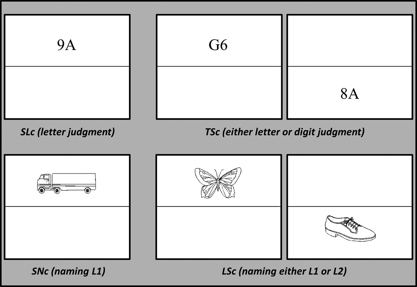
The experimental design used here was partially based on Abutalebi et al., Reference Abutalebi2008 and has been already used by Magezi et al., Reference Magezi, Khateb, Mouthon, Spierer and Annoni2012. This design, completed during one acquisition session, was composed of two experiments, one alphanumeric (control task) and one linguistic (experimental task), that all subjects performed in the same order. Both experiments required overt oral responses whilst in the scanner. The suitability of this design for the study was established by a previous electroencephalographic study that used this paradigm and reported differences in brain responses between linguistic and alphanumeric control task mechanisms (Magezi et al., Reference Magezi, Khateb, Mouthon, Spierer and Annoni2012). Each experiment comprised a simple non-selection context and a mixed selection context, which used a spatial cueing procedure based on Rogers and Monsell's work (Rogers & Monsell, Reference Rogers and Monsell1995). The stimuli were presented on a white screen divided by a horizontal black line aligned to the middle of the screen. The stimuli (see below) were presented above or below the line as illustrated in Fig. 1.
Examples of stimuli used during the experiment. The upper part shows the alphanumeric experiment and the bottom part shows the language experiment (see details in the text). SLc = simple letter context (run 1); TSc = task selection context (runs 2 and 3); SNc = simple naming context (run 4); LSc = language selection context (run 5 and 6). Each run was composed of 48 stimuli and lasted 4 min.

Alphanumeric experiment
The stimuli consisted of couples of letter-digit combinations with a counterbalanced position between letters and digits (e.g., 4B, B4). In the simple (non-selection) letter context (SLc), participants were asked to categorize orally the letter as a vowel or a consonant as fast and accurately as possible. This SLc was completed in one experimental run (~4 min) in which all stimuli appeared in the same half-screen position for the same subject (either the upper or the lower half of the screen). Having completed the SLc, the subject had to complete the experiment under the task selection context (TSc) conditions. In the TSc, the letter-digit couples appeared randomly either in the upper or lower half of the screen. For each stimulus presented, the participant was asked to categorize the letter (as vowel or consonant, exactly as in the SLc) and the digit as even or odd, depending on the position of the stimulus on the screen (these conditions are referred to hereinafter as “TSc-Let” and “TSc-Dig”). This TSc had a duration twice as long as the SLc and was thus completed over two experimental runs (2 × ~4 min).
Linguistic experiment
The language selection experiment respected the same principle and used black-and-white drawings of objects (manufactured objects: tools, clothes, kitchen objects, electric apparatus, vehicles, etc, and natural objects: animals, vegetables, fruits, body parts, etc). These pictures were selected from the databases of Alario et al. (Alario & Ferrand, Reference Alario and Ferrand1999), regrouping pictures from Cycowicz et al. (Snodgrass & Vanderwart, Reference Snodgrass and Vanderwart1980; Cycowicz, Friedman, Rothstein & Snodgrass, Reference Cycowicz, Friedman, Rothstein and Snodgrass1997). The images were chosen according to the highest possible name agreement value to avoid competition between several words within a language. Also, particular attention was paid to excluding images that elicited cognate names in the two languages of each combination (for instance French–German or French–English). In the simple naming context (SNc), participants were presented with pictures that they had to name in their L1. For the same participant, the pictures presented in the SNc always appeared in the same position (upper or lower half of the screen) as the letter–digit stimuli in the SLc. This SNc was completed over one experimental run (~4 min). After this, the participant undertook the experiment under the language selection context (LSc) conditions. Here, the pictures appeared randomly in either the upper or lower half of the screen; and, for each stimulus presented, the participant was asked to name the image in his/her L1 (exactly as in the SNc) or in his/her L2, depending on the position of the stimulus on the screen (conditions referred to hereinafter as “LSc-L1” and “LSc-L2”). The duration of the LSc was also twice as long as the SNc and hence was completed over two experimental runs (2 × ~4 min).
The stimuli within each context were presented after an automatic randomization for each subject to avoid any order effect. Notably, the experimental succession of conditions was identical between participants to force simple contexts to precede selection contexts, as well as to force the alphanumeric selection task behavioural register to precede the linguistic/language selection behavioural register. The simple contexts (SLc and SNc) were each composed of 48 different items and the selection/mixed contexts (TSc and LSc) were each composed of 96 different items (48 for TSc-Let and 48 TSc-Dig in TSc, and 48 for LSc-L1 and 48 for LSc-L2 in LSc). The subsets of 48 pictures in LSc were counterbalanced between L1 and L2 conditions across participants and group. This permitted us to avoid differences in naming difficulty due to lexical frequency known to affect the performance in picture naming (Mousikou & Rastle, Reference Mousikou and Rastle2015). The judgment associated with one spatial position of the stimuli was the same in the simple and mixed conditions for each participant to avoid spatial interference, and was counterbalanced across subjects. Thus, a subject who saw the letter–digit couple in the upper half of the screen during the SLc also had to categorize the letter in the upper part of the screen in the mixed TSc and the digit in the lower part of the screen. Inversely, if the subject saw the letter–digit couple in the lower half of the screen during the SLc, in the mixed TSc he/she had to categorize the letter in the lower part of the screen and the digit in the upper part of the screen. The same procedure was used for naming in the SNc and LSc.
Each trial started with a central fixation cross of 500 ms, followed by the presentation of the visual stimulus, which appeared for 300 ms, and finally by a blank screen with a pseudo-randomly varying duration of 2350, 2750, 3150, 3550, or 3950 ms, which yielded a mean trial duration of 3950 ms. After each trial, participants were instructed to speak softly and with minimal articulation to avoid head movements and to minimize distortion artifacts due to air flow during acquisition.
As already indicated, the whole experiment was composed of six experimental runs of approximately 4 min each with the same succession of contexts and experimental conditions for all subjects: one run for SLc followed by two runs for TSc, and one run for SNc in L1 followed by two runs for LSc. Each stimulus in each run was considered a separate event in the fMRI analysis. This was necessary because the analysis aimed to differentiate between letter and digit in the TSc runs, and between L1 and L2 naming in the LSc runs. In these mixed selection conditions, the stimuli were presented in random order, which was essential to characterize the brain activation related to each selection process. The task was explained at the beginning of each run, with a training session for the simple contexts, and a break of around 1 min was given after each experimental run. The whole experimental session lasted approximately 30 min in the scanner. The oral responses were recorded to assess the participants' accuracy during the experiment, and they were asked to articulate as quietly as possible with the microphone close to the mouth to avoid head and mouth movements during MRI acquisition known to blurred images (Jezzard & Clare, Reference Jezzard and Clare1999). The subjects participated in two experimental sessions with exactly the same protocol at an interval of at least one month. One session with MRI acquisition and another one outside of the MRI scanner with a EEG recording (see Magezi et al., Reference Magezi, Khateb, Mouthon, Spierer and Annoni2012). The EEG session permitted the collection of the participants' response times for the same tasks, as these were impossible to collect during fMRI acquisition (due to technical constraints). Half of the subjects started with the MRI session and the other half started with the EEG session.
All subjects were first tested in a monolingual context and then in the bilingual context. In detail, the succession of the conditions was: SLc, TSc, SNc, LSc. This choice was performed according to the “bilingual mode” theory. Grosjean distinguished two types of bilingual behaviour: when speaking to a monolingual person or when speaking to another bilingual person (Grosjean, Reference Grosjean1998). He proposed that bilinguals choose one language and deactivate (not totally) the other in monolingual mode (Grosjean, Reference Grosjean1985). In a bilingual context, they choose one language of reference and activate the other as a function of their need. When a bilingual speaks to a monolingual person, he forces himself to stay in the language of the receiver. These bilingual or monolingual modes could have an impact on the cognitive control mechanisms. This is why we decided always to perform the tasks in the same order: to have a comparable mental state between participants.
Experimental material
Visual stimuli were displayed on a screen using a video projector (Hitachi CP-X1200 with long focal distance, Hitashi LL-504, Hitachi Ltd., Tokyo, Japan) through a mirror system with a refresh rate of 60 Hz. The pictures were presented with a visual angle extending between 2.4° and 3.6° for the height and between 2.4° and 3.6° for the length. The stimuli were presented centrally at 2.71 cm above or below the middle of the screen. The E-Prime software (Psychology Software Tools, Inc, PA, USA) was used for stimulus presentation. Participants used a headphone linked to a microphone to give their oral responses. These were recorded via the audio system “DAP Center” from MRConfon (Magdeburg, Germany), which is equipped with a dual-channel noise cancellation device. Behavioural analyses were performed using the Statistica 12.0 software (StatSoft Inc, Tulsa, Okla).
MRI Acquisition
The experiments were conducted using a 3T MRI scanner (Siemens Magneton Trio, Siemens Medical Solutions, Erlangen, Germany) at the “Centre d'Imagerie BioMédicale” (CIBM, www.cibm.ch) of the University Hospital of Geneva (HUG). A high-resolution T1-weighted anatomical scan was recorded for each participant. Images were recorded on the coronal plane in a posterior to anterior direction (voxel size: 0.9 x 0.9 x 1.1 mm, number of slices: 192, repetition time (TR) = 2500 ms, echo time (TE) = 2.92 ms). Functional T2*-weighted images were acquired using a gradient-echo echo-planar imaging (GE-EPI) sequence. The blood oxygenation level-dependent contrast (BOLD) (Kwong, Belliveau, Chesler, Goldberg, Weisskoff, Poncelet, Kennedy, Hoppel, Cohen, Turner & et al., Reference Kwong, Belliveau, Chesler, Goldberg, Weisskoff, Poncelet, Kennedy, Hoppel, Cohen and Turner1992) was used as an index of local increases in brain activity. For each experimental run, 120 dynamic volumes with axial contiguous ascending acquisitions were obtained (voxel size: 2 x 2 x 3.5, matrix size: 120 × 120, number of slices: 29, interslice gap: 10%, TR = 2000 ms, TE = 30 ms, field of view: 240, flip angle = 85°) of 4 minutes. For each run, the functional scanning was always preceded by 6 s of dummy scans to ensure tissue steady-state magnetization. A B0 field inhomogeneity mapping sequence was applied to correct for geometrical distortion occurring along the phase-encoding direction with the same acquisition dimension parameters as the functional scan (# slices = 29, slice thickness = 3.5 mm, interslice gap = 10%, TR= 400 ms TE1 = 5.19 ms, TE2= 7.65 ms) (Jezzard & Clare, Reference Jezzard and Clare1999).
Data analysis
The behavioural results recorded for the LP and the HP during the experiment were compared for each experimental condition, using two sample t-tests for the reaction time (RT) and nonparametric Mann–Whitney U tests for response accuracy (as the latter was not normally distributed, checked with a Shapiro-Wilk test). A 2 × 2 × 2 ANOVA analysis of reaction time was performed using the behavioural register (alphanumeric vs. linguistic) and the context (simple vs. selection context) as within-subject factors (this analysis contains only the letter judgment and the L1 naming conditions: SLc, TSc-Let, SNc, LSc-L1), and the proficiency level in L2 as the between-subjects factor (i.e., grouping variable LP vs. HP). In addition, the effect of L2 proficiency was investigated in more detail with two separate 2 × 2 ANOVA for each selection register (TSc-Let vs. TSc-Dig for the first and LSc-L1 vs. LSc-L2 for the second). Finally, the language evaluation score was correlated with the behavioural results (using Pearson correlation for reaction time and Spearman's rank correlation for accuracy). Statistical significance was set at p < 0.05.
The MRI data were analyzed using the Statistical Parametric Mapping SPM12 software. All functional volumes were subjected to standard preprocessing procedures (Friston et al., Reference Friston, Ashburner, Kiebel, Nichols and Penny2007), including: unwarping (using the FieldMap2.1 toolbox; Andersson et al., Reference Andersson, Hutton, Ashburner, Turner and Friston2001), spatial realignment, slice timing, normalization (on the Montreal Neurological Institute (MNI) space with 2 × 2 × 2 mm3 voxel size), and smoothing with an isotropic 6 mm full-width at half-maximum (FWHM) Gaussian kernel. The ArtRepair toolbox was used on the preprocessed images to detect and interpolate volumes with fast motion (>0.5 mm/TR). One participant was omitted from the data analysis because more than 10% of the fMRI images needed to be interpolated. The combination of unwarping with interpolation of bad volumes with rapid movement has been validated by Birn et al. (Birn, Cox & Bandettini, Reference Birn, Cox and Bandettini2004) for the processing of fMRI involving verbal responses. Time series from each voxel were high-pass filtered (1/128 Hz cutoff) and the preprocessed functional volumes were then submitted to fixed-effect analysis (i.e., first-level analysis, FFX) using the general linear model applied to each voxel (Friston, Holmes, Worsley, Poline, Frith & Frackowiak, Reference Friston, Holmes, Worsley, Poline, Frith and Frackowiak1995; Worsley & Friston, Reference Worsley and Friston1995) and an auto-regressive [AR(1)] function to account for temporal correlations between them across the whole brain. Each stimulus onset was modelled as an event encoded in condition-specific “stick functions.” These stimulus functions were convolved with a canonical hemodynamic response function (with temporal and dispersion derivatives to increase the flexibility of the canonical hemodynamic response function modelled) (Friston et al., Reference Friston, Ashburner, Kiebel, Nichols and Penny2007).
To describe the brain activity induced by the task selection, a direct comparison was performed between TSc-Let and SLc (referred to hereinafter as the TSc contrast). A direct comparison was also made between LSc-L1 and SNc (hereinafter the LSc contrast) to determine brain activation related to language selection. To investigate differences between the two types of selection processes, an interaction analysis was performed between the LSc and TSc contrasts (referred to here as the LSc–TSc interaction). Indeed, this particular contrast highlighted regions that were more involved in language selection (by removing task selection components). These three contrasts were computed at the individual level (FFX). Afterwards, the contrasts of interest were sent to different second-level analysis (random-effects analysis, RFX) to generalize the results in the population (Friston et al., Reference Friston, Holmes, Price, Buchel and Worsley1999) with a ROI mask. This ROI included the bilateral PFC (including all Frontal_Sup, Frontal_Mid and Frontal_Inf masks), ACC, CN defined by the AAL atlas (Tzourio-Mazoyer, Landeau, Papathanassiou, Crivello, Etard, Delcroix, Mazoyer & Joliot, Reference Tzourio-Mazoyer, Landeau, Papathanassiou, Crivello, Etard, Delcroix, Mazoyer and Joliot2002) using WFU pickAtlas toolbox (Maldjian, Laurienti, Kraft & Burdette, Reference Maldjian, Laurienti, Kraft and Burdette2003). First of all, the LSc–TSc interaction was studied separately for each group (LP and HP) for illustration. This RFX model used a paired t-test between the two selection contexts. Then, this LSc–TSc interaction was compared between the LP vs. HP groups to study statistically the difference between them. The MRI statistics were studied at p FWE<0.05 family-wise error rate corrected for multiple comparisons at the peak level within the ROI. However, activations showed in figures were inflated using a p < 0.001 uncorrected statistic at peak level for visualization purpose. Anatomical locations were checked with the neuromorphometrics probabilistic atlas (Marcus, Wang, Parker, Csernansky, Morris & Buckner, Reference Marcus, Wang, Parker, Csernansky, Morris and Buckner2007; Worth & Tourville, Reference Worth and Tourville2014) provided in SPM12 and in the Talairach Daemon atlas (Lancaster, Rainey, Summerlin, Freitas, Fox, Evans, Toga & Mazziotta, Reference Lancaster, Rainey, Summerlin, Freitas, Fox, Evans, Toga and Mazziotta1997; Lancaster, Woldorff, Parsons, Liotti, Freitas, Rainey, Kochunov, Nickerson, Mikiten & Fox, Reference Lancaster, Woldorff, Parsons, Liotti, Freitas, Rainey, Kochunov, Nickerson, Mikiten and Fox2000) for Brodmann area labels. Contrast estimate illustrations were based on the voxel of local maxima of each cluster. All the coordinates derived from these analyses are given in the MNI space, with all illustrations using the neurological convention.
Results
Behavioural results
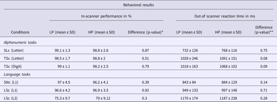
The subjects' in-scanner performance (rate of correct responses) during the fMRI session and their mean response times (RTs, i.e., voice onset time relative to the stimulus onset) collected outside the MRI session are presented in Table 1. Despite some small differences in L2 in favour of HP participants, the nonparametric Mann–Whitney U tests conducted on performance showed no significant difference between proficiency groups across the experimental conditions. In addition, the distribution of the accuracy in L2 naming for each group was normal (Shapiro–Wilk test p = 0.6 for the LP and HP groups) and no ceiling effect was observed. For the response times recorded outside the MRI scanner, parametric two-sample t-tests showed no difference between the LP and HP groups across experimental conditions, confirming that the two groups had quite good proficiency in L2.
Mean ± standard deviation (SD) of in-scanner performance and out-of-scanner response times for the different experimental conditions. The results are split according to group: low proficiency (LP) and high proficiency (HP). The results of statistical tests of difference between the groups are also reported (*Mann–Whitney U test, ** two-sample t-test). SLc = simple letter context; TSc = alphanumeric task selection context, including letter and digit conditions; SNc = simple naming context in L1; LSc = language selection context, including naming in L1 and L2.

The 2 × 2×2 ANOVA performed on the subjects' median reaction times during letter judgment and L1 naming used the behavioural register (alphanumeric vs. linguistic) and context (simple vs. selection) as within-subjects factors, and proficiency groups as a between-subjects factor. This revealed no effect of register, but a highly significant main effect of context (F(1,27) = 153.7, p < 0.05) driven by longer RTs in the selection/mixed contexts. In addition, a highly significant interaction (F(1,27) = 67.4, p < 0.05) was found between these factors, which could be attributed to the presence of a greater effect for mixing in the alphanumeric register (~310 ms) than in the linguistic one (~110 ms). No main effect or interaction with the proficiency groups was observed. In addition, the 2 × 2 ANOVA performed on the subjects' RTs during TSc, using the proficiency level as between-subjects factor and the condition (TSc-Let vs. TSc-Dig) as within-subjects factor, revealed no significant main factor or interaction between the factors. Finally, the 2 × 2 ANOVA conducted on the subjects' response times during LSc, again using proficiency as the between-subjects factor and condition (LSc-L1 vs. LSc-L2) as within-subjects factor, showed a highly significant main effect of conditions only (F(1, 27) = 48.5, p < 0.05) due to longer naming times in L2 than in L1, independent of language proficiency (see Table 1).
Of note here is the fact that the individual scores in the language assessment correlated positively with the subjects' in-scanner performance during LSc-L2 naming (Spearman ρ = 0.4, p = 0.04). No other correlations were found between these scores and the other (in and out of scanner) behavioural measures.
Functional results
Difference between LSc and TSc contrasts as an effect of proficiency
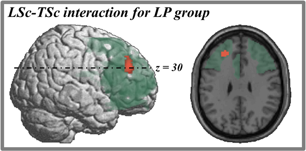
The main aim of this study was to describe the modulation by the level of proficiency of brain areas that play a role in language selection processes. These brain areas were highlighted by the comparison of brain activation induced during language selection (LSc contrast) and the alphanumeric task selection (TSc contrast), referred to as the LSc–TSc interaction. This comparison was studied separately for each proficiency group. For LP, the region localized in the left middle/superior frontal gyrus (x, y, z = −26, 38, 30; Z-score= 4.65) (around the Brodmann area 9, BA9) showed greater activation for LSc than TSc (Fig. 2). In the case of the HP group, no difference in brain activation was observed for this comparison.
In red color, activation map for the comparison between LSc and TSc contrasts only for low L2 proficiency (LP) participants in the middle frontal gyrus. In green color, illustration of the ROI region considered for statistical analysis. The axial section are located at the slices z = 30 (MNI coordinates). The activation map displayed are inflated regarding to studied statistical threshold (p < 0.05, FWE corrected at the peak level) for better visualization.

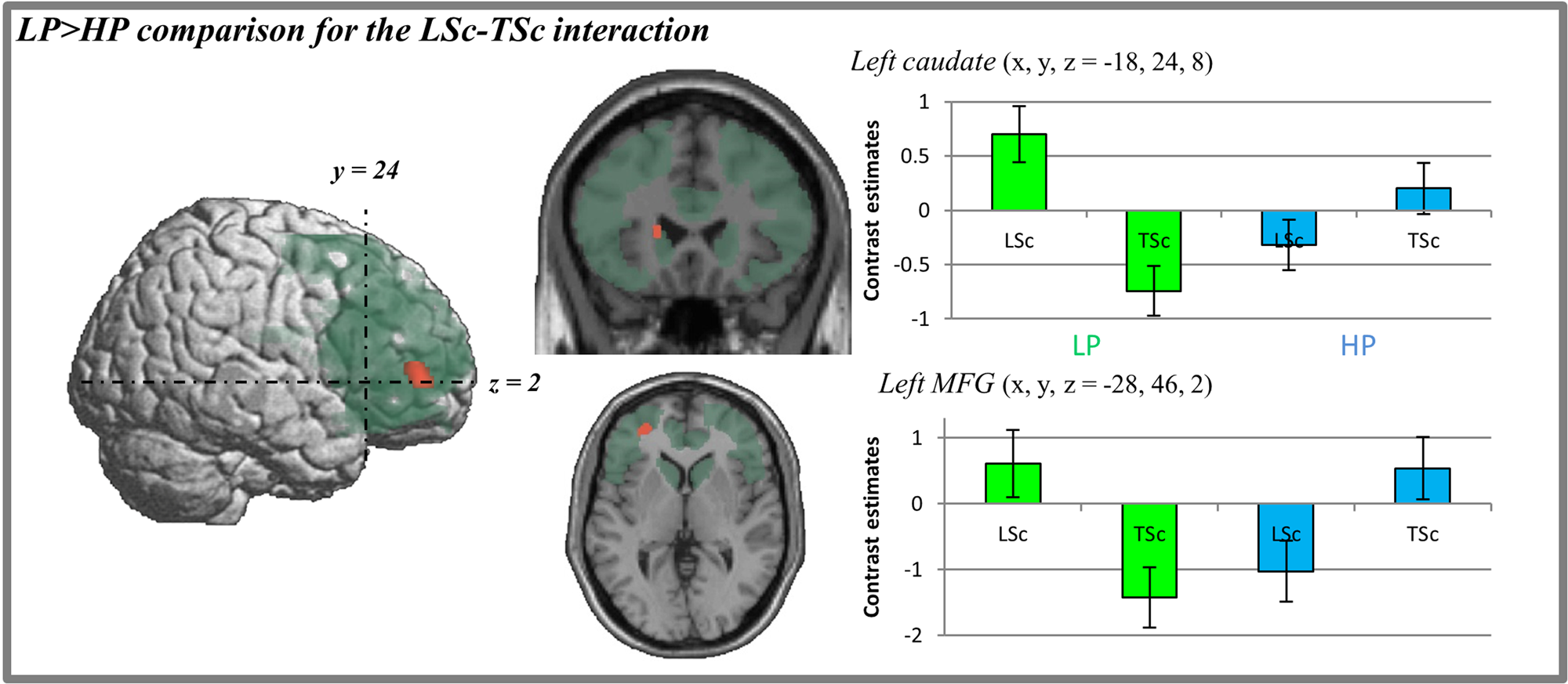
Direct comparison of the LSc–TSc interaction between the proficiency groups showed two brain regions to be more involved in the LP group than the HP group: the left caudate (x, y, z = −18, 24, 8; Z-score = 4.89) and the inferior part of the left middle frontal gyrus (x, y, z = −28, 46, 2; Z-score = 4.64) (around BA10) (Fig. 3). The right panel of Fig. 3 illustrates the contrast estimates (amplitude) in these regions. The opposite comparison did not show any significant brain area to be more involved for the HP group relative to the LP group.
In red color, activation map for the comparison between low- (LP) and high- (HP) proficiency bilinguals for the LSc–TSc interaction. In green color, illustration of the ROI region considered for statistical analysis. The right part of the figure illustrates the contrast estimates (amplitude) with standard error bars for each cluster's local maxima (localization provided in MNI coordinates). Green bars = LP group; Blue bars = HP group. The activation map displayed are inflated regarding to studied statistical threshold (p < 0.05, FWE corrected at the peak level) for better visualization. MFG = middle frontal gyrus.

Discussion
In this study, we sought to identify possible changes of activation in the bilingual brain during language selection processes according to the level of L2 proficiency. For this purpose, we compared fMRI responses in language selection contrast (LSc) and task selection contrast (TSc) experiments to reveal brain areas, the activity of which increases during the LSc relative to the TSc, and we compared activation in both proficiency groups. The particular strength of this study was the experimental population investigated, which was composed of university students studying translation. This allowed us to employ a population within a controlled language-training environment, in addition to a precise evaluation of proficiency by experts in the field. In addition, the split of participants according to their education level permitted us to have populations with distinct L2 proficiency. Indeed, the education level in translation improves proficiency in the second language and increases cognitive reserve (Lee-Jahnke, Reference Lee-Jahnke2005).
At the behavioural level, the subjects' performances in the language assessment confirmed a significant difference in L2 proficiency between the LP and HP groups. There were no differences in behavioural performance between the two groups, and the performance was near ceiling level (mean accuracy greater than 95%) in the alphanumeric conditions and in the L1 naming conditions. The L2 naming showed a normal distribution (no ceiling effect) and a slight difference between the groups but it failed to reach significance. However, individual performances in the LSc-L2 condition correlated positively with the participants' language scores. These findings suggest that the subjects' in-scanner performance and language assessment were consistent with the initial separation of the participants into LP and HP groups, and thus made this grouping suitable for assessing the effect of proficiency in functional analysis.
In terms of response speed, the subjects' out-of-scanner RTs during naming in L2 (LSc-L2) were significantly longer than in L1 (LSc-L1), but did not differ between the two proficiency groups (LP and HP). In contrast, the judgments of letters (TSc-Let) and digits (TSc-Dig) were performed at a similar speed by all participants. Response time analysis also showed a significant increase in the selection contexts (TSc and LSc) relative to the non-selection simple contexts (SLc, SNc). These differences, referred to here as mixing costs, are at least partially due to continuous switching between the two conditions of each of the selection contexts (Swainson, Jackson & Jackson, Reference Swainson, Jackson and Jackson2006). Such switching costs have often been attributed to an inhibition mechanism (Green, Reference Green1998; Meuter & Allport, Reference Meuter and Allport1999; Costa & Santesteban, Reference Costa and Santesteban2004; Verhoef, Roelofs & Chwilla, Reference Verhoef, Roelofs and Chwilla2009). They were higher in the alphanumeric than in the linguistic register, which presumably reflects the fact that bilinguals are more used to switching between L1 and L2 than to switching between letter and digit categorization. This interpretation might find some support in the fact that L1 naming in the LSc still exhibited the pattern of a dominant response, both in performance and RTs, while TSc-Let appeared to be as difficult to perform as TSc-Dig, despite the fact that letter categorization was relatively overtrained during the SLc.
At the neurophysiological level, our main objective was to assess the modulation of the language control system between low and high L2 proficiency bilinguals. This language selection network was highlighted by the comparison of L1 selection (LSc contrast) with a task selection (TSc contrast) to focus on the specific additional brain activity induced by language selection compared to general cognitive task selection. There were two different patterns for the LP and HP groups. Activation was found in the middle frontal gyrus (MFG) for the LP group (around BA9) (see Fig. 2), while there was no difference between language and task selection for the HP group. This divergence in patterns was confirmed by the direct comparison of the LSc–TSc interaction for the two groups. This highlighted a greater implication of the language selection network for LP than HP participants (see Fig. 3). Thus in our study, the general control system network was strongly associated to language control only in highly proficient subjects. As has been discussed in other studies (Calabria, Hernandez, Branzi & Costa, Reference Calabria, Hernandez, Branzi and Costa2011), linguistic and domain-general cognitive control are highly correlated but do not completely overlap. For example, data in bilinguals with aphasia, who have potential language control deficit, show dissociations between the mechanisms of language control (word-pair relatedness judgment task) and cognitive control (Flanker's task), thus providing evidence for domain specific cognitive control (Gray & Kiran, Reference Gray and Kiran2015). However, in this last study, the language control was implicated in lexical access and not in language selection. Our results support the hypothesis of executive control within bilingual young adults, but only when they were highly proficient. Actually, this advantage seems to be age-depend. In fact, bilingual children develop the ability to solve problems that contain conflicting cues at an earlier age than monolinguals (Zelazo, Frye & Rapus, Reference Zelazo, Frye and Rapus1996). In younger subjects, Bialystok and colleagues found a more efficient control (executive/inhibitory) system in bilinguals, demonstrated for example with the Simon task, while this difference was more difficult to demonstrate in adult populations (Bialystok, Craik & Luk, Reference Bialystok, Craik and Luk2008). In general, classical studies found an advantage in executive control advantage in bilingual children and elderly compared to monolinguals (Bialystok, Martin & Viswanathan, Reference Bialystok, Martin and Viswanathan2005) but results with the young adults are less consistent (Costa, Hernandez, Costa-Faidella & Sebastian-Galles, Reference Costa, Hernandez, Costa-Faidella and Sebastian-Galles2009; Paap & Greenberg, Reference Paap and Greenberg2013). For example, Bialystok et al. (Reference Bialystok, Martin and Viswanathan2005) reported a reaction time advantage to be bilingual during the Simon task for 5-years-old and older adults (60–80-years-old), but not in the case of younger adults (30–59-years-old). Our data, suggesting that proficiency can modify the relationship between language control network and general executive network, can bring additional insight into how variations in language practices and exposure affect bilinguals executive control skills.
This network was composed of the left MFG regions (at a lower position compared to previous described result), in addition to the left CN. Our results show evidence of a modulation of parts of the language control network, depending on language proficiency. Ghazi Saidi, Perlbarg, Marrelec, Pelegrini-Issac, Benali and Ansaldo (Ghazi Saidi et al., Reference Ghazi Saidi, Perlbarg, Marrelec, Pelegrini-Issac, Benali and Ansaldo2013) proposed a specific connectivity study to demonstrate the modulation of the connectivity between language network (defined by Price, Reference Price2010) and control network areas (chosen according to Abutalebi & Green, Reference Abutalebi and Green2007b’s work). This large analysis revealed that connectivity between both networks was actually depending on L2 proficiency. The results showed that, between and within network, integration levels decreased as proficiency for L2 increased. The authors suggested that this decrease in connectivity was due to L2 proficiency levels entailing effortful processes and attentional/executive resources, which would require high connectivity between language and control networks. These circuits become disengaged with increased L2 proficiency (Segalowitz & Frenkiel-Fishman, Reference Segalowitz and Frenkiel-Fishman2005; Abutalebi et al., Reference Abutalebi and Green2007b). This could explain why the control system becomes less active during language selection in more proficient bilinguals.
Although a complete discussion of the functional role of each of these areas implicated specifically in language control in LP subjects is beyond the scope of this study, we provide some elements in what follows.
Role of the middle frontal cortex
The difference in the LSc–TSc interaction between the LP and HP groups showed a distinct activation in the middle frontal gyrus. This area (including BA9, BA10) has already been reported to participate in language control processes (Price et al., Reference Price, Green and von Studnitz1999; Hernandez et al., Reference Hernandez, Martinez and Kohnert2000; Rinne, Tommola, Laine, Krause, Schmidt, Kaasinen, Teras, Sipila & Sunnari, Reference Rinne, Tommola, Laine, Krause, Schmidt, Kaasinen, Teras, Sipila and Sunnari2000; Hernandez et al., Reference Hernandez, Dapretto, Mazziotta and Bookheimer2001; Rodriguez-Fornells et al., Reference Rodriguez-Fornells, van der Lugt, Rotte, Britti, Heinze and Munte2005), but also in domain-general cognitive control tasks, such as conflict resolution and reorienting attention (Lepsien & Pollmann, Reference Lepsien and Pollmann2002; Fan, Flombaum, McCandliss, Thomas & Posner, Reference Fan, Flombaum, McCandliss, Thomas and Posner2003; Badre & Wagner, Reference Badre and Wagner2004; Gilbert, Spengler, Simons, Steele, Lawrie, Frith & Burgess, Reference Gilbert, Spengler, Simons, Steele, Lawrie, Frith and Burgess2006). Recently, Branzi et al. found a similar left prefrontal activation for linguistic and domain-general cognitive control, but did not specifically question whether proficiency modulated the language control system (Branzi et al., Reference Branzi, Della Rosa, Canini, Costa and Abutalebi2016). Our data suggest a differential activation of this left prefrontal structure network used for linguistic and domain-general cognitive control, which depends on the language proficiency of bilinguals. Such a difference could be indicative of a higher need for cognitive resources in the case of LP to make the correct choice of language, although no differences were observed in the response times or accuracy of the two proficiency groups. In line with this interpretation, it has been shown that proficient bilinguals perform language switches with less cognitive effort and costs because they are more highly trained and efficient than monolinguals in performing switching tasks (Costa, Hernandez & Sebastian-Galles, Reference Costa, Hernandez and Sebastian-Galles2008). Also, mechanisms for lexical searching in the left middle frontal region (for a review, see Price, Reference Price2010) have shown greater activation when language proficiency is low and less automatic (Petrides, Reference Petrides, Roberts, Robbins and Weiskrantz1998; see also Wagner, Pare-Blagoev, Clark & Poldrack, Reference Wagner, Pare-Blagoev, Clark and Poldrack2001; Badre, Poldrack, Pare-Blagoev, Insler & Wagner, Reference Badre, Poldrack, Pare-Blagoev, Insler and Wagner2005; Kostopoulos & Petrides, Reference Kostopoulos and Petrides2008). Comparable results were reported by Stein et al. (Stein, Federspiel, Koenig, Wirth, Lehmann, Wiest, Strik, Brandeis & Dierks, Reference Stein, Federspiel, Koenig, Wirth, Lehmann, Wiest, Strik, Brandeis and Dierks2009). Differences related to proficiency in the activation of left prefrontal areas might also reflect the increased role played by the brain's executive network in solving cognitive conflicts caused by the challenging mixed context (Braver et al., Reference Braver, Barch, Gray, Molfese and Snyder2001; Carter & van Veen, Reference Carter and van Veen2007; Wang et al., Reference Wang, Xue, Chen, Xue and Dong2007; Friesen, Chung-Fat-Yim & Bialystok, Reference Friesen, Chung-Fat-Yim and Bialystok2015).
Roles of the caudate nucleus
The main functional result of this study (Fig. 3) highlighted that the left caudate was more activated in the LSc–TSc interaction for the LP group than the HP group. Such a result is not new. It has been proposed that the left caudate plays a critical role in the process of selecting the correct language (Abutalebi et al., Reference Abutalebi, Della Rosa, Ding, Weekes, Costa and Green2013). In line with this view, Friederici (Reference Friederici2006) proposed that the recruitment of the left caudate was related to the involvement of nonautomatic processes, and thus to increasing levels of control needs. Therefore, the caudate may not specifically be recruited by language processing, but more by important cognitive requirements: such as planning, selecting, and executing appropriate plans to achieve goals (Saint-Cyr, Taylor & Nicholson, Reference Saint-Cyr, Taylor and Nicholson1995; Grahn et al., Reference Grahn, Parkinson and Owen2008). The higher contribution of the left caudate in LP participants could come from higher competition/selection between the L1 and L2. More resources are needed to select the correct language in the LP case because there is a greater difference in proficiency between L1 and L2 than in the HP case. Several other studies have shown increased left caudate activity in challenging language selection contexts (Abutalebi et al., Reference Abutalebi, Brambati, Annoni, Moro, Cappa and Perani2007a; Abutalebi et al., Reference Abutalebi2008; van Heuven et al., Reference van Heuven, Schriefers, Dijkstra and Hagoort2008; Ali et al., Reference Ali, Green, Kherif, Devlin and Price2009). It has been proposed that the ACC is associated with left caudate activity during language selection (Kerns, Cohen, MacDonald, Cho, Stenger & Carter, Reference Kerns, Cohen, MacDonald, Cho, Stenger and Carter2004; van Veen & Carter, Reference van Veen and Carter2005; Abutalebi et al., Reference Abutalebi, Della Rosa, Ding, Weekes, Costa and Green2013; Branzi et al., Reference Branzi, Della Rosa, Canini, Costa and Abutalebi2016). Considering this interpretation, we were expecting to find greater activation for the LP than the HP in the ACC, which was not the case. This has also been noted in previous studies (Abutalebi et al., Reference Abutalebi, Della Rosa, Ding, Weekes, Costa and Green2013). Recent data-driven hypotheses suggest that the ACC has a specific role in monitoring the optimal allocation of control by determining the expected value of control needed (EVC hypothesis) (Shenhav, Cohen & Botvinick, Reference Shenhav, Cohen and Botvinick2016). Such expected value of control would not be specifically influenced by L2 proficiency, and this hypothesis would explain why ACC was not sensitive to L2 proficiency in language control processes.
It can be noted that the studies which compare bilingual interpreters with a lower L2 proficiency population showed modifications in functional activation (Hervais-Adelman, Moser-Mercer & Golestani, Reference Hervais-Adelman, Moser-Mercer and Golestani2015) and gray matter volume (Elmer et al., Reference Elmer, Hanggi, Meyer and Jancke2011) more localized on the right side of the caudate nucleus, instead of the present left result. This difference could be explained by the fact that simultaneous interpretation involves a more complex language selection than standard bilingual tasks. Such activity should train and demand additional brain networks.
Summary
The modulations with L2 proficiency of these three areas implicated in cognitive control and error monitoring suggest that the mechanisms of language control evolve with the increase in proficiency. During acquisition of a second language, the brain needs to recruit supplementary resources to manage the challenge. These resources rely on a neural network that is larger than that used to manage any general cognitive conflict. When proficiency becomes sufficiently high, the cognitive control network can manage language selection using the same cognitive resources as for any other domain-general cognitive selection task. This would suggest that these differences in patterns are due to a modulation of activity of language control networks within a more domain-general cognitive control system. Most previous neuroimaging studies (e.g., Khateb, Abutalebi, Michel, Pegna, Lee-Jahnke & Annoni, Reference Khateb, Abutalebi, Michel, Pegna, Lee-Jahnke and Annoni2007) compared brain activity during a linguistic task performed in mixed- and single-language contexts. As both conditions involve the same or similar tasks, such a design would not allow direct comparisons across cognitive control in different domains. In contrast, the present study included different “alphanumeric” tasks. If there were brain regions specialized for language control not involved in domain-general cognitive control, this difference would have remained even in the HP group. This idea has previously been proposed in the literature (for review, see Abutalebi, Reference Abutalebi2008) based on Green's convergence hypothesis (Green, Reference Green, van Hout, Hulk, Kuiken and Towell2003), which claims that language representations should converge and the neural differences should disappear as proficiency increases. Indeed, our results are coherent with this hypothesis and confirm that L2 proficiency has a major impact on the neural resources needed to manipulate two languages, in accordance with other studies (e.g., Green, Crinion & Price, Reference Green, Crinion and Price2006; Consonni, Cafiero, Marin, Tettamanti, Iadanza, Fabbro & Perani, Reference Consonni, Cafiero, Marin, Tettamanti, Iadanza, Fabbro and Perani2013; Goral, Naghibolhosseini & Conner, Reference Goral, Naghibolhosseini and Conner2013; Bice & Kroll, Reference Bice and Kroll2015; White, Malt & Storms, Reference White, Malt and Storms2016).
This study suffered from a number of limitations, such as the difference in terms of age between the two groups of participants. This issue could be of importance for the interpretation of results, because some previous works have suggested that certain brain structures might evolve between the ages of 20–30 years (Sowell, Peterson, Thompson, Welcome, Henkenius & Toga, Reference Sowell, Peterson, Thompson, Welcome, Henkenius and Toga2003; Westlye, Walhovd, Dale, Bjornerud, Due-Tonnessen, Engvig, Grydeland, Tamnes, Ostby & Fjell, Reference Westlye, Walhovd, Dale, Bjornerud, Due-Tonnessen, Engvig, Grydeland, Tamnes, Ostby and Fjell2010). However, this difference was inherent to our choice to investigate proficiency effects in a population of future professional translators at different stages of their academic formation (Bachelor/Master). This choice aimed to maximize the difference in proficiency between the two groups while at the same time allowing control of their academic environment. The fact that the age of the participants was not regressed out of the analysis due to the relatively small number of participants in each group (which per se is another limitation), although comparable to many other fMRI investigations, limits the power of the calculations. The missing proficiency evaluation for four participants is also an issue. However, excluding these four participants yielded similar results in the left caudate nucleus, in line with the main result presented in this paper. Unfortunately, the relatively small number of students in the translation school did not allow us to recruit more participants in each of the groups. This also explains why different L1–L2 combinations were used, but we cannot rule that this last point could interfere with our general results. A related issue is the possible effect of the “linguistic distance” between each of the L1-L2 pairs studied here on the engagement of the language control system, a topic that has to date been scarcely studied. A recent clinical observation might support the fact that the distance between languages may influence language control (Radman, Spierer, Laganaro, Annoni & Colombo, Reference Radman, Spierer, Laganaro, Annoni and Colombo2016); however, these data have been obtained with very distant language pairs (Persian–French) and not with nearer European languages. Moreover, it is of interest that despite the absence of differences between the groups at the behavioural level (performance and reaction time), the functional analysis was still able to show that activation patterns were influenced by the difference in proficiency between groups, and by their ability to retrieve lexical information and manage the use of their two languages. These findings indicate that the analysis of brain activity using fMRI enables the detection of differences in processing that are not evident from behavioural measures alone, and thus potentially leads to a more comprehensive understanding of the underlying neural processes. Indeed, even with equal behavioural performances, the changes in activation in anticipated ROI showed that different brain strategies are actually being used for a similar result. This type of result has also been reported in other bilingual studies (e.g., Ansaldo et al., Reference Ansaldo, Ghazi-Saidi and Adrover-Roig2015; Reverberi et al., Reference Reverberi, Kuhlen, Seyed-Allaei, Greulich, Costa, Abutalebi and Haynes2018). Measures of brain activity could be even more valuable in situations such as that encountered here, in which the processing of stimuli does not lead to differences in performance in spite of differences in language proficiency/formational level and age. Future studies assessing the effects of proficiency in L2 on language control processes, with larger samples and without other confounds, such as age, are still necessary to provide greater clarification on this issue. Finally, the choice of the picture to name should be better controlled regarding homophone and word frequency, which could have an effect on the lexical representation (Caramazza, Costa, Miozzo & Bi, Reference Caramazza, Costa, Miozzo and Bi2001; Mousikou & Rastle, Reference Mousikou and Rastle2015).
Conclusion
This study has implications for research in language selection. We showed that L2 language proficiency has a significant effect on the brain network recruited during language selection. The language selection network converged with the cognitive control network involved in domain-general cognitive selection when the difference between L1 and L2 proficiency decreased. Future research has to take this point into consideration by selecting homogeneous populations in terms of L2 proficiency. The results of this study lend some support to the hypothesis that convergence between language selection network and domain-general cognitive control system depends on numerous biographic and linguistic specificities among bilingual individuals.
Acknowledgements
This research was supported by the Swiss National Science Foundation grants no’ 325100-118362 and 320030-125196, by the Israeli Science Foundation grant no' 623/11 (to A.K.) and finally by the Center for Biomedical Imaging (CIBM) of the Geneva-Lausanne Universities, the Geneva-Lausanne University hospitals and the EPFL. We thank Dr Stéphane R. Simon for his contribution in MRI data acquisition and Dr. Tatiana Aboulafia-Brakha for her contribution to the task. We thank Dr. Sebastian Dieguez for his reading and his suggestions. Finally, we thank all the bilingual participants for their precious collaboration.