Introduction
Kartagener’s syndrome (KS) is a rare subgroup of primary ciliary dyskinesia, with an incidence of 1 per 20,000–30,000 live births. Reference Abbasi, Suleman and Ahmed1 It is an autosomal recessive hereditary disorder characterised by genetic heterogeneity. Reference Leigh, Pittman and Carson2 Clinically, KS is defined by the classic triad of chronic sinusitis, bronchiectasis, and situs inversus. Reference Knowles, Zariwala and Leigh3 The association between bronchiectasis, chronic sinusitis, and situs inversus was first described by Siewert in 1904. Reference Siewert4 Later, in 1933, Kartagener identified this congenital condition and established its clinical triad. Reference Kartagener5
Normal ciliary activity is essential for sperm motility, mucociliary clearance in the respiratory system, and correct left-right axis determination during embryonic development. Mutations in genes such as DNAI1 and DNAH5 in KS result in impaired ciliary motility, predisposing individuals to recurrent sinopulmonary infections, infertility, and left-right asymmetry defects. Reference Tadesse, Alemu, Silamsaw and Gebrewold6 Males and females are equally affected. Reference Casanova, Tuji, Yoo and Haiter-Neto7
The clinical manifestations of KS usually appear in childhood and include chronic rhinitis due to pansinusitis, persistent cough, wheezing, and dyspnoea due to recurrent respiratory infections and bronchiectasis. Otitis media-related hearing impairment is also common. Reference Shapiro, Zariwala and Ferkol8,Reference Mishra, Kumar and Jaiswal9
The recommended diagnostic approach for KS includes nasal nitric oxide measurement, ciliary biopsy with electron microscopy, high-speed videomicroscopy for ciliary beat/waveform analysis, and genetic testing. Reference Shapiro, Zariwala and Ferkol8
Management of KS primarily involves daily chest physiotherapy for airway clearance, antibiotics for acute respiratory exacerbations, and routine vaccinations. Additional therapies may include long-term suppressive antibiotics, inhaled hyperosmolar agents to enhance mucociliary clearance, and bronchodilators. Reference Shapiro, Zariwala and Ferkol8 Lung transplantation is considered in cases of end-stage KS with respiratory failure. Reference Leigh, Pitman and Carson10
While pulmonary hypertension (PH) is a recognised complication of chronic respiratory disease, severe forms are uncommon in patients with stable lung function. Recently, PH was reported in a patient with KS carrying a novel homozygous nonsense mutation in the CCDC40 gene. Reference Dai, Wan, Guang and Zhang11
To our knowledge, this is the first report describing the association of KS with both aortic coarctation (CoAo) and pulmonary hypertension.
Case report
We present the case of a 17-year-old female patient diagnosed with KS, situs inversus, and aortic coarctation, which was successfully repaired at two months of age.
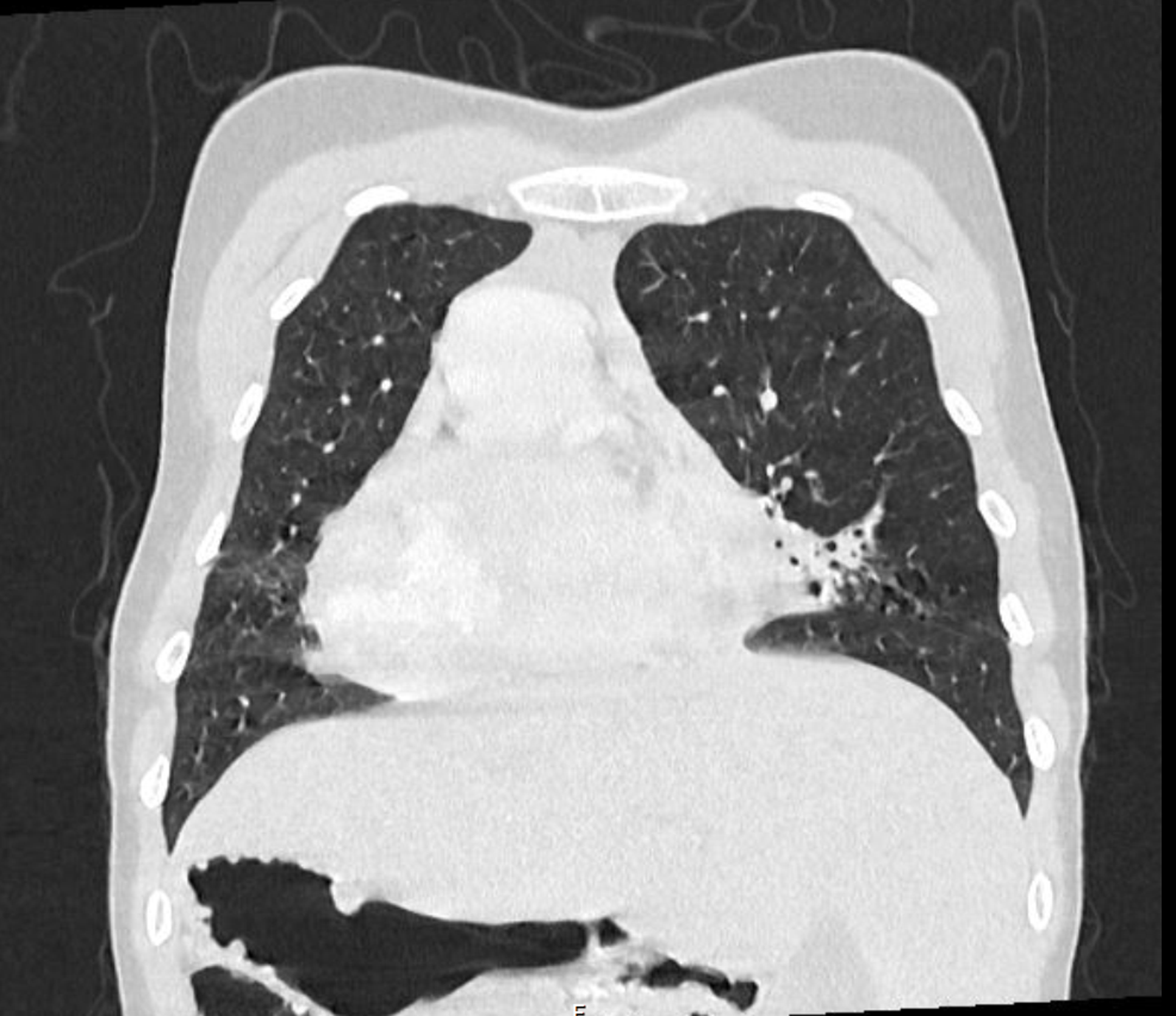
At the age of 12, she was asymptomatic except for a mild chronic cough and nasal congestion. During follow-up, PH was detected by echocardiography, with an estimated pulmonary artery systolic pressure (PASP) of 70 mmHg. Right heart catheterisation (RHC) revealed pulmonary artery pressure of 58/10/24 mmHg, without significant gradient at the aortic isthmus. Thoracic computed tomography (CT) (Figure 1 and 2) demonstrated multiple bronchiectasis in the upper lobes bilaterally, while pulmonary function tests (spirometry, plethysmography, and 24-hour oximetry) were normal. Genetic testing identified a pathogenic variant (c.1852C >T,p.(Arg618*)-rs778780449) and another variant of uncertain significance (c.5290T >C, p.(Ser1764Pro)-rs748763552) in the DNAH5 gene, confirming the diagnosis of KS with PH.
Thoracic CT scan: bronchiectasis.

Thoracic CT scan: bornchiectasis.

The patient started on inhaled glucocorticoids and chest physiotherapy. She remained clinically stable, with no episodes of pneumonia or acute exacerbations of bronchiectasis. Annual lung function tests remained within normal limits, and serial echocardiograms showed no significant changes. At the age of 16, thoracic CT demonstrated no progression of lung disease.
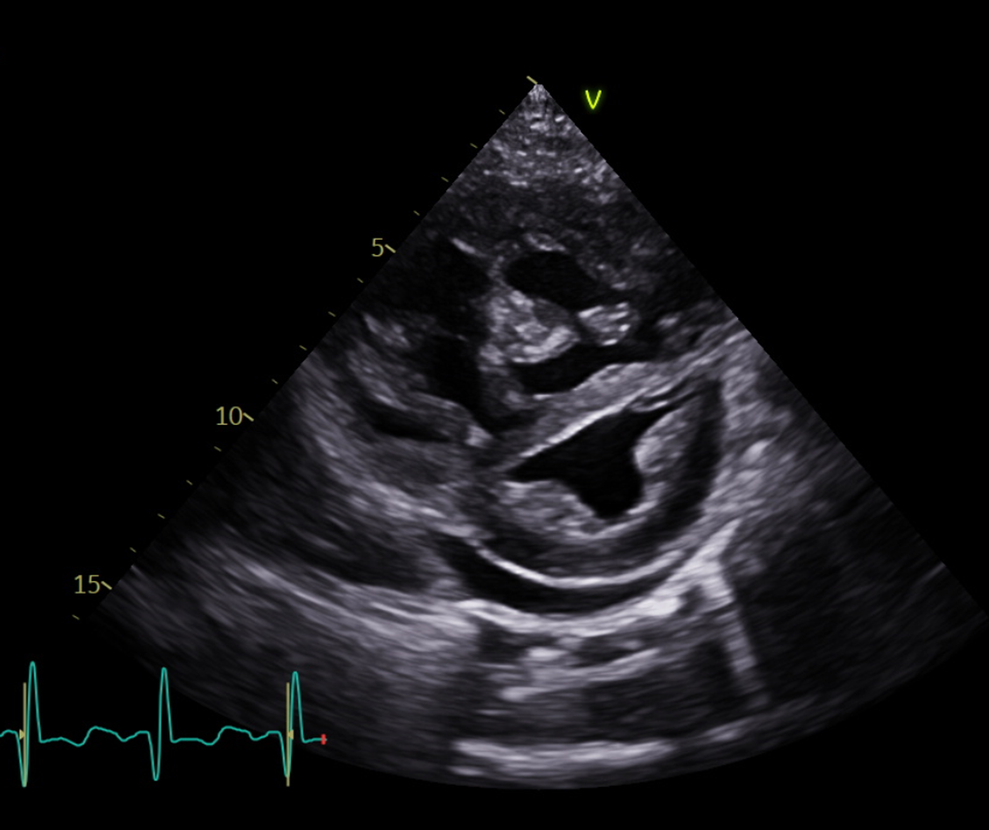
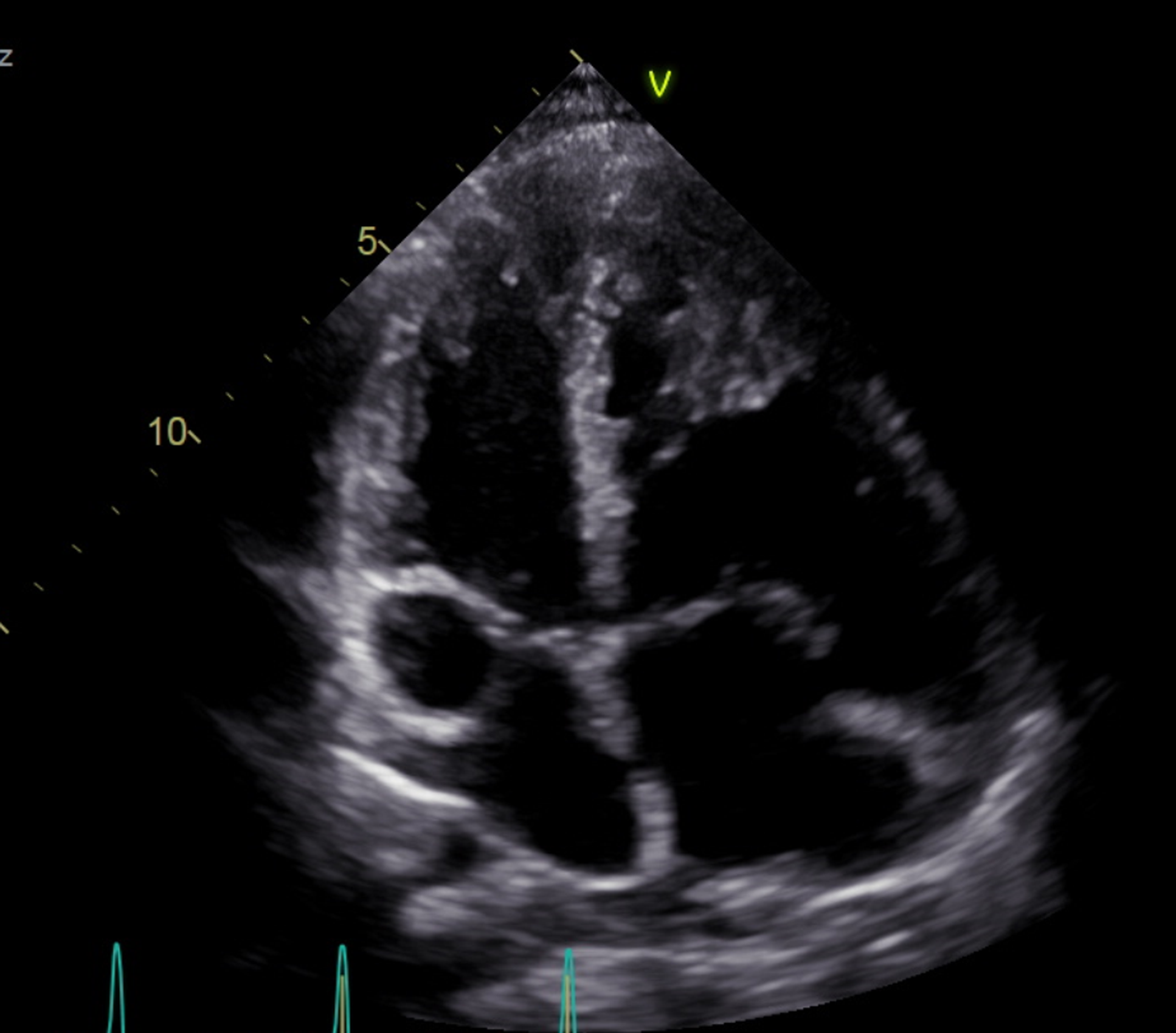
However, at 16 years of age, she developed significant physical activity limitation. Echocardiography revealed right ventricular (RV) dilatation (Figures 3 and 4), reduced longitudinal function (TAPSE 15 mm, Z-score -4.4), suprasystemic PASP (120 mmHg), an eccentricity index of 2.3, normal left ventricular (LV) function, and moderate pericardial effusion (PE). NT-proBNP was markedly elevated, 2292 pg/mL.
Echocardio: right ventricle hipertrophy.

Echocardio: situs inversus, RV hypertrophy.

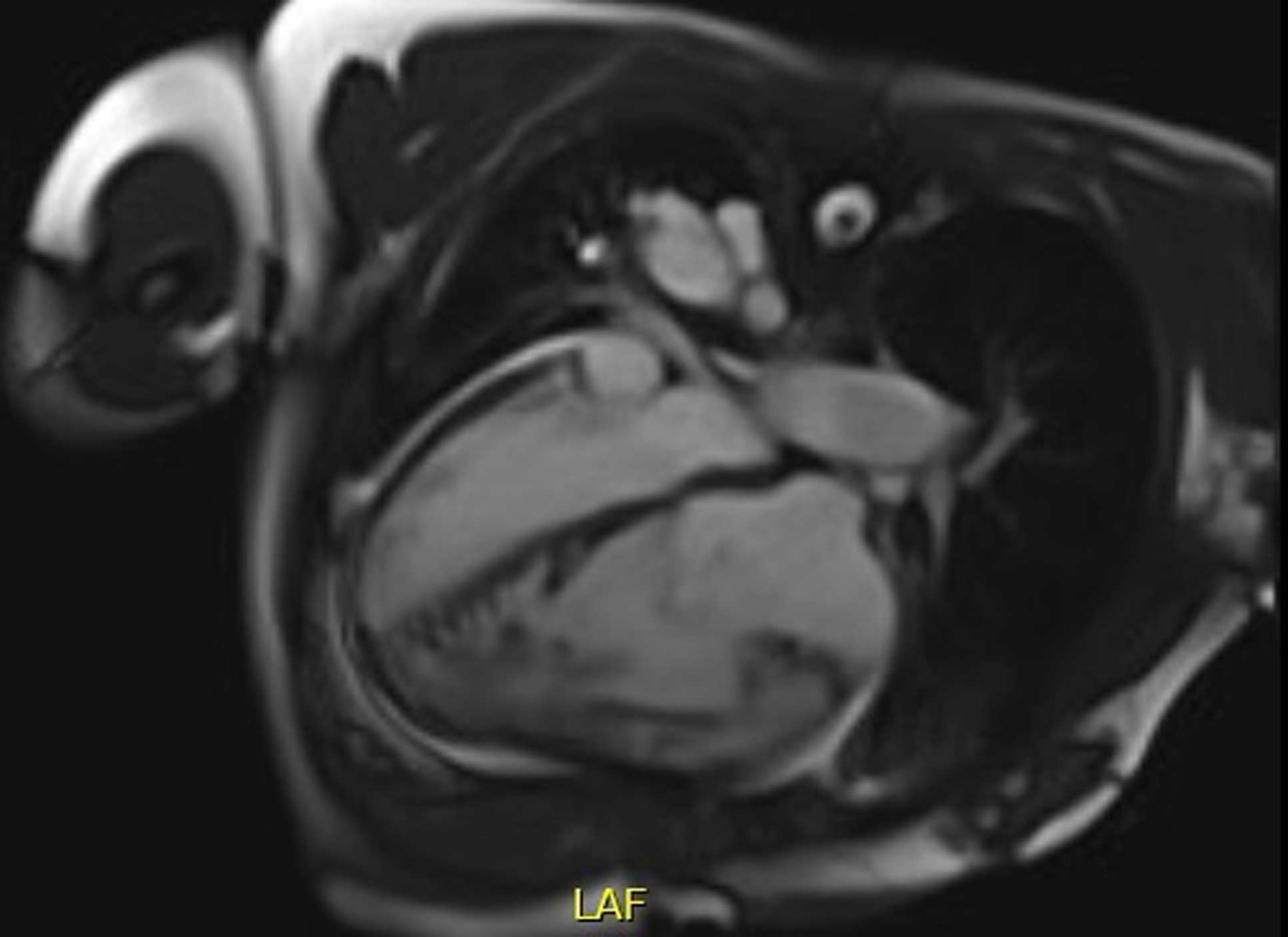
Cardiac magnetic resonance (MR) angiography (Figures 5, 6, and 7) confirmed situs inversus totalis and normal pulmonary venous return. A bilateral superior vena cava (SVC) was identified, with the right-sided SVC draining into a dilated coronary sinus (30 mm). Additionally, an interruption of the infrahepatic inferior vena cava (IVC) was observed, with venous return continuing via the azygos system, which drained superiorly into the right-sided SVC.
Cardiac MR—right ventricle hiperrophy.

Cardiac MRpulmonary artery (PA) dilatation.

Cardiac MR—central PA dilatation.

The pulmonary artery (PA) root and trunk were markedly dilated, measuring 45 x 42 mm and 50 x 50 mm, respectively. The main pulmonary arteries were confluent, approximately symmetrical, and both significantly dilated. A right aortic arch with a mirror-image branching pattern of the supra-aortic trunks was also identified. The thoracic aorta had a preserved calibre, with no evidence of recoarctation.
The RV was markedly dilated (end-diastolic volume index: 241 ml/m2), hypertrophied, and exhibited myocardial hypertrabeculation. There was a moderate reduction in systolic function (ejection fraction EF: 31%) due to global hypokinesia, which was most pronounced in the free wall at the basal and mid-segments. The LV was neither dilated (end-diastolic volume index: 71 mL/m2) nor hypertrophied, with systolic function at the lower limit of normal (EF: 45%).
A flattening of the interventricular septum was noted throughout the cardiac cycle, indicative of RV pressure overload. Late gadolinium enhancement was observed in the anterior and inferior junctional myocardium, consistent with the clinical presentation.
The right atrium was dilated (A4C 17cm2/m2), whereas the left atrium had normal dimensions. There was severe tricuspid regurgitation (regurgitant volume: 74 mL, regurgitant fraction: 57%) and a mild-to-moderate pericardial effusion, predominantly inferior, with a maximum thickness of 19 mm. The spleen could not be definitively identified. Additionally, traction bronchiectasis was observed in the left middle lobe, with associated subsegmental atelectasis.
The patient was initiated on combination target PH therapy, including an endothelin receptor antagonist, a phosphodiesterase type 5 inhibitor, diuretics, and supplemental oxygen. A significant clinical improvement was observed, with improvement in functional class (WHO-FC IIIa to I), echocardiographic parameters (PSAP 80 mmHg, TAPSE 16 mm, resolution of PE), and NT-proBNP reduction (171 pg/mL).
She remained clinically and haemodynamically stable for approximately one year. However, further deterioration occurred, prompting an angio-CT scan, which identified a partial anomalous pulmonary venous return (PAPVR), with the left superior lobar vein draining into the right atrium (Figure 8).
Angio CT scanpartial anomalous pulmonar venous return.

Electrocardiogram (ECG) (Figure 9) and Holter monitoring showed no arrhythmias.
Situs inversus (dextrocardia): ECG with reversed left-right leads and precordial scans performed on the right.

A new genetic panel for PH was performed, yielding negative results (Table 1).
Complete list of analysed gene

HGNC = Hugo Gene Nomenclature Committee; LGR = Locus Reference Genomic; RefSeq = NCBI Reference Sequence; EBI = European Bioinformatics Institute; OMIM = Online Mendelian Inheritance in Man.
Given the progressive nature of her condition, her PH therapy was intensified: bosentan was switched to macitentan, sildenafil was replaced with tadalafil, and a prostacyclin receptor agonist (selexipag) was added. Despite this, she met criteria for further escalation (NT-pro BNP: 1152 pg/mL; 6-minute walk test: 480 m), leading to repeat RHC.
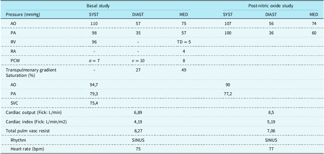
RHC (Table 2) confirmed precapillary PH with normal right atrial mean pressure and preserved cardiac index. Acute vasoreactivity testing with inhaled nitric oxide (30 p.p.m.) was negative.
RHC results

AO = Aorta; PA = Pulmonary Artery; RV = Right Ventricle; TD = end-dyastolic; RA = Right Atrium; PCW = Pulmonary Capillary Wedge Pressure; a = a wave; v = v wave; SVC = Superior Vena Cava; TOTAL PULM VASC RESIST = Total Pulmonary Vascular Resistance.
Discussion
The rapid progression of PH in this patient, despite normal lung function and unimpressive thoracic CT findings, is atypical for PH associated with lung disease. Extensive investigations for alternative causes of pulmonary arterial hypertension, including genetic testing, have thus far been negative.
Although a partial pulmonary venous return anomaly (PAPVR) was identified, its haemodynamic impact appeared minimal (Qp:Qs = 1.09, indicating low shunt flow).
Additionally, the patient had a history of successfully repaired aortic coarctation (CoAo). While CoAo is a congenital defect affecting 3–4 per 10,000 live births and is generally considered surgically correctable, long-term cardiovascular complications remain a concern. Despite successful repair, patients frequently develop systemic hypertension, increased risk of coronary artery disease, stroke, and overall higher mortality rates. Reference Choudhary, Canniffe, Jackson, Tanous, Walsh and Celermajer12,Reference Lee, Babu-Narayan and Kempny13 Emerging evidence suggests that these outcomes may be due to an underlying diffuse vasculopathy rather than solely residual aortic obstruction.
Even in cases with mild CoAo, increased arterial stiffness and systemic vascular dysfunction contribute to elevated left ventricle (LV) afterload, leading to LV hypertrophy, reduced compliance, and diastolic dysfunction. The extent of LV hypertrophy has been shown to correlate with echocardiographic markers of diastolic dysfunction. Reference Egbe, Reddy, Obokata and Borlaug14,Reference Egbe, Miranda and Connolly15 Furthermore, foetal LV remodelling may predispose these patients to a more globular LV shape and impaired diastolic function in later life. Reference Soveral, Crispi and Walter16
Increased LV filling pressures and decreased left atrial compliance ultimately contribute to elevated pulmonary resistance and pulmonary arterial pressure. Reference Egbe, Miranda, Connolly and Borlaug17 However, the impact of this type 2 PH on right ventricular function in post-CoAo repair patients remains poorly studied.
Although the newly identified PAPVR alone does not seem to fully explain the severe progression of PH in this case, its presence—when considered alongside other potential contributors, such as underlying lung disease and a history of CoAo—may have played a role in disease progression.
Given the aggressive course of the disease and the lack of an alternative definitive aetiology, idiopathic PAH remains a possible diagnosis.
Few clinical reports have described an association between KS and PH, and those that exist primarily involve adult patients. Reference Dai, Wan, Guang and Zhang11,Reference Shen, Wen and Yi18,Reference Ghewade, Cladius, Chaudhari and Bhake19,Reference Wang, Zhang and Jiang20
Conclusion
A more aggressive monitoring strategy for PH should be considered in young patients with Kartagener’s syndrome, congenital heart defects, and coincidental PH to enable early and effective therapeutic escalation when needed, while carefully considering emerging treatment options.
A multidisciplinary approach, involving primary care physicians, pulmonologists, cardiologists, radiologists, nurses, physiotherapists, and psychologists, is essential to optimise patient outcome.