The relationship between acute haemorrhagic encephalomyelitis (AHEM) also known as acute haemorrhagic leukoencephalitis (AHLE) and myelin oligodendrocyte glycoprotein antibody-associated disease (MOGAD) is garnering increasing attention. Reference Skarsta, Nicoletti and Frick1,Reference Bang, Kim and Seok2 AHEM is a severe, rapidly progressive demyelinating disease affecting the white matter within the central nervous system (CNS) and is often considered a severe variant of acute disseminated encephalomyelitis (ADEM). Reference Grzonka, Scholz and De Marchis3 Unlike typical ADEM, AHEM predominantly affects adults and confers significantly poorer outcomes. Reference Grzonka, Scholz and De Marchis3 Its precise aetiology remains unclear but is presumed to be of autoimmune origin. Reference Grzonka, Scholz and De Marchis3
Recent studies have detected anti-myelin oligodendrocyte glycoprotein auto-antibodies (MOG-Abs) in the serum of AHEM patients, sparking discussions about a potential link between MOGAD and AHEM. Reference Skarsta, Nicoletti and Frick1,Reference Bang, Kim and Seok2 MOGAD constitutes a syndrome of acquired CNS autoimmune demyelinating disorders purportedly driven by MOG-Abs, distinct from multiple sclerosis and aquaporin-4 (AqP4)-seropositive neuromyelitis optica spectrum disorder (NMOSD). Reference Banwell, Bennett and Marignier4 Clinical manifestations of MOGAD are heterogeneous but are classically associated with ADEM (in children), optic neuritis or transverse myelitis. Reference Banwell, Bennett and Marignier4 Patients may also experience brainstem, cerebellar or cortical encephalitis symptoms. Reference Banwell, Bennett and Marignier4 More recently, adult-onset AHEM has been suggested as an emerging MOGAD phenotype. Reference Skarsta, Nicoletti and Frick1,Reference Bang, Kim and Seok2
Here we report a case of adult-onset AHEM in a patient with serum MOG-Ab positivity. Our findings add to the developing discussion between AHEM and MOGAD while also delving into the pathogenic relevance of MOG-Abs and the diagnostic complexities of MOGAD.
A previously healthy 35-year-old man presented to a rural hospital with sudden-onset paraplegia, sensory loss up to T10 and urinary retention, following an upper respiratory tract infection (URTI) one week prior. Within three days of admission, he developed bulbar symptoms, respiratory distress and encephalopathy, necessitating intubation and ventilation.
Three days post-onset, CSF analysis revealed pleocytosis (270 WBC/μL [lymphocytes: 11%, neutrophils: 89%]) and elevated protein levels (2.5 g/L). Investigations into infective causes including bacterial cultures, screening for tuberculosis, cryptococcus, syphilis, Polymerase chain reaction (PCR) analysis for Japanese encephalitis, herpes simplex viruses 1 and 2, enteroviruses, human herpes virus 6, varicella-zoster virus and human parechovirus were negative. CSF oligoclonal bands and IgG index were not tested due to a temporary suspension of testing services by our national laboratory. His C-reactive protein level was initially mildly raised (33.9 mg/L), while his ANA, p-ANCA and c-ANCA tests were negative. Other routine blood tests were unremarkable. He was empirically commenced on intravenous ceftriaxone and acyclovir.
Due to the lack of MRI services at the rural hospital where he was initially treated, our patient required transfer to our centre for further evaluation and care. However, logistical challenges and unstable haemodynamics resulting from nosocomial sepsis caused significant delays in his transfer.
Brain and whole spine MRIs, performed 30 days from symptom onset, revealed T2-weighted fluid-attenuated inversion recovery hyperintensities in the midbrain, pons, bilateral internal capsules, cerebellar peduncles, globus pallidi and thalami. Additionally, a centrally located, longitudinally extensive transverse myelitis (LETM) was observed, involving the cervical and thoracic cord and conus medullaris. Grey matter involvement was also noted, creating the characteristic “H sign” (Figure 1 – Panel 1 [a–e], Panel 2 [a–c]). These lesions were not diffusion-restricted and non-enhancing. Multiple T1-weighted imaging hyperintensities suggestive of haemorrhages with concurrent susceptibility weighted imaging blooming artefacts indicative of microhaemorrhages were observed over the corpus callosum, both globus pallidi, thalami, pons, midbrain, left cerebellum, bilateral cerebellar peduncles and spinal cord (Figure 1 – Panel 1 [f–l], Panel 2 [d,e]). Collectively, these clinico-radiological features characterise AHEM.
Brain and spine MRI performed 30 days from symptom onset. Panel 1: MR brain – (a–e) axial/T2-weighted fluid-attenuated inversion recovery (T2-FLAIR) brain images revealing (non-enhancing) hyperintensities in (a–b) bilateral internal capsules, (c) midbrain, (d) pons and (e) middle cerebellar peduncles. Note also the presence of ventriculomegaly along with symmetrical areas of perilesional hypointensities surrounding the bilateral (b) internal capsules and (c) medial lemniscus signifying extensive destructive processes. (f–j) Axial/susceptibility weighted imaging (SWI) brain images revealing multiple blooming artefacts suggestive of microhaemorrhages. (k–l) Axial/T1-weighted imaging (T1W) brain images revealing hyperintensities with concurrent blooming artefacts on SWI, signifying haemorrhages. Panel 2: MR spine – (a–c) sagittal/T2-FLAIR spine images (and their corresponding axial views in red boxes) revealing a longitudinally extensive transverse myelitis involving the cervical (a), thoracic (b) and lower thoracic (c) spinal cord along with “H sign” on the corresponding axial views. (d–e) Sagittal/T1W spine images revealing hyperintensities suggestive of haemorrhages along with their corresponding axial views.

Serum MOG-IgG-Ab testing using a fixed-cell-based assay (fixed-CBA) (EUROIMMUN) performed 30 days post-onset yielded a low-positive result (cut-off 1:10), while serum anti-AqP4-IgG auto-antibodies were not detected. Despite concurrent treatment with intravenous methylprednisolone (1 g/day; 5 days) and plasma exchange (5 cycles over 10 days), the patient showed no improvement. A repeated lumbar puncture, performed 40 days post-onset, demonstrated persistent pleocytosis and hyperproteinorachia, albeit to a milder degree, but CSF testing for MOG-IgG-Ab was negative. Regrettably, his condition deteriorated, and he succumbed 13 days after being transferred to our centre (43 days from symptom onset) due to an overwhelming nosocomial sepsis, prior to initiation of further immunotherapies. A post-mortem brain biopsy was declined by his kin due to cultural considerations.
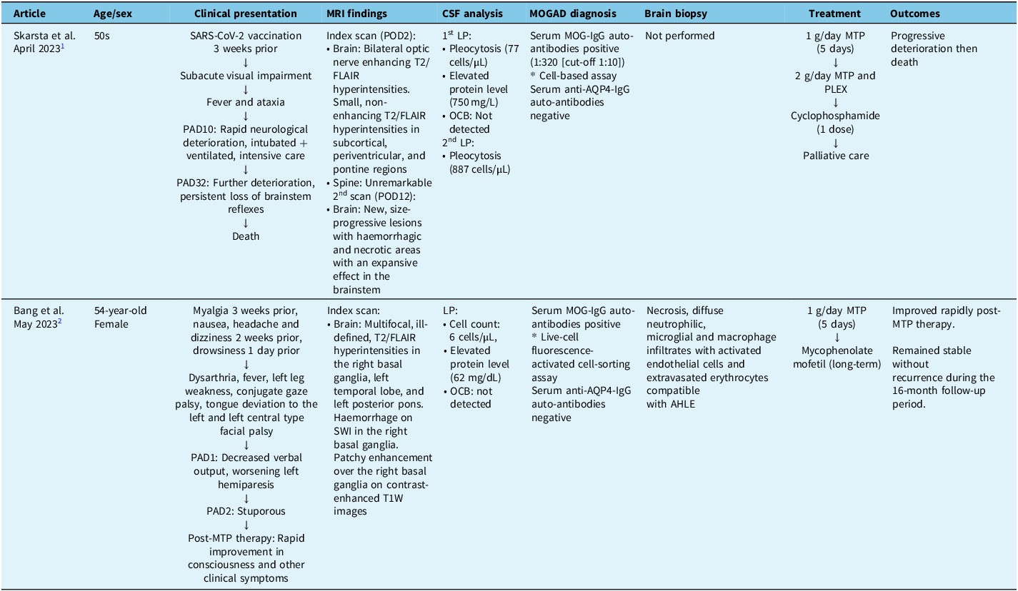
Two cases of adult-onset AHEM with positive MOG-Abs have been reported to date. Reference Skarsta, Nicoletti and Frick1,Reference Bang, Kim and Seok2 They are summarised in Table 1.
Summary of two recent cases of adult-onset acute haemorrhagic encephalomyelitis with anti-MOG auto-antibody positivity

AHLE = acute haemorrhagic leukoencephalitis; AqP4 = aquaporin 4; LP = lumbar puncture; MOG = myelin oligodendrocyte glycoprotein; MOGAD = myelin oligodendrocyte glycoprotein antibody-associated disease; MTP = methylprednisolone; OCB = oligoclonal bands; PAD = post-onset day; PLEX = plasma exchange; POD = post-onset day; SARS-CoV-2 = severe acute respiratory syndrome coronavirus 2; SWI = susceptibility weighted imaging; T1W = T1-weighted imaging; T2/FLAIR = T2-weighted fluid-attenuated inversion recovery; MTP = methylprednisolone.
As per the 2023 consensus diagnostic criteria, our patient’s clinical, laboratory and radiological features are consistent with the diagnosis of MOGAD. Reference Banwell, Bennett and Marignier4 He experienced a core clinical demyelinating event, having presented with ADEM (in this case AHEM) and myelitis, while testing positive for serum MOG-IgG-Ab through a fixed-CBA at a low titre. Additionally, he exhibited supporting features including LETM, conus involvement, the “H sign” and the presence of multiple supra- and infratentorial lesions while also testing negative for anti-AqP4-IgG auto-antibodies. Interestingly, the low-titre MOG-Abs seropositivity raises several questions and affords us the unique position to discuss the potential pathogenicity of MOG-Abs in AHEM.
The timing of testing for MOG-Abs is crucial in MOGAD, as antibody titres significantly decline over time. Reference Forcadela, Rocchi and San Martin5 A study found that only 55% of patients tested within 30 days of symptoms had positive MOG-Abs results, with half of them becoming negative and 28% becoming low positive 6–12 months later. Reference Forcadela, Rocchi and San Martin5 This temporal dynamic could explain the low-titre MOG-Ab positivity observed in our patient, as there was a significant delay in testing. Given the inherent complexity in diagnosing AHEM, it is plausible that some patients who would otherwise be MOG-Abs seropositive might fall outside the diagnostic window when tested, contributing to the relative rarity of reported MOG-Ab seropositivity in AHEM cases.
The possibility of a false-positive MOG-Ab result warrants careful consideration, especially given our patient’s low titre. While he met the consensus criteria for MOGAD and possessed classical features (LETM, conus involvement and “H sign”), his URTI and the moderately symmetrical brain MRI findings raise concerns for alternative diagnoses, such as viral, parainfectious or autoimmune encephalitides. Although we addressed key differential diagnoses, resource limitations hindered a more thorough investigation of all possible causes.
In a similar vein, we emphasise that caution is essential when interpreting the potential pathogenicity of MOG-Abs in patients with neurological symptoms. Furthermore, MOG-Abs are also found in conditions like seropositive NMOSD, Reference Gou, Yang and Feng6 glioblastoma Reference Amin, Mays, Polston, Flanagan, Prayson and Kunchok7 and anti-NMDA-receptor antibody encephalitis, Reference Ding, Li and Tian8 where they are not deemed primary pathogenic drivers. This observation raises the question of whether the presence of MOG-Abs in AHEM may merely represent an epiphenomenon, potentially complicating the understanding of their role in the disease process.
Histopathological disparities between MOGAD and AHEM further complicate their association. MOGAD is characterised by a primary immune attack on myelin, manifesting with perivenous and confluent demyelinating lesions. Reference Takai, Misu, Fujihara and Aoki9 Conversely, AHEM reveals a broader spectrum of pathology, including prominent haemorrhages, oedema, axonal injury, fibrinoid vessel necrosis and complement deposition. Reference Skarsta, Nicoletti and Frick1,Reference Grzonka, Scholz and De Marchis3 Early and extensive astrocyte injury is also reported, suggesting that demyelination in AHEM could be secondary to astrocyte damage, akin to observations in NMOSD. Reference Skarsta, Nicoletti and Frick1,Reference Grzonka, Scholz and De Marchis3 This contrast prompts consideration of whether MOG-Abs actively participate in the primary pathogenic process of AHEM or are instead secondary to extensive neuronal destruction. These differences may also explain why patients with MOGAD generally respond to immunosuppression, while AHEM does not.
However, treatment resistance does not entirely rule out MOGAD. Some patients with MOGAD respond poorly to steroids and plasma exchange but demonstrate dramatic improvement with third-line anti-IL-6R therapy. Reference McLendon, Gambrah-Lyles and Viaene10 Similarly, AHEM might represent a form of MOGAD that necessitates rapid escalation to third-line therapies.
In the absence of a convincing alternative diagnosis, we proceeded with a MOGAD-directed treatment course. Unfortunately, our ability to initiate tocilizumab was hindered by the patient’s rapid decline and recurrent sepsis. Diagnostic delays, compounded by healthcare disparities in the rural setting, significantly reduced the window for intervention, and earlier diagnosis along with treatment escalation could have potentially changed the outcome.
Our unique case prompts more nuanced explorations of MOGAD’s potential contribution to the pathogenesis of AHEM, highlighting that current data are still insufficient to establish a definitive aetiological link. The temporal dynamics of MOG-Abs titres, histopathological disparities and variable treatment outcomes further underscore the necessity for cautious interpretation of MOG-Abs seropositivity in AHEM. Moving forward, prospective studies are needed to better understand the potential aetiological association between MOGAD and AHEM. Retrospective cohorts face challenges due to the dynamic decline of MOG-Abs titres over time. Increased clinician awareness and timely testing are crucial for early diagnosis and evaluation of second- and third-line immunotherapy, potentially improving outcomes.
Acknowledgements
This research was not funded by any agencies in the public or private sectors.
Funding statement
This research was not funded by any agencies in the public or private sectors.
Competing interests
All authors have no competing interests to declare.
Ethics statement
The patient’s next of kin provided informed consent for the use of de-identified data for publication purposes.
Statement of authorship
SYT is the main author of this manuscript. PP, JSW, PJK, YKC and JCEO contributed equally to the management of the patient and drafting/editing of the manuscript.