Introduction
Clinical neuroimaging is increasingly being used in the diagnostic work-up of patients presenting to a memory clinic. Structural neuroimaging methods such as computed tomography (CT) and, more sensitively, magnetic resonance imaging (MRI) are well suited to exclude possibly (surgically) treatable cause of dementia (e.g. tumors, subdural hematomas, hydrocephalus, etc.). However, these patients represent the vast minority (<8%) of patients presenting with memory complaints to a memory clinic (Scheltens et al., Reference Scheltens, Fox, Barkhof and Carli2002). More importantly, structural brain imaging can demonstrate certain patterns of atrophy, vascular pathology or inflammatory changes, which can substantially support the clinical diagnosis of dementia. In other words, during the past decade, the role of structural neuroimaging as a diagnostic tool in a memory clinic setting has changed from an exclusionary to a more inclusionary approach. In addition, MRI is increasingly being used for disease monitoring (e.g. progression of atrophy and vascular changes) and has therefore been established as a valuable method for efficacy and safety monitoring in clinical treatment trials (Frisoni et al., Reference Frisoni, Fox, Jack, Scheltens and Thompson2010). As a consequence, this has led to the incorporation of clinical neuroimaging into current diagnostic guidelines of dementia, which state that neuroimaging should be performed at least once during the diagnostic work-up of patients with suspected or definite dementia (Hort et al., Reference Hort2010).
Structural MRI protocol in dementia
Multidetector CT is a suitable diagnostic method for the exclusion of possible treatable causes of dementia, as well as for the assessment and visual rating of focal and global cortical atrophy, atrophy of the medial temporal lobe and vascular white matter changes (Wattjes et al., Reference Wattjes2009). However, structural MRI is the imaging modality of choice due to the lack of radiation, better gray matter/white matter contrast, and the ability to manipulate tissue contrast with different pulse sequences.
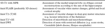
The MRI protocol should include pulse sequences which address the following issues: assessment of focal and global cortical atrophy as well as atrophy of the medial temporal lobe (particularly the hippocampus); vascular white matter changes (small and large vessel disease); vascular changes in deep gray matter structures (in particular thalamus infarction); cerebral microbleeds (MBs) and macrohemorrhages (including post-traumatic changes). An example of a possible MRI multisequence protocol is given in Table 1.
Example of a multisequence MRI protocol for patients presenting to a memory clinic

D = dimensional; DWI = diffusion-weighted imaging; FLAIR = fluid-attenuated inversion recovery; MPR = multiplanar reconstructions; TSE = turbo spin echo; GE = gradient echo.
How to read MRI scans in memory clinic patients
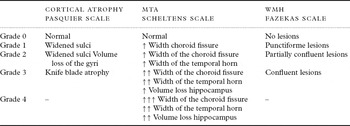
MRI scans in a clinical routine setting should be read in a structured and reproducible way. Regarding the two most relevant pathological features in dementia (cortical atrophy including medial temporal lobe atrophy and vascular changes), visual rating scales have been established, validated and are widely used in a clinical routine setting. Table 2 summarizes the most established rating scales for focal and global cortical atrophy, hippocampal atrophy and vascular white matter changes. In addition, certain aspects of MRI pathology associated with cognition and normal aging, such as the presence of MBs, should be assessed and classified.
Overview of the most established visual rating scales for the assessment of cortical atrophy, hippocampal atrophy (medial temporal lobe atrophy, MTA) and white matter hyperintensities (WMH)

Cortical atrophy
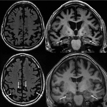
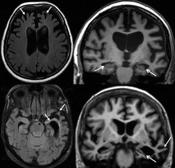
Visual rating of cortical atrophy can be easily performed using the four-point (0–3) Pasquier scale (Pasquier et al., Reference Pasquier1996) based on the width of the sulci and volume of the gyri (Figure 1). The Pasquier scale can be applied either globally (global cortical atrophy) or regionally for certain anatomic regions (temporal, parietal, etc.).
MRI findings in patients presenting with Alzheimer's disease (AD). Top row: axial FLAIR (left) and oblique coronal reconstructions (according to the axis of the hippocampus) of a 3D T1-weighted sequence (right) obtained from an 81-year-old male presenting with a late-onset (senile) type of AD. On the FLAIR images, a severe global cortical atrophy (grade 2) can be observed. The cortical atrophy is more prominent but not restricted to the parietal lobes. In addition, substantial (grade 2) atrophy of the hippocampus (widening of the choroid fissure in combination with a volume loss of the hippocampus) is present (arrows). Bottom row: comparable MR images obtained from a 50-year-old male presenting with an early-onset (presenile) type of AD. In these patients a rather focal atrophy pattern involving the parietal lobe (left, arrows), including the precuneus and the posterior cingulate, can be frequently observed, while the medial temporal lobe structures are relatively spared (right, arrows).

Medial temporal lobe atrophy
The visual rating of medial temporal lobe atrophy is more sophisticated since we are dealing with a complex interaction of rather small anatomic structures and landmarks. The five-point Scheltens scale, considering the width of the choroid fissure, temporal horn as well as the volume of the hippocampus, is a well-established rating scale for the assessment of the medial temporal lobe, which correlates well with histopathology and volumetric measurements (Scheltens et al., Reference Scheltens1992) (Figure 1).
Vascular pathology
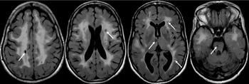
The assessment of vascular changes is crucial in the radiological evaluation of dementia patients, particularly for the diagnosis of vascular dementia (VaD) (van Straaten et al., Reference van Straaten, Scheltens and Barkhof2004). However, small vessel white matter changes which do not formally fulfill the diagnostic criteria for VaD act in a synergistic way with neurodegenerative changes with relevant impact on clinical outcome measures (Inzitari et al., Reference Inzitari2009). FLAIR is the most sensitive MRI sequence for the detection of white matter hyperintensities (WMH) as a marker of small vessel vascular damage. An easily applicable and highly reproducible four-point rating scale (Fazekas scale) is widely used in clinical practice and corresponds well with more detailed ratings scales and histopathology (Fazekas et al., Reference Fazekas1987, Scheltens et al., Reference Scheltens1998). Another important marker of small vessel disease, which has been included into the NINDS-AIREN criteria for VaD, is the presence of lacunar infarcts (Román et al., Reference Román1993). These are strongly associated with the presence and amount of WMH and can be frequently found in the cerebral deep white matter, basal ganglia and crucial anatomic structures such as the thalamus. Lacunes in the white matter can also be sensitively detected and differentiated from perivascular spaces on FLAIR sequences (Figure 2). However, lacunes in the infratentorial white matter and deep gray matter structures (particularly the thalamus) are frequently missed on FLAIR and therefore T2-weighted (turbo) spin-echo sequences are mandatory for these purposes (Bastos-Leite et al., Reference Bastos-Leite, van Straaten, Scheltens, Lycklama and Barkhof2004).
Axial FLAIR images obtained from a 47-year-old female diagnosed with vascular dementia according to the NINDS-AIREN criteria. On MRI, confluent WMH (Fazakas grade 3) can be observed involving more than 25% of the white matter. Multiple lacunes (arrows) are present in the deep white matter but also in the basal ganglia region. Note the severe involvement of the white matter of the anterior temporal lobe suggestive of CADASIL (cerebral autosomal dominant arteriopathy with subcortical infarcts and leukoencephalopathy). In fact, a mutation in the Notch-3 gene was found in this patient.

Besides the assessment of small vessel disease, the detection of large vessel disease, including territorial infracts and watershed infarcts are of importance since they have been incorporated into the NINDS-AIREN criteria for VaD (Román et al., Reference Román1993).
Cerebral microbleeds
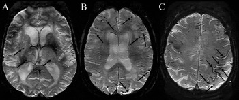
MBs are defined as small hypointense dot-like lesions in the brain parenchyma on T2*-weighted or susceptibility weighted MRI (SWI). Pathologically, MBs represent perivascular hemosiderin deposits stored in surrounding macrophages, presumably due to pathological amyloid deposition in the vascular wall leading to permeability and leakage. Alternatively, MBs can also be seen in association with small vessel disease, i.e. with hyperintense vasculopathy. In terms of anatomic distribution and the underlying pathophysiology, MBs can be subdivided in lobar MBs located in the cortical gray–white matter interface and non-lobar MBs located in deep brain structures (e.g. basal ganglia) and the posterior fossa (Figure 3). Lobar MBs are related to cerebral amyloid angiopathy (CAA), and almost invariably present in patients with Alzheimer's disease (AD), whereas non-lobar MBs are more related to hypertensive vasculopathy (Cordonnier and van der Flier, Reference Cordonnier and van der Flier2011). These two different vasculopathies may co-occur in dementia patients.
Axial SWI images presenting different types of microbleeds (MBs). (A) MBs (arrows) located in the deep white and gray matter structures (non-lobar) are associated with hypertensive encephalopathy whereas (B) MBs (arrows) located in the white matter–cortcial gray matter interface are rather associated with cerebral amyloid angiopathy (CAA). (C) Superficial siderosis (open head arrows) represents a leptomeningeal manifestation of CAA. In this patient, lobar MBs (closed head arrows) are located close to the areas of superficial siderosis.

Depending on the sensitivity of the imaging technique (magnetic field strengths, pulse sequences, echo time, spatial resolution), the prevalence of MB in a normal elderly healthy population is almost 25% (Poels et al., Reference Poels2011). In a memory clinic population, the prevalence of MB is approximately 17%, ranging from 10% in patients with subjective memory complaints, 18–20% in AD, to 65% in patients with VaD (Cordonnier et al., Reference Cordonnier, van der Flier, Sluimer, Leys, Barkhof and Scheltens2006). In general, MBs are associated with clinical outcome measures, such as cognitive function and mortality, age, apolipoprotein E genotype, cerebrospinal fluid biomarkers, as well as vascular damage seen on MRI, including WMH and lacunes (Goos et al., Reference Goos2009; Reference Goos2010). In addition, there is emerging evidence that lobar cortical MBs might have predictive value in terms of the future development of AD (Kirsch et al., Reference Kirsch2009). It is also conceivable that non-lobar MB might be a predictive factor of VaD, but this remains to be investigated.
Amyloid-associated leakage from leptomeningeal vessel leads to the pathological hemosiderin deposition in the subpial space, which has coined the term of superficial siderosis. On T2*-weighted and susceptibility-weighted MRI, superficial siderosis can be identified by low signal abnormalities in the subpial space lining the surface of the brain. Similar to lobar hemorrhage and MBs, superficial siderosis seems to be a marker for CAA (Linn et al., Reference Linn2010) (Figure 3). Its prevalence is rather lower than 1% among a normal healthy population and there is strong co-incidence with lobar MBs (Vernooij et al., Reference Vernooij, Ikram, Hofman, Krestin, Breteler and van der Lugt2009). However, compared to MBs, the clinical and prognostic role of superficial siderosis in dementia patients is rather unclear and needs further investigation.
Use of MRI to support the diagnosis of neurological disease associated with dementia
Beside the assessment of normal aging findings (mild global cortical and hippocampal atrophy and WMH), MRI is helpful to support the clinical diagnosis of neurodegenerative, inflammatory and metabolic disease associated with dementia, by showing specific atrophy patterns and white and/or gray matter changes. Here we describe the MRI pathology of the most frequent diseases associated with dementia.
Alzheimer's disease
AD is the most common cause of dementia and is characterized by the histopathological hallmarks of amyloid-β and hyperphophorylated tau accumulation (McKhann et al., Reference McKhann, Drachman, Folstein, Katzman, Price and Stadlan1984). The role of neuroimaging in the diagnosis of AD is becoming increasingly important. This is reflected in new proposed diagnostic criteria in which, as well as cerebrospinal fluid (CSF) (amyloid-β42, tau or phospho-tau) and positron emission tomography (PET) findings (temporo-parietal hypometabolism on FDG-PET, amyloid imaging), a structural imaging marker (medial temporal lobe atrophy) has been included as a key criterion for the diagnosis of AD (Dubois et al., Reference Dubois2007).
Corresponding to the neuropathological stages described by Braak and Braak (Reference Braak and Braak1991), the neurodegenerative changes in terms of atrophy in senile AD patients can be detected first in the medial temporal lobe, particularly in the hippocampus and best seen on oblique coronal T1-weighted images. Although MTA is very characteristic for AD, a normal volume of the medial temporal lobe does not exclude AD. In addition, MTA is a common feature in other neurodegenerative disorders and is therefore not a specific marker to confidently exclude other neurodegenerative disease associated with dementia. Besides the diagnostic hallmark MTA, cortical atrophy predominantly located in the parietal lobes is a common radiological feature and may be helpful in differentiating AD from other neurodegenerative diseases associated with dementia (Jones et al., Reference Jones2006; Frisoni et al., Reference Frisoni2007) (Figure 1). The degree of cortical atrophy and MTA can be easily assessed by visual rating, as described above, and corresponds well with histopathology. Visual rating of MTA in a memory clinic population in order to distinguish AD patients from those without cognitive complaints provides sensitivity and specificity values of 80–85% (Frisoni et al., Reference Frisoni, Fox, Jack, Scheltens and Thompson2010).
Compared to classical senile AD manifestations, early-onset (pre-senile) AD may present with a different distinct atrophy pattern predominantly involving the parietal cortex, precuneus and posterior cingulum and relatively sparing the medial temporal lobe (Figure 1). In advanced stages, the medial temporal lobe becomes more and more affected (Karas et al., Reference Karas2007; Shiino et al., Reference Shiino2008).
In patients with mild cognitive impairment (MCI) who present with cognitive impairment not fulfilling the diagnostic criteria for dementia, the degree of cortical and hippocampal atrophy measured by visual rating has a strong predictive value for further cognitive decline and development of AD (Likeman et al., Reference Likeman2005; Bouwman et al., Reference Bouwman2007; Davatzikos et al.; Reference Davatzikos, Bhatt, Shaw, Batmanghelich and Trojanowski2010).
It is important to realize that the vast majority of patients with AD pathology present with a combination of neurodegenerative changes and vascular pathology, such as WMH and lacunar infarcts. This vascular co-morbidity has prompted the pathophysiological concept that AD patients do not usually present with a “pure Alzheimer pathology,” which has coined the term of “mixed pathology” (de la Torre, Reference de la Torre2004; DeCarli, Reference DeCarli2006). This concept is further supported by the distribution of MBs in AD patients. The vast majority of MBs is located in lobar regions in the cortical white–gray matter interface, suggesting CAA pathology which is strongly associated with AD. However, a substantial proportion of MBs is located in non-lobar regions, suggesting underlying microangiopathy due to vasculo-ischemic damage. In fact, these findings further support the concept of “mixed pathology” and the clinical impression that amyloid pathology and cerebrovascular pathology are likely acting in a synergistic way (Kalaria and Ballard, Reference Kalaria and Ballard1999).
Fronto-temporal lobe degeneration
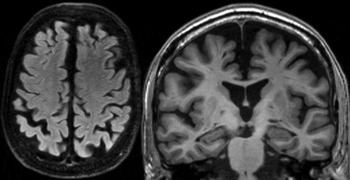
The disease entity of fronto-temporal lobe degeneration (FTLD) consists of three different and distinct clinical subtypes: the behavioral variant of fronto-temporal dementia (bvFTD), progressive aphasia (PA) and semantic dementia (SD). These subtypes present with different clinical manifestations and also different atrophy patterns on MRI (Neary et al., Reference Neary1998). The bvFTD is radiologically characterized by (bilateral) atrophy of the frontal lobes, including the dorsolateral, frontobasal and mesofrontal cortical areas, as well as of the anterior parts of the temporal lobes (Figure 4). PA is a non-fluent aphasia characterized by left-sided (most often) atrophy of the lateral parts of the temporal lobe, basal parts of the frontal lobe and the insular cortex, resulting in a unilateral widening of the Silvian fissure (Gorno-Tempini et al., Reference Gorno-Tempini2011). Semantic aphasia is a progressive form of fluent-aphasia and shows focal cortical atrophy of the anterior parts of the temporal lobes (temporal pole), more frequently on the left side than on the right side (Figure 4). These atrophy patterns correlate well with quantitative MRI methods, such as voxel-based morphometry (VBM), and with pathological studies.
Axial FLAIR (left column) and oblique coronal reconstruction of a 3D T1-weighted sequence (right column) presenting different types of fronto-temporal lobe degeneration (FTLD). Top row: a 56-year-old female presenting with bvFTD. Note the focal atrophy of the frontal lobes (particularly of the mesofrontal area) (arrows). Atrophy of the medial temporal lobe structures (open head arrows) can also be observed in these patients and definitely does not exclude FTLD. Bottom row: a 76-year-old female presenting with semantic dementia. On MRI, a severe focal and asymmetric atrophy of the left temporal lobe can be observed in the vast majority of cases (open head arrows). In particular the temporal pole is affected. Ballooning of the temporal horn and a small rim of high signal on the FLAIR/T2-weighted images can be seen around the temporal horn, which is probably a gliotic reaction as a consequence of end-stage atrophy (arrows).

In early disease stages, structural neuroimaging can be normal and/or shows unspecific global cortical atrophy, as seen in other dementias. In these stages, PET imaging may already reveal relevant glucose hypometabolism in fronto-temporal regions. The focal cortical atrophy pattern may develop slightly over time and become more evident in later disease stages. Also, medial temporal lobe structures such as the hippocampus and the amygdala are frequently affected. In patients with end-stage atrophy, a “ballooning” of the temporal horn can be observed, which is surrounded by a small confluent area of high signal in the white matter on T2-weighted images (Rosen et al., Reference Rosen2002; Vitali et al., Reference Vitali2008) (Figure 4).
Dementia associated with parkinsonian dementias
Most of the parkinsonian syndromes are associated with cognitive decline and dementia, including idiopathic Parkinson's disease, dementia with Lewy bodies (DLB), multisystem atrophy (MSA), corticobasal degeneration (CBD) and progressive supranuclear palsy (PSP).
Dementia with Lewy bodies
DLB is a frequent neurodegenerative disease associated with dementia, which is pathologically characterized by α-synuclein-positive Lewy body inclusions in the cortical gray matter. Clinically, DLB patients present with progressive cognitive impairment mainly affecting memory as well as visuo-spatial and executive domains, with fluctuations of symptoms combined with visual hallucinations and often sleeping disorders. Structural neuroimaging has limited value in the diagnosis of DLB. Most patients show mild to moderate global cortical atrophy, while the medial temporal lobe is relatively preserved (Figure 5). However, considering this atrophy pattern, there is substantial overlap between DLB and AD as well as normal aging. This overlap can be overcome by more specific imaging techniques, such as molecular imaging of the dopaminergic system (e.g. dopamine transporter imaging, DAT), allowing a more specific diagnosis of DLB.
Axial FLAIR (left) and oblique coronal reconstruction of a 3D T1-weighted sequence (right) obtained from a 75-year-old male with DLB. Note the mild to moderate global cortical atrophy and the relative sparing of the hippocampus.

Multisystem atrophy
Clinical characteristics of MSA include orthostatic hypotension, urine incontinence, and cerebellar symptoms. Imaging criteria of MSA consist of focal atrophy of the basal ganglia, in particular of the putamen, as well as the cerebellar peduncles (most obvious in the middle cerebellar peduncle), brain stem and cerebellum. On T2-weighted images of some MSA patients, linear signal abnormalities can be observed in white matter lateral to the putamen (“putaminal rim sign”), pons (“hot cross bun sign”) and cerebellar peduncles, which can further support the clinical diagnosis of MSA (Figure 6) (Vitali et al., Reference Vitali2008).
Axial T2-weighted (left) and sagittal T1-weighted MR images (middle and right) in a patient presenting with multisystem atrophy. Note the severe atrophy of the brain stem and the cerebellum, including the middle cerebellar peduncle (arrows). In the pons, two linear areas with high signal (“hot cross bun sign”) can be detected on the T2-weighted image.

Cortical degeneration
Patients with CBD typically present with asymmetric cortical symptoms, including language and speech disturbances, visuo-spatial deficits and hemineglect, apraxia and occasionally myclonus, together with extra pyramidal signs. On MRI, CBD patients frequently show an asymmetric cortical atrophy of the parietal and frontal cortex, while the temporal lobe, including the medial temporal lobe, is relatively spared. The left side seems to be involved more frequently, and VBM analysis has shown that the neurodegenerative changes are most pronounced at the interface between the superior frontal and precentral sulci (Boxer et al., Reference Boxer2006)
Progressive supranuclear palsy
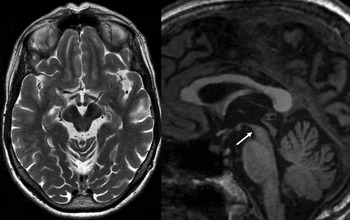
PSP is clinically characterized by vertical eye movement disturbances and postural instability. MRI frequently shows indirect and direct evidence of mid-brain atrophy (Figure 7). An often used and reproducible MRI parameter is the anterior–posterior diameter of the mid-brain, which is significantly reduced in PSP patients. Different cut-off values of the anterior–posterior midbrain diameter have been reported. A diameter shorter than 15 mm measured in the mid-sagittal view has been well established and validated in a PSP population. In addition, a reduced volume of the cerebral peduncles and an increased inter-peduncular angle can be observed (Warmuth-Metz et al., Reference Warmuth-Metz2001; Oba et al., Reference Oba2005). However, mid-brain atrophy is a rather non-specific feature in the diagnosis of PSP since other neurodegenerative diseases, including CBD and MSA, can present with mid-brain atrophy, resulting in a considerable overlap. Studies also considering other key structures such as the middle and superior cerebellar peduncles were able to better differentiate PSP from MSA and Parkinson's disease.
Axial T2-weighted and sagittal T1-weighted MR images of a patients with the diagnosis of progressive supranuclear palsy demonstrating mesencephalic atrophy (reduced gap diameter, increased interpeduncular angle), while the pons is relatively preserved. This atrophy pattern resembles the head and body of a humming bird, which has coined the term “humming bird” sign (arrow).

Vascular dementia
Vascular dementia (VaD) is probably the most common cause of dementia after AD. Clinically, VaD is characterized by memory impairment and dysfunction of other cognitive domains, particularly frontal-executive functions. Neuroimaging plays a key role in the diagnosis of VaD. CT and, more sensitively, MRI are able to make the diagnosis of VaD according to diagnostic criteria and can distinguish pure VaD pathology from vascular comorbidity in terms of “mixed pathology.” Within the group of VaD patients, the distinct pattern of distribution of vascular lesions can lead to a specific diagnosis of the underlying disease causing VaD, e.g. CADASIL (cerebral autosomal dominant arteriopathy with subcortical infarcts and leukoencephalopathy) (Chabriat et al., Reference Chabriat1998) (Figure 2).
Diagnostic criteria for VaD have been proposed with differing sensitivity and sensibility (Pohjasvaara et al., Reference Pohjasvaara, Mätyla, Ylikoski, Kaste and Erkinjunti2000). The NINDS-AIREN criteria have been developed for establishing the diagnosis of VaD in research studies and clearly state that neuroimaging is an essential part of the diagnostic process of VaD. According to these criteria, patients can be classified as possible, probable and definite VaD. The NINDS-AIREN criteria are based on three main features: the presence of dementia, evidence of cerebro-vascular disease either clinically or in MRI/CT, and the temporal relationship between the clinical onset of dementia and the cerebrovascular disease. Radiologically, VaD can be diagnosed based on the presence of small vessel disease (WMH affecting at least 25% of the white matter, multiple basal ganglia and frontal white matter lacunes, bilateral thalamic lesions) and large vessel disease (territorial and watershed infarcts) (Guermazi et al., Reference Guermazi2007). Regarding the latter, both the severity (e.g. bilateral) and location (e.g. dominant hemisphere and involving association cortex) are crucial parameters. However, the diagnosis within strict diagnostic criteria remains a matter for debate. The temporal relationship between vascular damage (e.g. stroke) and cognitive decline is difficult to determine in many cases since a substantial number of strokes and, moreover, progression of WMH is silent. In addition, chronic hemodynamic conditions leading to cerebral hypoperfusion (e.g. carotid stenosis) may cause cognitive impairment and dementia without any vascular changes stated in the diagnostic criteria (Johnston et al., Reference Johnston2004). Elderly patients with dementia who fulfill the clinico-radiological diagnostic criteria of VaD in particular also show changes consistent with AD pathology, which again reinforces the concept of “mixed dementia.” Another possible limitation of VaD diagnostic criteria is that MBs, which are associated with cognitive deficits in several populations, are currently not being considered.
Despite the difficulties and problems in the diagnosis of VaD, neuroimaging has become crucial in identifying certain patterns of vascular damage causing VaD, as well as identifying underlying vascular pathology in the early disease course, which might have therapeutic consequences (Tzourio et al., Reference Tzourio2003).
Dementia secondary to inflammation
Besides primary neurodegenerative diseases associated with dementia, cognitive decline is commonly associated with neuroinflammatory and infectious disease of the central nervous system, including multiple sclerosis, herpes simplex infections, progressive multifocal leukoencephalopathy (PML) and prion diseases. Concerning the diagnosis of prion diseases such as Creutzfeld-Jakob disease (CJD), MRI has become one of the most important diagnostic methods. Depending on the clinical subtype of CJD, different patterns of high signal abnormalities on FLAIR and T2-weighted imaging can be observed in the cortical gray matter and the basal ganglia, particularly the striatum and thalamus (Figure 8). The abnormalities show features of restricted extracellular diffusion with a high signal on diffusion-weighted imaging and corresponding low apparent diffusion coefficient (ADC) values (Shiga et al., Reference Shiga2004). Different patterns of gray matter involvement on MRI in sporadic (sCJD), genetic and variant (vCJD) forms of CJD have been described (Zeidler et al., Reference Zeidler2000; Tschampa et al., Reference Tschampa, Zerr and Urbach2007). Based on pattern recognition, MRI diagnosis of CJD can be made with a high sensitivity and specificity beyond 90%. Therefore, MRI may not only support the clinical diagnosis of CJD, but also allow a rather specific diagnosis of CJD in combination with other biomarkers, including the CSF (14–3-3 protein) and electroencephalogram.
A 54-year-old female presenting with a cortical variant of Creutzfeldt-Jakob disease. The axial FLAIR (left) and more sensitively the DWI sequence (right) shows high signal abnormalities in the cortical gray matter of both hemispheres, more pronounced in the left parietal lobe (arrows).

Serial MRI
The atrophy patterns in neurodegenerative disease described above are based on cross-sectional MRI. In neurodegenerative diseases such as AD, the atrophy rate is more accelerated when compared to normal aging brains. Therefore, serial MRI examinations can assess atrophy progression during follow-up, which can further support the clinical diagnosis, particularly in early disease stages when the neurodegenerative findings might be rather discrete (Jack et al., Reference Jack1998; Riidha et al., Reference Riidha2006). With the advent of more specific imaging methods such as FDG-PET and amyloid-PET for these purposes, the need for serial MRI in order to support the diagnosis is substantially decreasing. However, atrophy progression assessed by MRI is increasingly used as a safety and efficacy outcome measure in clinical trials (Frisoni et al., Reference Frisoni, Fox, Jack, Scheltens and Thompson2010).
Conclusion
Structural MRI has become a crucial part of the diagnostic evaluation of memory clinic patients, which is reflected by current guidelines dealing with the diagnosis of dementia. Similar to CT, MRI can exclude possibly (surgically) treatable causes of dementia and also show distinct atrophy patterns and cerebrovascular changes that can support the clinical diagnosis of dementia. For these purposes, visual rating scales have been established and allow a fast, reliable and reproducible assessment. However, MRI has substantial advantages over CT and can demonstrate certain aspects of pathology associated with dementia, such as MBs as a marker of CAA associated with AD pathology and/or vascular damage. In addition, MRI is able to further characterize the amount and distribution of cerebrovascular pathology, which impacts the diagnosis, prognosis and therapeutic strategy in these patients. In some disease entities, such as prion disease, MRI has become the key diagnostic modality for a sensitive and specific diagnosis.
Despite the high value of structural MRI in the diagnostic scheme of memory clinic patients, advanced and quantitative imaging modalities, including PET and quantitative MRI techniques discussed elsewhere in this special issue, will shed further light on the dynamic field of the possibilities and challenges of neuroimaging in dementia.
Conflict of interest
None.
Acknowledgments
The author wishes to thank Professor Frederik Barkhof and Dr. Jeroen D.C. Goos for their support and critical reading of the manuscript.