Introduction
Breast cancer is the most commonly occurring form of cancer among women around the world. Reference Sung, Ferlay and Siegel1 Postmastectomy radiotherapy (PMRT) is recognised as an important form of treatment for advanced incidences of breast cancer. It can improve the chances of patients remaining disease-free and has been found to increase the overall survival rates of cancer patients. Reference Recht, Edge and Solin2 Many current treatment techniques employ PMRT such as three-dimensional radiotherapy (3DRT), Reference Yousefi Kashi, Karimi, Rakhsha, Javadzadegan and Taghizadeh-Hesary3–Reference Zhang, Dilworth, Marina, Chen, Benedetti and Liu7 ntensity-modulated radiotherapy Reference Wang, Jie, Liang, Wu and Cheng8,Reference Koshy, Zhang, Naqvi, Liu and Mohiuddin9 and volumetric arc radiotherapy. Reference Iorio, Franco and Gallio10,Reference Nichols, Fontenot, Gibbons and Sanders11 Sophisticated techniques provide a high radiation dose to the target while preserving organs at risk (OARs); however, they require long periods of time for preparation and treatment. Reference Foroudi, Wilson and Bressel12 In reality, the number of patients per treatment room is limited in contrast to the actual patient population. As has been previously mentioned with regard to sophisticated techniques, 3DRT is still a necessity in the treatment of this disease because of the short overall amount of treatment time involved and the low doses of radiation that are delivered to the OARs. Reference Liu, Chen, He and Li13,Reference Haertl, Pohl, Weidner, Groeger, Koelbl and Dobler14
3DRT is the basic technique employed to address this scourge. The three-fields technique, which consists of opposing tangential of the chest wall (CW) and anterior field of the supraclavicular (SC), is normally employed for the treatment planning. Various techniques are utilised such as the dual-isocenter technique (DIT), the single-isocenter technique (SIT) with various beam geometry methods. The DIT has been commonly used in the treatment planning. The problem associated with this method arises at the junction between opposing tangential fields (OTFs) and SC field (SCF). Consequently, the uncertainty associated with the setup process may impact the degree of dose homogeneity delivered at the junction. Reference Mahmoud, Somayeh, Mahbod, Mehdi, Vahid and Ghazale15 Many methods have been proposed in an attempt to address this problem. Reference Banaei, Hashemi and Bakhshandeh4–Reference Zhang, Dilworth, Marina, Chen, Benedetti and Liu7 The SIT is just one of the techniques that has been proposed for effective treatment planning. SIT employing the half beam block (HBB) method Reference Banaei, Hashemi and Bakhshandeh4,Reference Wadasadawala, Lewis and Gaikwad5 is one of the approaches used in the PMRT. Each half of the treatment field was utilised for the CW and SC; thus, the limitation of field size is transpired. The geometrical arrangement Reference Romeo6,Reference Zhang, Dilworth, Marina, Chen, Benedetti and Liu7 is another approach for the dose homogeneity improvement. A formula is constructed to determine the angles of beam geometry, especially the couch rotation. A perfect dose homogeneity is found by utilising this approach. However, the degree of the uncertainty may raise by the rotating couch.
The immobilisation is one of the considerations in the PMRT. The Breast-board is the main device of various immobilisations. The disadvantages of this device are found not only a large setup error in the craniocaudal direction Reference Nalder, Bidmead, Mubata, Tait and Beardmore16 but also an inconvenient and long setup time. Reference Raza, Agarwal, Das, Kumar and Lal17 A Wing-board is the alternative device for immobilisation. The replacement of this device impacts the currently treatment method in our centre. Utilising this method with the Wing-board, a underdose is found at the junction between the two treatment fields.
Various methods have been proposed to solve the problem of field matching; however, there are various issues associated with the insufficient matching methods such as dose inhomogeneity at the junction, field size limitations and uncertainty of setup errors in the geometrical arrangements. This study, then, has proposed an alternative method of field matching that would improve dose homogeneity and minimise the limitations associated with the treatment field size. The proposed method uses the coplanar plane between the superior border of the medial tangential field and the inferior border of the SCF. To evaluate the performance of this approach, the SITHBB method was benchmarked with not only the proposed method but also the currently identified method (Traditional three-fields monoisocenter). The dosimetric comparison among the three methods has been analysed with the use of relevant statistics.
Materials and Methods
Ethical statement
In accordance with the requirements for prospective studies, the concept and study design of this study were submitted for ethical approval. Ethical clearance was granted by the Chiang Mai University Ethics Committee (study code: RAD-2563–07760).
Data preparation and sample size
All image sets of the patients were acquired by SOMATOM DefinitionAS (Siemens Healthineers, Germany) at 5 mm of slice thickness. A Wing-board (Standard BoardTM, CIVCO Radiotherapy, USA) was used for the immobilisation. Twenty-two patients of the left-side postmastectomy were randomly selected during the period of January 2019 to December 2020. The sample size was calculated according to the work of Naing et al. Reference Naing, Winn and Rusli18
Treatment area and dose prescriptions
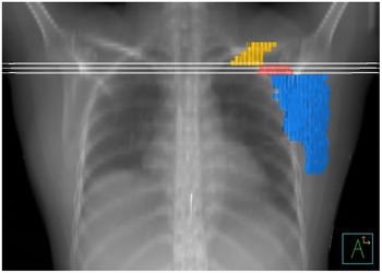
All plans were created with the use of the Pinnacle3 treatment planning system v.16.0 (Philips Radiation Oncology Systems, USA), and doses were delivered by PrimusTM linear accelerator (Siemens Healthcare, Germany). The treatment area was specified according to Halperin et. al. Reference Halperin, Perez and Brady19 guideline. The medial, lateral, superior and inferior borders were drawn on the images of the CW and SC by radiation oncologists. The ipsilateral lung and heart were denealiated. LN I, II and III were delineated in order to evaluate the performance of various methods according to NRG Oncology. 20 The region of interest was created to investigate the dose at the junction area (so called ‘ROIg’). ROIg was constructed from the body surface on CT image at the junction between the tangential and SC fields including the adjacent upper and lower slices, as is shown in Figure 1. Doses (2·67Gy and 2·65Gy in 15 and 16 fractions, respectively) were prescribed at the isocentre/prescribed point on the CW and the prescribed point on the SC.
Region of interest at the matching area (ROIg) presents in the three white lines. Lymph node levels I, II and III are present in blue, red and yellow, respectively.

Beam geometry methods
All plans employed the SIT, but beam arrangements were prepared differently. All three different methods were created on an image set of each patient and described as follows.
Coplanar field border (CFB) method
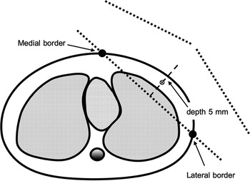
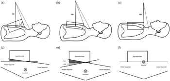
This proposed method was created to provide an alternative choice for PMRT. Accordingly, the slice at the centre between the superior and inferior border of the CW was selected. The isocenter was then placed on the perpendicular line of the midplane between the medial and lateral borders. This point was located 5 mm under the surface as is shown in Figure 2. The OTFs were then created. Instead of employing the parallel the chest curve, the collimator angle was determined as follow and demonstrated in Figure 3a:
 $$\theta = 90 - \cos^{ - 1}\left( {{x \over {100}}} \right)$$
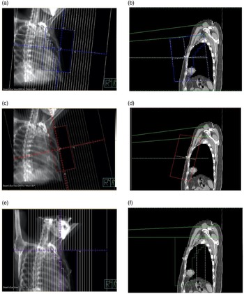
where θ represents the collimator angle in the degree and x represents the upper half of the tangential field in centimeters. Figure 3d demonstrates the beam direction used in this method. Multi-leaf collimators blocked the lung at 2 cm from the rib cage. With this block, the partial heart was also blocked. The lateral tangential field was created with the use of opposed beam geometry. The SCF was then prepared by sharing the isocenter with the OTFs. This field was limited by radiation oncologists. The inferior border of the SCF and the superior border of the OTFs were along the same line. The rectangular open field is normally used so as to exclude the head of the humerus. The prescribed point was neccesarilly used for this off-axis field. The point was placed on the slice of the mid-thoraxic vertebral body level I. On this slice, this point was located 3 cm from the midline and at a depth of 3.5 cm from the surface on average. Figure 4a and 4b present an example of beam geometry of this method.
Diagram of the isocenter location of the opposing tangential fields.

Diagram of various beam geometry method. top line shows the collimator direction, whereas the bottom line is the beam geometry. The CFB method demonstrates in (a) and (d). Figure (a) also shows the parameters for the calculation of the collimator angle (θ). The TTM method is in (b) and (e). Finally, the HBB method is in (c) and (f).

Demonstrates the example of three different methods in the 3DRT technique. (a) and (b) illustrate the CFB method. (a) shows the medial tangential field with the blocked lung, whereas Figure 4B demonstrates the off-axis SC field and the coplanar between the superior border of the medial tangential and inferior border of the SC field. (c) and (d) present the example of beam geometries by utilising the TTM method. The opposing tangential fields were placed on the chest wall as illustrated in (c). The off-axis SC field was created as shown in (d–f) present the beam geometries by using the HBB method. (e) reveals the medial tangential of the upper half beam block field. The beam uses the MLC for lung block. (f) demonstrates the SC field of the lower half beam block field.

Traditional three-field monoisocenter (TTM) method
All patients had been previously treated with this method. The isocenter was placed at the same location as was used in the CFB method. The OTFs were then prepared as well. Collimators were used to rotate the treatment fields parallel to the chest curve, as is demonstrated in the Figure 3b. This tangential fields involved the lung no further than 2 cm at the isocenter plane. The SCF and the prescribed point were created by employing the same method used in the CFB. A diagram of this method is illustrated in Figure 3b and 3e. An example of the beam geometry is demonstrated in Figure 4c and 4d.
Half beam block (HBB) method
HBB is commonly used in conjunction with 3DRT in many treatment regions. The slice of the superior border of the CW was selected for this approach. The isocenter was placed in the same method used in the CFB. The lower half of the beam was used for the OTFs, whereas the upper half was used for the SC. The collimator angle was paralleled on the longitudinal axis. The lung and heart were blocked in the same way they were in the CFB. The SCF was also created in the same manner as was employed in the previous method. Figure 3c and f present a diagram of this method. Because the isocenter of this method was partially blocked, the prescribed point had to be created on the OTFs and the SCF. The prescribed points were placed at the same location as in the CFB. Accordingly, placements were designated at not only the isocenter of the OTFs but also at the prescribed point of the SCF. An example of this method is shown in Figure 4e and f.
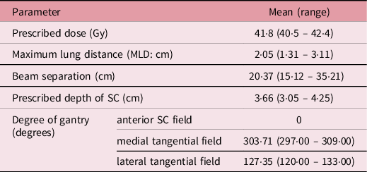
To evaluate the performance among different methods, all beam angles of the three different approaches were fixed as were the radiation doses prescribed to the same points for all methods. The plan parameters of the patients are shown in Table 1. The maximum lung distance was the distance between the tangential field border and the rib cage. Beam separation was measured between the medial and lateral borders. The depth of the prescribed point on the SC was set and measured from the skin.
Treatment plan parameters

Plan evaluations
All treatment plans were accepted at 95% of the prescribed dose, whereas the dose coverage was evaluated by radiation oncologists. The acceptable criteria was based on the dose coverage and overdose volume. This dose coverage did not exceed the area of lung tissue by more than 2 cm from the rib cage. The dose of 2cc (D2cc) also did not exceed 115% of the prescribed dose. The dosimetric parameters were evaluated on the ipsilateral lung and heart and investigated on LN I, II, II and ROIg. All levels of LNs were analysed on the mean dose (Dmean) and dose at 95% volume (D95%). The OARs were analysed on Dmean, percent volume at 5Gy (V5Gy), 20Gy (V20Gy) and 30Gy (V30Gy) for the ipsilateral lung and Dmean, V10Gy and V40Gy for the heart. Furthermore, ROIg was analysed on D2%, D2cc and D10cc.
Statistical analysis
The results were analysed at 95% confidence intervals of the statistics. The Shapiro–Wilk test was used to evaluate the normal distribution of the dosimetric parameters. Repeated ANOVA test was used to evaluate the normal distribution of the results, whereas Friedmen’s two-way ANOVA was used for the other group.
Decision score
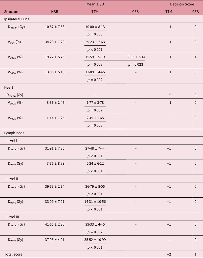
Decision score was used to evaluate the performance of each method. The score was considered based on the results that exhibited a significant difference when using the HBB benchmarks. Either plus one or minus one indicated that a result was either better or lesser, respectively, than the HBB method. No significant differences were observed between HBB and each of the other methods and, thus, they received no score.
Results
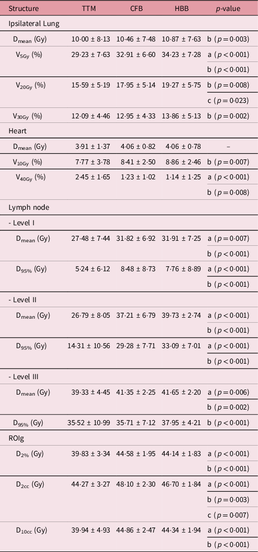
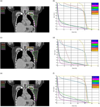
The test results indicate that V5Gy, V20Gy and Dmean values of LN I were of normal distribution, whereas the others revealed non-normal distribution. Table 2 presents the mean and standard deviation (Mean ± SD) values of the parameters when using various methods. The TTM method delivered significantly lower Dmean and V5Gy, V20Gy and V30Gy values on the ipsilateral lung than the HBB method (p = 0·003, p = 0·001, p = 0·008 and p = 0·002, respectively). The CFB method presented a significantly higher V5Gy value than the TTM (p = 0·001), but a significantly lower V20Gy value than the HBB method (p = 0·023). The heart is one of the primary considerations when a lesion occurs in the left breast. The Dmean value of the heart revealed no significant differences among the three methods. The HBB method exhibited a significantly higher V10Gy value on the heart than the TTM method (p = 0·007). With regard to the V40Gy value of the heart, the TTM method provided a significantly higher value than the CFB and HBB methods (p < 0·001 and p = 0·008, respectively). The LN coverage can present at the junction. The TTM method exhibited significantly lower Dmean and D95% values on LNs I and II than both the CFB (p ≤ 0·007) and the HBB (p ≤ 0·001) methods. LN III revealed parallel results to other LNs with the exception of Dmean (p ≤ 0·006). The D95% value of LN III was found to be significantly higher when the HBB method was used rather than the CFB method (p = 0·001). ROIg was created to determine the overdose/underdose specifications at the field junction The TTM method exhibited a significantly lower dose on ROIg than either the CFB (p ≤ 0·001) or the HBB (p ≤ 0·003) methods for all dosimetric parameters. However, the CFB method revealed a significantly higher D2cc value on ROIg than the HBB method (p = 0·007). Figure 5 illustrates the dose distribution and dose volume histogram of an example case with the use of the CFB, TTM and HBB methods.
Mean and standard deviation (Mean ± SD) of dosimetric parameters by using various beam geometries

*a is TTM versus CFB methods, b is TTM versus HBB methods and c is CFB versus HBB methods.
Dose distribution and dose volume histogram (DVH) of the example case by utilising three different methods. Left column is the dose distribution where the evaluated dose presents in light green line (40·3 Gy). Right column is the histogram between the proportion of normalised organ volume versus the absolute dose in Gy. Top, middle and bottom are the example results by employing the CFB, TTM and HBB, respectively.

Table 3 presents the significant difference parameters and decision scores of various methods by applying the HBB benchmark. The scores indicate only the significant differences between the methods. The Dmean value of the heart is the only parameter that indicated no significant differences among the various methods. The V20Gy value of the ipsilateral lung when using the CFB method was also the only parameter that exhibited a significant difference when compared with the HBB method.
Mean and standard deviation (Mean ± SD) of the dosimetric parameters using HBB benchmark, including the decision score

Discussion
The SIT with CFB method is an alternative beam geometry of our centre. The proposed method focused to improve the dose homogeneity at the junction. Performance was then evaluated using the dosimetric parameters on various levels of LN, OARs and ROIg. The TTM method is not widely used in many centres. The underdose was found at the junction by utilising these methods with Wing-board.
Various LN levels were not necessarily treated in the PMRT. These organs lie within the region of the matching fields that indicate dose homogeneity between the OTFs and SCF. Reference Banaei, Hashemi and Bakhshandeh4,Reference Nadi, Abedi-Firouzjah, Banaei, Bijari and Elahi21 To ensure the evaluation of the overdose/underdose area only at the junction, ROIg was created. The results indicate slightly equivalent dose levels between D2% and D10cc. The CFB method focused on the coplanar plan only the medial tangential field. This proposed method would slightly increase the dose at the junction by the divergent of the lateral tangential field as is shown in Figure 3e. This overdose was increased to 106·6%, 115·1% and 107·3% of the mean prescribed dose for D2%, D2cc and D10cc, respectively.
By using the SIT without HBB method, the outcomes of this study were almost inline with the study of Zhang et al. Reference Zhang, Dilworth, Marina, Chen, Benedetti and Liu7 However, hybrid planning was utilised in their work but not for this study. A perfect dose homogeneity was revealed in their research when the rotations of the couch were required. The couch rotation was unnecessarily used in this proposed method resulting in a high dose on the medial border of the SCF; however, the overdose volume was determined to be acceptable.
The decision score clearly showed that the OARs of the TTM method received a lower dose than the others. However, this method also revealed a low dose by the observation of the LNs. The CFB method exhibited a good level of performance, but the D2cc value of the ROIg received a significantly higher dose than in the HBB method (p = 0·007). The ROIg was not included in the decision score because the HBB method also provided a high dose at the junction (D2cc = 111·7% of the mean prescribed dose). The degree of dose homogeneity in the large volume, however, revealed no significant differences between the CFB and HBB methods.
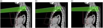
The CFB method provided a full range of field size for the CW and improved dose homogeneity at the junction. The degree of dose homogeneity was not as optimal as the HBB method, but the volume of overdose was acceptable. The ipsilateral lung of the CFB method received a lower dose than with the HBB method because of the beam divergence. The off-axis SCF from the CFB method projected obliquely up to the apex of the lung as is shown in Figure 6. As a consequence of the rotation of the collimator, the dose was fulfilled at the junction when using the CFB method, but not when using the TTM method. The proposed method increased the dose on OARs when compared with the TTM method, but these outcomes were determined to be acceptable. The proposed method, however, provided an advantage for the matching fields, but this is related to the accuracy of the treatment unit as all beam patterns. Thus, the uncertainty of the relevant mechanics requires further investigation. The clinical outcome is one of the consideration issues. The skin reaction at the junction and the tumor recurrence required an exploration.
Impact of SC field direction by utilising three different method. The off-axis beam shows the divergence out of the lung apex on the CFB (a) and TTM (b) methods, whereas no divergence at the central beam is on the HBB (c) method.

Conclusion
PMRT has recently required 3DRT. The number of patients and treatment times were most impacted when sophisticated techniques were used. This study has proposed a alternative beam geometry for PMRT. Consequently, the CFB method improved the degree of dose homogeneity at the junction between the tangential and SC fields, whereas the dose delivered to OARs remained low. This method was found to not only produce comparable results but also minimised the limitations of the HBB method.
Conflict of Interest
None.