

Introduction
The guano deposit situated on Pabellón de Pica Mountain, 1.5 km south of Chanabaya village, Iquique Province, Tarapacá Region, Chile (20°55’S, 70°08’W) belongs to the famous belt of guano occurrences of the Atacama Desert stretching along the northern coast of Chile (Ericksen, Reference Ericksen1981; Pankhurst and Herve, Reference Pankhurst, Herve, Moreno and Gibbons2007; Appelton and Nothold, Reference Appleton and Nothold2002; Bojar et al., Reference Bojar, Walter, Baumgartner and Färber2010). Pabellón de Pica is one of the most interesting mineralogical occurrences in this area being the type locality of several nitrogen-bearing and organic minerals: ammineite, CuCl2(NH3)2 (Bojar et al., Reference Bojar, Walter, Baumgartner and Färber2010), joanneumite, Cu(C3N3O3H2)2(NH3)2 (Bojar et al., Reference Bojar, Walter and Baumgartner2017), chanabayaite, Cu4(N3C2H2)4(NH3)4Cl2(Cl,OH)2⋅H2O (Chukanov et al., Reference Chukanov, Zubkova, Möhn, Pekov, Pushcharovsky and Zadov2015a), shilovite, Cu(NH3)4(NO3)2 (Chukanov et al., Reference Chukanov, Britvin, Möhn, Pekov, Zubkova, Nestola, Kasatkin and Dini2015b), antipinite, KNa3Cu2(C2O4)4 (Chukanov et al., Reference Chukanov, Aksenov, Rastsvetaeva, Lysenko, Belakovskiy, Färber, Möhn and Van2015c), möhnite, (NH4)K2Na(SO4)2 (Chukanov et al., Reference Chukanov, Aksenov, Rastsvetaeva, Pekov, Belakovskiy and Britvin2015d) and triazolite, NaCu2(N3C2H2)2(NH3)2Cl3⋅4H2O (Chukanov et al., Reference Chukanov, Zubkova, Möhn, Pekov, Belakovskiy, Van, Britvin and Pushcharovsky2018).
Specimens with the new mineral ammoniotinsleyite, ideally (NH4)Al2(PO4)2(OH)⋅2H2O, were collected at Pabellón de Pica in 2019 by one of the authors (G.M.). Ammoniotinsleyite is named as the ammonium analogue of tinsleyite, KAl2(PO4)2(OH)⋅2H2O, with prevailing of NH4+ over K+. The new mineral and its name were approved by the Commission on New Minerals, Nomenclature and Classification of the International Mineralogical Association (IMA2019-128, Chukanov et al., Reference Chukanov, Möhn, Pekov, Zubkova, Ksenofontov, Belakovskiy, Vozchikova, Britvin and Desor2020). The holotype specimen is deposited in the collection of the Fersman Mineralogical Museum of the Russian Academy of Sciences, Moscow, Russia, with the registration number 5510/1.
Occurrence, general appearance and physical properties
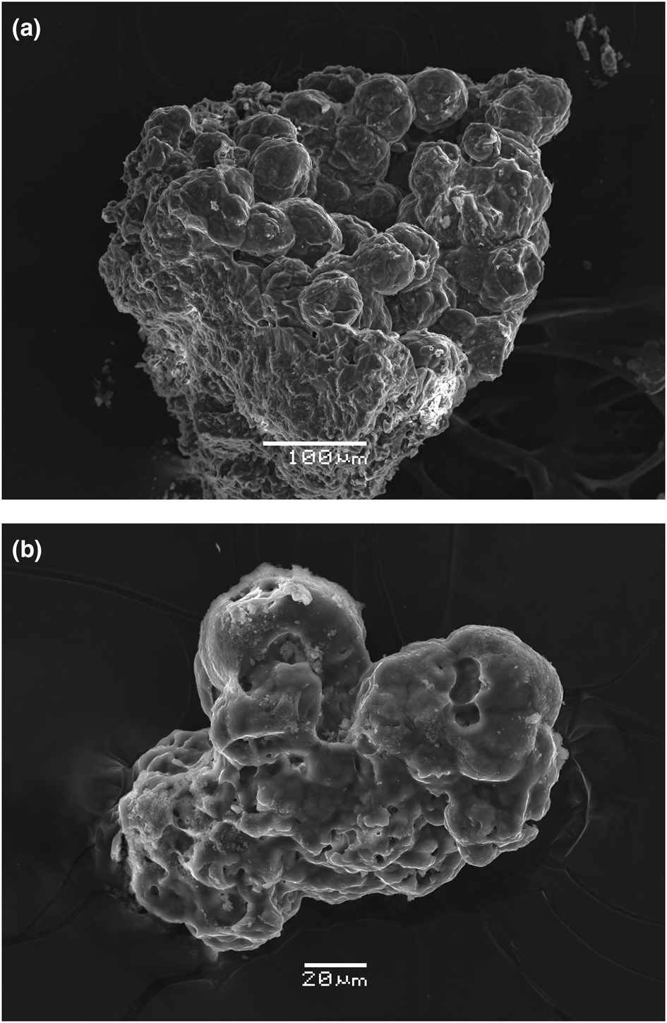
Ammoniotinsleyite occurs in a guano deposit on the lower part of the steep southern slope of Pabellón de Pica Mountain. It forms globular, partly porous polycrystalline aggregates up to 3 mm across in cavities of granular aggregates of associated minerals (Figs 1 and 2). Sizes of elongate single-crystal fragments are below 10–15 μm. Associated minerals are halite, gypsum, salammoniac and clay minerals.
Aggregates of ammoniotinsleyite (pink) in association with gypsum and halite. FOV 0.7 mm. Photo: A.V. Kasatkin, cotype specimen.

(a,b) Scanning electron microscopy (secondary electron) images of aggregates of ammoniotinsleyite, holotype specimen.

Aggregates of ammoniotinsleyite are translucent to transparent. The colour is pink to pale violet. The lustre is vitreous. The streak is white. Mohs hardness is 4. Ammoniotinsleyite is brittle. Cleavage was not observed and the fracture is uneven. Density measured by flotation in heavy liquids (mixtures of bromoform with benzene) is 2.42(2) g cm–3 while the density calculated using the empirical formula is 2.451 g cm–3.
Ammoniotinsleyite is optically biaxial (+), α = 1.557(2), β = 1.559 (calculated using 2Vmeas.), γ = 1.563(2) (λ = 589 nm); 2Vmeas. = 75(10)°. Dispersion of optical axes is not observed. Under the polarising microscope, the mineral is pale pink and non-pleochroic. The Gladstone-Dale compatibility index (Mandarino, Reference Mandarino1981) is: 1 – (K p/K c) = 0.042 (good) with D calc. and 0.030 (excellent) with D meas.
Infrared spectroscopy
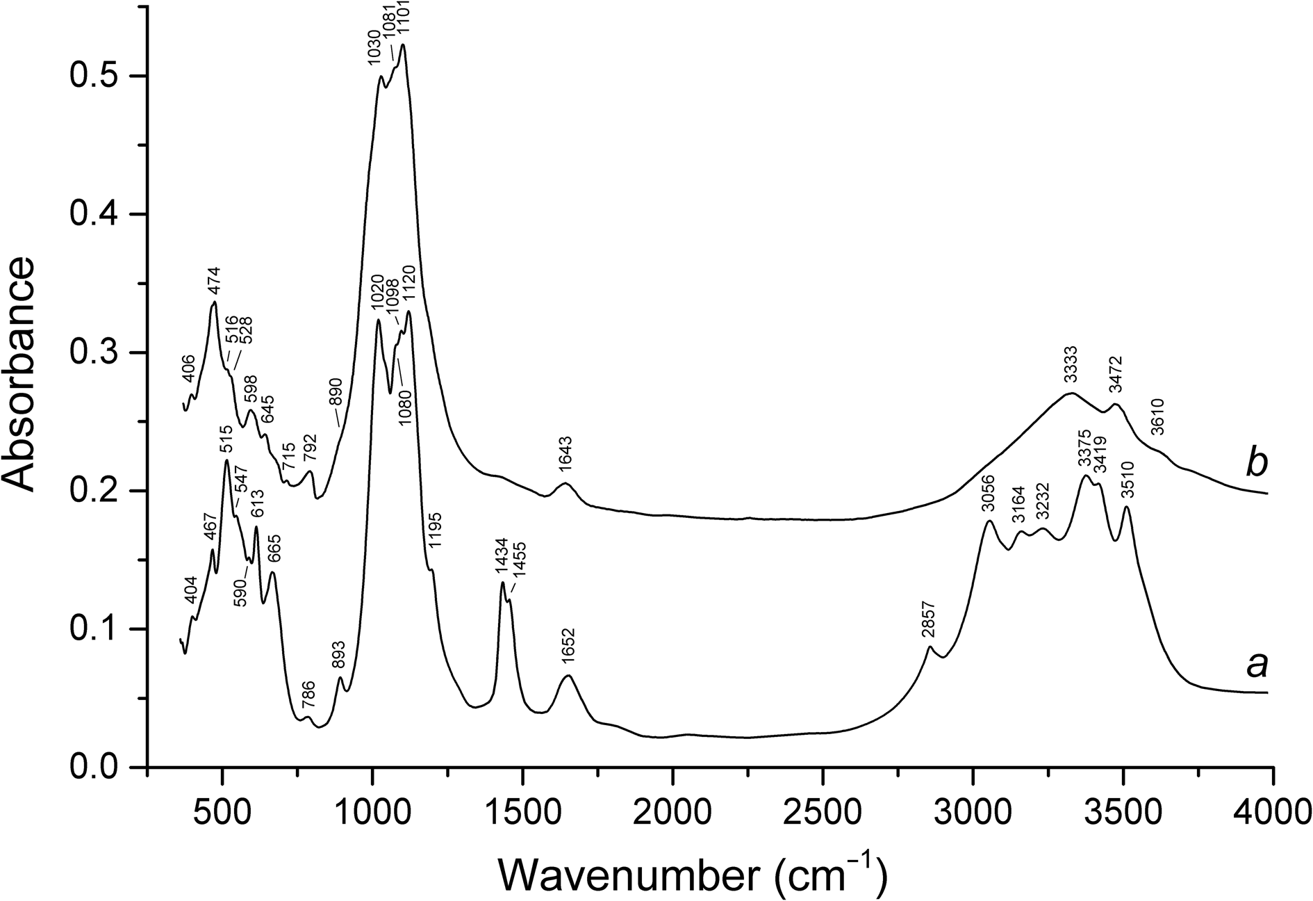
In order to obtain infrared (IR) absorption spectra (Fig. 3), powdered samples of ammoniotinsleyite and tinsleyite (used for comparison) were mixed with dried KBr, pelletised, and analysed using an ALPHA FTIR spectrometer (Bruker Optics) in the range 360–4000 cm–1 with a resolution of 4 cm–1. A total of 16 scans were collected for each spectrum. The IR spectrum of an analogous pellet of pure KBr was used as a reference.
Powder infrared absorption spectra of (a) ammoniotinsleyite and (b) Fe-rich tinsleyite with the composition (K0.86Ca0.10Na0.04)(Al1.36Fe0.57Mg0.05)(PO4)2.00(OH)⋅2H2O from Mejillones Peninsula, Mejillones, Antofagasta, Chile.

Wavenumbers of IR absorption bands of ammoniotinsleyite and their assignments are (cm–1; s – strong band, w – weak band, sh – shoulder): 3510s, 3419s, 3375s (O–H stretching vibrations of OH groups and H2O molecules), 3232, 3164, 3056s, 2857 (N–H stretching vibrations of NH4+ cations), 1652, (bending vibrations of H2O molecules), 1455, 1434 (bending vibrations of NH4+ cations),1195, 1120s, 1098s, 1080sh, 1020s [ν3(F 2) asymmetric stretching vibrations of PO43– ions], 893 [ν1(A 1) symmetric stretching vibrations of PO43– ions], 786w (Al⋅⋅⋅O–H bending vibrations), 665, 613, 590 [ν4(F 2) – asymmetric bending vibrations of PO43– ions], 547sh, 515s (Al–O stretching vibrations), 467, 404 (lattice modes, possibly involving Fe3+–O and Mg–O stretching vibrations, librational vibrations of H2O molecules and ν2(E) symmetric bending vibrations of PO43– ions]. The band assignment was carried out in accordance with Nakamoto (Reference Nakamoto2008, Reference Nakamoto2009), Chukanov (Reference Chukanov2014), and Chukanov and Chervonnyi (Reference Chukanov and Chervonnyi2016).
The IR spectrum of ammoniotinsleyite differs from that of tinsleyite by the presence of distinct bands of NH4+ in the ranges 1400–1500 and 2800–3300 cm–1. In addition, most bands in the IR spectrum of tinsleyite are observed at lower frequencies than analogous bands of ammoniotinsleyite which is due to the differences in masses of the NH4+ and K+ cations as well as hydrogen bonds formed by NH4+.
Chemical data
Four chemical analyses were carried-out using a Tescan VEGA-II XMU scanning electron microscope equipped with an energy dispersive spectrometer INCA Energy 450 (EDS mode, 20 kV, 600 pA, beam diameter 0.16–0.18 μm) for K, Mg, Ca, Al, Fe and P. The beam was rasterised on an area 16 μm × 16 μm to minimise unstable sample damage.
Gas chromatography on products of ignition in an oxygen stream at 1200°C was used to determine H, N and C. Contents of carbon and other elements with atomic numbers higher than 6 are below detection limits. Analytical data are given in Table 1.
Chemical composition of ammoniotinsleyite.

Total iron was measured as Fe2O3; S.D. = standard deviation.
*The value of 7.25 wt.% for (NH4)2O is calculated from 3.9 wt.% N determined by gas chromatography.
**The value of 14.89 wt.% for H2O is calculated from the rest of hydrogen after subtraction of H in NH4+ cations. The total H content determined by gas chromatography is 2.79 wt.% which corresponds to 24.9 wt.% H2O.
The charge-balanced empirical formula (based on 11 O apfu) is [(NH4)0.88K0.10Ca0.02)]Σ1.00(Al1.86Fe3+0.09Mg0.03)Σ1.98(PO4)1.96(OH)1.05⋅2.11H2O. Minor excess of H2O relative to the idealised formula may be due to adsorbed water. The idealised formula is (NH4)Al2(PO4)2(OH)⋅2H2O which requires (NH4)2O 8.27, Al2O3 32.37, P2O5 45.06, H2O 14.30, total 100.00 wt.%.
Evolution of NH3 from ammoniotinsleyite on moderate heating in air was detected organoleptically (by smell) and with a wet pH-paper. No gas evolution was observed in 20% HCl, confirming the absence of CO32– groups.
X-ray diffraction and crystal structure
Single-crystal X-ray diffraction studies of ammoniotinsleyite could not be carried-out due to the absence of suitable single crystals: aggregates of ammoniotinsleyite (Figs 1 and 2) are polycrystalline and consist of very small imperfect individuals. For this reason, the crystal structure of ammoniotinsleyite was refined based on powder X-ray diffraction data, using the Rietveld method.
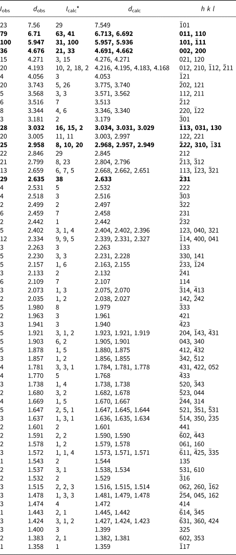
Powder X-ray diffraction data (Table 2) were collected with a Rigaku R-AXIS Rapid II single-crystal diffractometer equipped with cylindrical image plate detector (radius 127.4 mm) using Debye-Scherrer geometry, CoKα radiation (rotating anode with VariMAX microfocus optics), 40 kV, 15 mA and 15 min exposure. Angular resolution of the detector is 0.045° (for 2θ) and pixel size is 0.1 mm. The data were integrated using the software package Osc2Tab (Britvin et al., Reference Britvin, Dolivo-Dobrovolsky and Krzhizhanovskaya2017).
Powder X-ray diffraction data (d in Å) of ammoniotinsleyite.

*For the calculated pattern, only reflections with intensities ≥ 1% are given.
The strongest lines are given in bold.
Diffraction peaks are well indexed in the monoclinic unit cell, space group P21/n. The unit-cell parameters calculated from the powder data are: a = 9.5871(1), b = 9.6089(1), c = 9.6467(2) Å, β = 103.4461(8)° and V = 864.31(2) Å3.
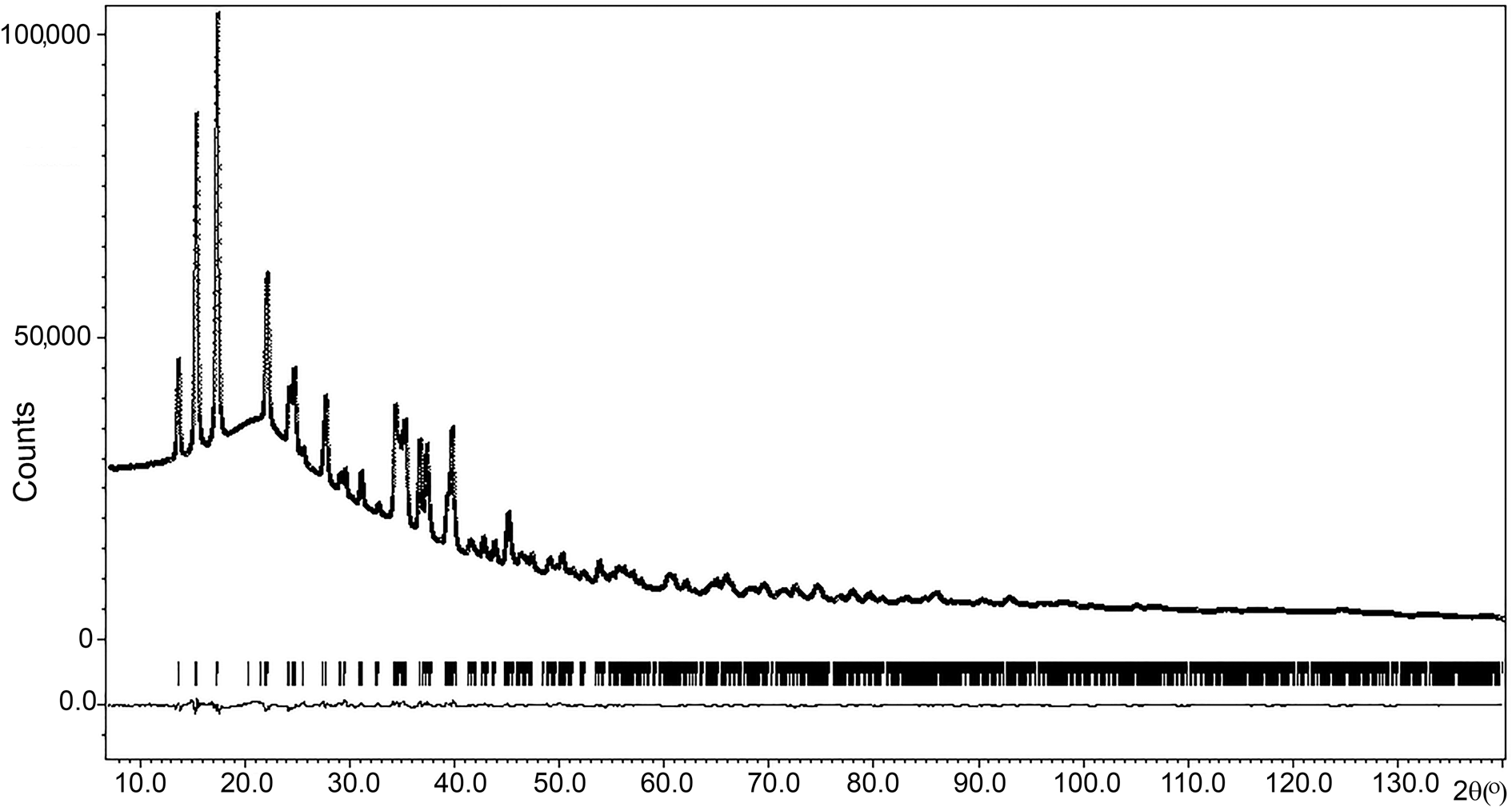
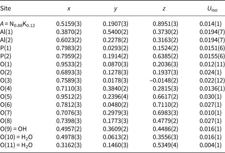
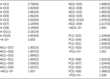
Refinement of the crystal structure of ammoniotinsleyite was performed using the Rietveld method using the structure of tinsleyite (Dick, Reference Dick1999) as the starting model except for H atoms which were excluded from the refinement. Data treatment and the Rietveld structure analysis were carried-our using the JANA2006 program package (Petříček et al., Reference Petříček, Dušek and Palatinus2006). The profiles were modelled using a pseudo-Voigt function. The structure was refined in isotropic approximation of atomic displacements. The P–O and Al–O interatomic distances were softly restricted nearby the values obtained for the synthetic tinsleyite (Dick, Reference Dick1999). A small amount of K (12%) was added to the N site and fixed during the refinement assuming the K + Ca admixture according to chemical data. The final agreement factors are: R p = 0.0071, Rw p = 0.0093, R obs = 0.0167. The observed and calculated powder X-ray diffraction diagrams demonstrate a very good agreement (Fig. 4). Coordinates and thermal displacement parameters of atoms are given in Table 3 and selected interatomic distances in Table 4. The crystallographic information files have been deposited with the Principal Editor of Mineralogical Magazine and are available as Supplementary material (see below).
Observed and calculated powder X-ray diffraction patterns of ammoniotinsleyite. The solid line corresponds to calculated data, the crosses correspond to the observed pattern and vertical bars mark all possible Bragg reflections. The difference between the observed and calculated patterns is shown by the curve at the bottom.

Coordinates and isotropic displacement parameters (U iso, in Å2) of atoms for ammoniotinsleyite.

Selected interatomic distances (Å) in the structure of ammoniotinsleyite.

Discussion
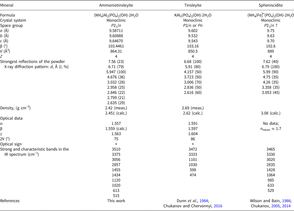
Ammoniotinsleyite, (NH4)Al2(PO4)2(OH)⋅2H2O, is a member of the leucophosphite group with the general formula AB 2[PO4]2(OH)⋅2H2O (A = NH4 or K; B = Al or Fe3+). This new mineral is the ammonium analogue of tinsleyite, KAl2(PO4)2(OH)⋅2H2O, with prevailing NH4+ over K+ and the aluminium analogue of spheniscidite, (NH4)Fe3+2(PO4)2(OH)⋅2H2O, with prevailing Al3+ over Fe3+ (Table 5). The synthetic analogue of ammoniotinsleyite is well-known (Parise, Reference Parise1984; Pluth et al., Reference Pluth, Smith, Bennett and Cohen1984; Aubert et al., Reference Aubert, Porcher, Souhassou and Lecomte2003; Parnham et al., Reference Parnham, Drylie, Wheatley, Slawin and Morris2006; Vaughan et al., Reference Vaughan, Yennawar and Perrotta2012). A crystal chemical review on minerals of the leucophosphite group and related synthetic compounds was reported by Yakubovich et al. (Reference Yakubovich, Massa and Dimitrova2012).
Comparative data for ammoniotinsleyite, tinsleyite and spheniscidite.

In the structure of ammoniotinsleyite (Fig. 5), Al cations occupy two crystallographically non-equivalent sites with octahedral coordination. As mentioned in previous work (Yakubovich et al., Reference Yakubovich, Massa and Dimitrova2012 and references therein), the remarkable feature of the crystal structure of members of the leucophosphite group is the occurrence of tetrameric units of four AlO6 octahedra built by two Al(1) octahedra sharing an edge and two additional Al(2) octahedra attached at each side of this common edge via common corners. These octahedral units are cross-linked by PO4 tetrahedra, thus forming a heteropolyhedral pseudo-framework with channels running along the a, b and c axes which in ammoniotinsleyite host large NH4+ ions (with K admixture) and H2O molecules. There are two crystallographically non-equivalent sites of water molecules [O(10) and O(11)]; O(10) participates in the coordination polyhedra of Al(2) and NH4 cations whereas O(11) is only involved in the coordination polyhedron of NH4+.
The crystal structure of ammoniotinsleyite: view along the b axis (positions of O atoms of OH groups are shown as green circles, O atoms of H2O molecules are given as blue circles). The unit cell is outlined.

Ammoniotinsleyite is a product of a deep alteration of bird guano over the course of geologically long time processes. Obviously, guano was the primary source of N and P in this mineral, and the host rock was the source of Al.
Comparative data for ammoniotinsleyite and related minerals are given in Table 5. As one can see from these data, wavenumbers of IR bands can be considered as a reliable diagnostic characteristic of ammoniotinsleyite.
Acknowledgements
The authors are grateful to Béla Fehér and two anonymous reviewers for valuable comments. This work was supported by the Russian Foundation for Basic Research, grants nos. 18-29-12007-mk. A part of this work (IR spectroscopy and chemical analyses) was carried-out in accordance with the state task, state registration number ААAА-А19-119092390076-7. The authors thank the X-ray Diffraction Centre of Saint-Petersburg State University for instrumental and computational resources.
Supplementary material
To view supplementary material for this article, please visit https://doi.org/10.1180/mgm.2020.45