Introduction
Cucullanidae Cobbold, 1864 is a family of nematodes that includes parasites of marine, brackish and freshwater fish, a few species being reported in aquatic chelonians (Petter, Reference Petter1974; Anderson et al., Reference Anderson, Chabaud and Willmott2009). Six genera are assigned to this family, namely Campanarougetia Le Van Hoa & Pham-Ngoc-Khue, 1967; Cucullanus Müller, 1777; Dichelyne Jägerskiöld, 1902; Neocucullanus Travassos, Artigas & Pereira, 1928; Oceanicucullanus Schmidt & Kuntz, 1969; and Truttaedacnitis Petter, 1974 (sensu Petter, Reference Petter1974; Anderson et al., Reference Anderson, Chabaud and Willmott2009; Gibbons, Reference Gibbons2010). Nevertheless, some authors consider Truttaedacnitis as a subgenus of Cucullanus (e.g. Moravec, Reference Moravec1979, Reference Moravec1998).
Cucullanus and Dichelyne are the most representative genera within Cucullanidae considering the number of valid species, and both are taxonomically problematic. The boundaries between these genera have been demonstrated to be blurry, since their main distinctive traits (e.g. presence/absence of an intestinal caecum and cuticular frame in the oesophastome) are easy to overlook or to be misinterpreted, as well as being inconsistent in some cases (see Li et al., Reference Li, Du, Xu, Guo, Wang and Zhang2014). Furthermore, the high number of poor descriptions and the rather uniform morphology of cucullanids, represent a real challenge for taxonomists when dealing with these parasites (Vieira et al., Reference Vieira, Pereira, Pantoja, Soares, Pereira, Timi, Scholz and Luque2015).
Phylogenetic relationships within Cucullanidae are still unresolved, the family is assumed to be monophyletic, but the real relationships among its genera are poorly understood, since molecular data are scarce and the systematic problems are numerous (Choudhury & Nadler, Reference Choudhury and Nadler2016). Thus, integrative approaches using both morphology and molecular data are necessary to shed light on the phylogeny of the Cucullanidae and resolve the existing systematic issues.
During a parasitological study, some cucullanids were recovered from three different species of freshwater fishes from northern Brazil. Morphological examination of these nematodes has revealed that the specimens found in Oxydoras niger (Valenciennes, 1821) (Actinopterygii: Doradidae) belong to Cucullanus grandistomis (Ferraz & Thatcher, 1988) and those from Cichla melaniae Kullander & Ferreira, 2006 (Actinopterygii: Cichlidae) and C. pinima Kullander & Ferreira, 2006 represent a new species. Parasites were described in detail using light and scanning electron microscopy (SEM), and genetically characterized. A preliminary phylogeny of Cucullanidae was also performed.
Materials and methods
Collection and examination of nematodes
Fish were collected by hook and trawler nets, with the help of local fisherman. One specimen of C. melaniae (total body length 24 cm) and one of O. niger (total body length 1.06 m) were collected in River Xingu, Municipality of Altamira, State of Pará, Brazil, during September 2013. One specimen of C. pinima (total body length 49.5 cm) was collected in River Jamarí, Municipality of Ariquemes, State of Rondônia, Brazil during October 2015. Host nomenclature and classification follows Froese & Pauly (Reference Froese and Pauly2016). Nematodes found alive were washed in saline, fixed in hot 4% formalin and preserved in 70% ethanol. For morphological examinations, nematodes were cleared in glycerine. The middle body parts of some specimens were excised and fixed in molecular-grade 96–99% ethanol for genetic studies; the anterior and posterior parts were fixed in 4% formalin for morphological identification. Anterior and posterior ends of these specimens, i.e. hologenophores (the voucher specimens from which the molecular sample is directly derived; for more details see Astrin et al., Reference Astrin, Zhoun and Misof2013) were identified based on their morphology.
Drawings were made using a drawing tube attached to a microscope Olympus BX51 (Olympus, Center Valley, Pennsylvania, USA). Measurements are given in micrometres, unless otherwise stated. Specimens used for SEM were dehydrated through a graded ethanol series, dried in hexamethyl disilazane, coated with gold and examined in a JEOL JSM-740 1F (JEOL Inc., Peabody, Massachusetts, USA), at an accelerating voltage of 4 kV. The system adopted for describing male caudal papillae is according to Moravec & Justine (Reference Moravec and Justine2011), Yooyen et al. (Reference Yooyen, Moravec and Wongsawad2011) and Pereira et al. (Reference Pereira, Vieira and Luque2014, Reference Pereira, Vieira and Luque2015b). The terminology used to describe and classify the cuticular frame in the oesophastome follows that of Berland (Reference Berland1970).
Specimens deposited in the Coleção Helmintológica do Instituto Oswaldo Cruz (acronym CHIOC) of Cucullanus tucunarensis Lacerda, Takemoto, Marchiori, Martins & Pavanelli, 2013 (holotype male and allotype female, CHIOC 35077a, b) and C. grandistomis (Ferraz & Thatcher, 1988) (two female paratypes, CHIOC 32456), were re-examined. Newly collected specimens were deposited in CHIOC.
Molecular and phylogenetic analyses
Genomic DNA was isolated from small tissue samples, using DNeasy Blood & Tissue Kit (QIAGEN, Hilden, Germany), following the manufacturer's instructions. The small subunit (SSU) rRNA gene (18S) was amplified by polymerase chain reactions (PCR) (volume 25 μl, consisting of 2.5 μl of 10 × PCR buffer minus magnesium, 1.0 μl of MgCl2 (50 mm), 2 μl of each deoxynucleoside triphosphate (dNTP) (2.5 mm), 0.25 μl of each oligonucleotide primer (10 mm), 0.2 μl of Platinum Taq DNA polymerase (5 U/μl) (Invitrogen, Carlsbad, California), 0.25 μl of bovine serum albumin (BSA), 16.5 μl of H2O and 2.0 μl of genomic DNA) using the PCR conditions and the primers Philonema F + PhilPCRr described in Černotíková et al. (Reference Černotíková, Horák and Moravec2011). The large subunit (LSU) rRNA gene (28S) was amplified by PCR (volume 25 μl, consisting of 2.5 μl of 10 × PCR buffer minus magnesium, 1.5 μl of MgCl2 (50 mm), 2 μl of each dNTP (2.5 mm), 0.25 μl of each oligonucleotide primer (10 mm), 0.2 μl of Platinum Taq DNA polymerase (5 U/μl) (Invitrogen), 0.25 μl of BSA, 16.0 μl of H2O and 2.0 μl of genomic DNA) using the PCR primers D2A (5′-ACA AGT ACC GTG AGG GAA AGT-3′) + D3B (5′-TGC GAA GGA ACC AGC TAC TA-3′) of Nunn (Reference Nunn1992) and cycling conditions of Pereira et al. (Reference Pereira, Tavares, Scholz and Luque2015a). PCR products were purified by enzymatic treatment with exonuclease I and shrimp alkaline phosphatase (Werle et al., Reference Werle, Schneider, Renner, Völker and Fiehn1994) and Sanger sequenced at GATC Biotech (Konstanz, Germany), using the PCR primers and two internal primers in the case of 18S (WF760 + WR800, see Černotíková et al., Reference Černotíková, Horák and Moravec2011). Contiguous sequences were assembled in Geneious (Geneious v. 9.1.5 created by Biomatters, available from http://www.geneious.com/) and deposited in the GenBank database under accession numbers KX752092, KX752094, KX752096 (SSU rDNA); KX752091, KX752093, KX752095 (LSU rDNA).
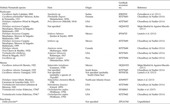
The taxa, for which sequences have been retrieved from GenBank for phylogenetic analyses, are listed in table 1. Due to data availability, only the 18S rDNA sequences were used to infer the phylogeny of Cucullanidae; they were selected according to a minimum length of 1286 bp (minus gaps) in gene superposition area of alignment for maximization of the number of informative characters. The 28S rRNA gene was used to evaluate genetic relationships within the newly collected samples. Datasets were aligned separately using the E-INS-i algorithm of the program MAFFT (Katoh et al., Reference Katoh, Misawa, Kuma and Myiata2002) implemented in Geneious. Gene alignments were then subjected to maximum likelihood (ML) and Bayesian inference (BI) analyses. ML and BI trees were generated under the Tamura–Nei model (TrN) of evolution, using PHYML (Guindon & Gascuel, Reference Guindon and Gascuel2003) and MrBayes (Huelsenbeck & Ronquist, Reference Huelsenbeck and Ronquist2001) Geneious plug-ins, respectively. The model of evolution was chosen under the Akaike informative criterion (AIC) using jModelTest 2 (Guindon & Gascuel, Reference Guindon and Gascuel2003; Darriba et al., Reference Darriba, Taboada, Doallo and Posada2012). Bayesian posterior probability (BPP) values were determined after running the Markov chain Monte Carlo (2 runs, 4 chains) for 4 × 10⁶ generations, with sampling frequency every 4 × 10³ generations and discarding the initial 25% of sampled trees (1 × 10⁶) as burn-in. For ML analysis, bootstrap resampling was performed with 1000 replications. Trees were rooted by Zeldia punctata (Thorne, 1925) according to previous phylogenies of Cucullanidae (Choudhury & Nadler, Reference Choudhury and Nadler2016).
List of nematodes whose sequences of small subunit rDNA were used for analyses, associated with hosts and geographical origin.

*Tentative identification of species according to Choudhury & Nadler (Reference Choudhury and Nadler2016); **used as the outgroup; †brackish-water fish; ‡anadromous fish.
Results
Cucullanidae Cobbold, 1864; Cucullanus Müller, 1777; Cucullanus opisthoporus n. sp.
Description
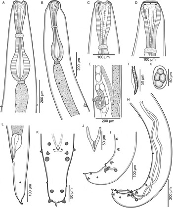
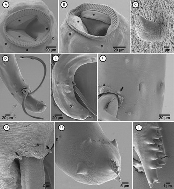
Small, whitish nematodes (figs 1, 2). Body elongate, tapering at both ends; cuticle smooth with inconspicuous transverse striations (fig, 2F–H). Lateral alae absent. Cephalic end rounded, with smooth constriction in females (fig. 1B). Oral opening dorsoventrally elongate, surrounded by wide membranous alae (collarette) armed with row of c. 68 conical and elongated basal teeth (fig. 2A, B). Four large submedian cephalic papillae and a pair of small lateral amphids surrounding the oral opening (fig. 2A, B). Inner circle of papillae absent. Oesophagus muscular, expanded at both ends, forming anterior narrow pseudobuccal capsule (oesophastome) and posterior wide basal dilatation (fig. 1A, B). Anterior part of inner wall of oesophastome strongly cuticularized, forming small spines; visible in lateral view (fig. 1D). Cuticular frame in oesophastome visible only in SEM micrographs (fig. 2A, B). Oesophageal valve well-developed, non-sclerotized, connecting oesophagus and intestine (fig. 1A, B). Nerve ring encircling oesophagus at 41.9–45.2% of its length (fig. 1A, B). Deirids drop-shaped (fig. 2C), anterior to excretory pore; both far posterior to oesophagus–intestine junction (fig. 1A, B). Postdeirids not observed. Cloacal and anal lips not protruded (figs 1H, I, L, and 2E–G). Apical appendage in tail tip, with small conical spines on its ventral surface (figs. 1H, I, K, L and 2D, E, H, I).
The morphology of Cucullanus opisthoporus n. sp., showing: (A) male and (B) female anterior ends, dorsoventral and lateral views, respectively; (C) male and (D) female cephalic ends, dorsoventral and lateral views, respectively; (E) vulva; (F) spicule distal end; (G) egg; (H) and (I) posterior end of male, lateral views; (J) gubernaculum, ventral view; (K) tail of male, ventral view; (L) tail of female, lateral view.

SEM of Cucullanus opisthoporus n. sp., showing: (A) and (B) cephalic end, apical and subapical views, respectively (asterisks indicate oesophastome cuticular frame); (C) deirid; (D) and (E) male posterior end, subventral and lateral views, respectively; (F) and (G) male cloacal region, ventral and subventral views, respectively (arrows indicate papillae on lip); (H) male tail tip, apical view; (I) caudal appendix of female. Abbreviations: a, amphid; c, cephalic papilla; p, phasmid; u, unpaired papilla.

Male – based on holotype with measurements inside parentheses and three paratypes with range of measurements from C. melaniae; one paratype from C. pinima with measurements inside brackets; all adult specimens. Body length (5.3) 4.6–5.9 [5.9] mm, maximum width 161–208 (208) [246]. Oesophagus (472) 382–472 [590] long, (119) 116–135 [111] wide at posterior end, representing (8.9) 6.7–8.9 [10]% of total body length. Oesophastome (75) 72–75 [128] long, (75) 75–87 [97] wide. Nerve ring, deirids and excretory pore located at (205) 205–264 [255], (662) 557–689 [768] and (809) 735–925 [983], respectively, from anterior body end. Posterior end of body ventrally bent. Spicules slightly sub-equal, slender, poorly sclerotized, with broad ala, ending in narrow tip (figs 1F, H and 2D), left and right (885) 739–927 [882] and (999) 954–1180 [961] long, respectively; representing (16.2) 12.9–17.2 [14.9]% and (18.9) 18.4–21.0 [16.3]% of total body length, respectively. Gubernaculum V-shaped in ventral view, (57) 48–65 [69] long (fig. 1J). Ventral pseudosucker absent. Caudal papillae mammilliform (figs. 1H, I, K and 2D–H). Five pairs of subventral precloacal papillae: first pair far anterior to second, third and fourth pairs close together, fifth pair near cloacal region; fourth and fifth pairs somewhat laterally displaced (fig. 1H, I). Anterior cloacal lip bearing pair of small papillae and conspicuous unpaired one (figs 1H, I, K and 2E–G). Two pairs of adcloacal papillae, one subventral and one lateral (figs 1H, I, K and 2E, F). Three pairs of postcloacal papillae: first pair subventral slightly posterior to cloacal opening, second and third pairs near end of tail, subventrally and subdorsally placed, respectively (figs 1H, I, K and 2E–H). Pair of lateral papilla-like phasmids between first and second postcloacal pairs (figs 1H, I, K and 2E, H). Tail (228) 138–228 [252] long.
Female – based on allotype with measurements in parentheses and one paratype from C. melaniae; one paratype from C. pinima with measurements inside brackets; all gravid specimens. Body length (5.6) 5.1 [7.5] mm, maximum width (237) 216 [331]. Oesophagus (507) 477 [660] long, (101) 89 [154] wide at posterior end, representing (9.0) 9.3 [8.8]% of total body length. Oesophastome (80) 85 [173] long, (69) 67 [120] wide. Nerve ring, deirids and excretory pore located at (229) 200 [263], (701) 646 [748] and (984) 942 [1043], respectively, from anterior body end. Vulva postequatorial, (3.1) 2.7 [4.3] mm from anterior end, at (55.4) 52.1 [57.6]% of body length. Vulval lips slightly elevated (fig. 1E). Vagina and ovijector muscular, directed anteriorly from vulva (fig. 1E). Uteri amphidelphic. Thin-shelled eggs, usually with large inner cells 66–76 × 44–50 (n = 10) (fig. 1G). Pointed papilla-like phasmids located somewhat in mid length of tail (fig. 1L). Tail (205) 201 [246] long.
Taxonomic summary
Type host
Cichla melaniae Kullander & Ferreira, 2006 (Actinopterygii: Cichlidae).
Other host
Cichla pinima Kullander & Ferreira, 2006 (Actinopterygii: Cichlidae).
Site of infection
Intestine.
Type locality
River Xingu near municipality of Altamira, State of Pará, Brazil (3°14′S; 52°06′W).
Other locality
River Jamarí, municipality of Ariquemes, State of Rondônia, Brazil (9°55′S; 63°0.4′W).
Specimens deposited
Holotype male, CHIOC 36756a; allotype female, CHIOC 36756b; paratype female, CHIOC 36756c; paratypes male and female, CHIOC 38327.
Etymology
The specific name refers to the particular far-posterior position of the excretory pore in the specimens.
Remarks
The newly collected specimens belong to the genus Cucullanus because their mouth is perpendicular to the body axis, the cuticle in the oesophageal region is thin and, especially, because they lack an intestinal caecum (Petter, Reference Petter1974; Moravec, Reference Moravec1998; Anderson et al., Reference Anderson, Chabaud and Willmott2009).
Males and females of C. opisthoporus n. sp. possess a caudal appendage covered by small spines on its ventral surface. This quite particular feature has been reported in only one congener, namely C. tucunarensis, a parasite of Cichla piquiti Kullander & Ferraz, 2006 (Actinopterygii: Cichlidae) from the River Tocantins, State of Tocantins, Brazil (Lacerda et al., Reference Lacerda, Takemoto, Marchiori, Martins and Pavanelli2013). Even though both species parasitize congeneric species of fish, the Rivers Xingu and Jamarí belong to the basin of River Amazonas, whereas the River Tocantins belongs to the basin of Tocantins–Araguaia, these basins not being physically connected. Furthermore, despite the similar morphology shared by C. opisthoporus n. sp. and C. tucunarensis, they differ as follows.
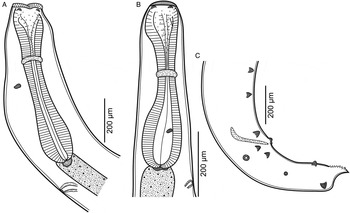
A major distinction was noted in the position of deirids and the excretory pore. In the new species both structures are located far posterior to the oesophagus–intestine junction (fig. 1A, B), whereas in the type material of C. tucunarensis (CHIOC 35077a, b) deirids and excretory pore were observed well anterior and slightly posterior to this junction, respectively (fig. 3A, B). Since C. opisthoporus n. sp. and C. tucunarensis are similar in body length (males measuring 4.6–5.9 mm vs. 4.5–5.9 mm; females measuring 5.1–7.5 mm vs. 4.8–7.0 mm), their substantial differences in the location of deirids and excretory pore were considered as interspecific variation. Furthermore, in C. opisthoporus n. sp. the following measurements are smaller than those of C. tucunarensis: oesophagus to body length ratio (6.7–10.0% vs. 13–15% in males; 9.0–9.3% vs. 12–16% in females) and gubernaculum (48–69 μm vs. 80–100 μm) (Lacerda et al., Reference Lacerda, Takemoto, Marchiori, Martins and Pavanelli2013).
The morphology of type specimens of Cucullanus tucunarensis (CHIOC 35077a, b), showing: (A) female anterior end, sublateral view (the specimen was twisted); (B) male anterior end, lateral view; and (C) male posterior end, lateral view.

Another difference was in the number of precloacal papillae: C. opisthoporus n. sp. has five pairs whereas C. tucunarensis has six. Nevertheless, it should be noted that the arrangement of caudal papillae in both species is quite similar (see figs 1H, I and 3C).
It is worth noting that the presence of spines in the tail tip has also been reported in some species of Dichelyne (see Moravec et al., Reference Moravec, Wolter and Körting1999, Reference Moravec, Khosheghbal and Pazooki2014; Moravec & Justine, Reference Moravec and Justine2011). However, these representatives of Dichelyne clearly differ from C. opisthoporus n. sp. in many features, such as the presence of an intestinal ceacum or caeca (vs. absence), arrangement of caudal papillae, and different morphology and pattern of the spines on the tail tip.
Remarks on Cucullanus tucunarensis Lacerda, Takemoto, Marchiori, Martins & Pavanelli, 2013
Observations on holotype male (CHIOC 35077a) and allotype female (CHIOC 35077b) of C. tucunarensis have revealed some features inaccurately reported and/or overlooked in the original description, even though the specimens are poorly preserved.
Lacerda et al. (2013) reported the excretory pore ‘situated between the second half and the distal end of oesophagus’; however, it is in fact slightly posterior to the oesophagus–intestine junction (fig. 3A, B). Furthermore, the authors had not observed the deirids that are located well anterior to the posterior end of the oesophagus (fig. 3A, B). Most likely, Lacerda et al. (Reference Lacerda, Takemoto, Marchiori, Martins and Pavanelli2013) confused a deirid with the excretory pore (see Fig. 1A in Lacerda et al., Reference Lacerda, Takemoto, Marchiori, Martins and Pavanelli2013).
The gubernaculum of C. tucunarensis was described as ‘spoon-shaped’ (Lacerda et al., Reference Lacerda, Takemoto, Marchiori, Martins and Pavanelli2013), probably because it was observed only in lateral view (fig. 3C). However, in ventral view the gubernaculum is somewhat Y-shaped (this was hard to observe because of the poor condition of the material and caution required not to break the specimen when trying to move it into the correct position in the mount under the microscope).
The cuticular frame in the oesophastome was not observed in the type material of C. tucunarensis (we observed the specimens using only light microscopy), but the entire inner wall of the oesophastome was strongly cuticularized.
Lacerda et al. (Reference Lacerda, Takemoto, Marchiori, Martins and Pavanelli2013) reported the males of C. tucunarensis as having four pairs of postcloacal papillae, but they illustrated just three pairs. We observed three pairs of postcloacal papillae and one pair of papilla-like phasmids in the holotype (not reported in the original description) (fig. 3C). Furthermore, the presence of an unpaired papilla and a pair of small mammilliform papillae in the anterior cloacal lip, not originally reported, were observed for the first time in the holotype (fig. 3C).
Cucullanidae Cobbold, 1864; Dichelyne Jägerskiöld, 1902; Dichelyne grandistomis n. comb. (Ferraz & Thatcher, 1988) (= Cucullanus grandistomis)
Redescription
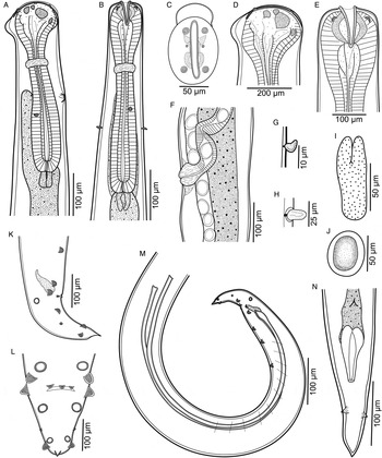
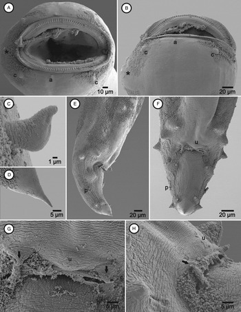
Medium-sized, whitish nematodes (figs 4 and 5). Body elongate; cuticle with fine transverse striations (fig. 5E–H). Lateral alae absent. Cephalic end rounded, with broad cuticular protuberance located dorsally (figs 4A, C, D and 5A, B). Oral opening dorsoventrally elongate, surrounded by wide membranous alae (collarette) armed with a row of c. 140 small, triangular, thin, pointed teeth (figs 4C and 5A, B). Four medium-sized submedian cephalic papillae and a pair of small lateral amphids surrounding oral opening (figs 4C and 5A, B). Oesophagus muscular, stout, expanded at both ends, forming an anterior, wide, pseudobuccal capsule (oesophastome), and posterior narrower dilatation (fig. 4A, B). Cuticular frame of oesophastome composed of three paired and one unpaired well-sclerotized plates: small lateral and small arrow-shaped structures, one pair each; one pair of large reniform structures, elongate in lateral view; unpaired small transverse plate ventrally located (fig. 4A–E). Oesophageal valve muscular, well-developed, non-sclerotized, opening into intestine (fig. 4A, B). Dorsal intestinal caecum present, extending somewhat between nerve ring and excretory pore (fig. 4A, B). Nerve ring encircling oesophagus at 33.9–37.4% of its length (fig. 4A, B). Deirids ‘boot-shaped’ (figs 4G and 5C), posterior to excretory pore, both far anterior to oesophagus–intestine junction (fig. 4A, B). Postdeirids elongate and rounded at tip (fig. 4H). Cloacal and anal regions not protruded (figs 4K–N and 5E–H). Tail conical, thin, pointed (figs 4K–M and 5D–F).
The morphology of Dichelyne grandistomis, showing: (A) and (B) female anterior end, lateral and dorsoventral views; (C), (D) and (E) cephalic end, apical, lateral and dorsoventral views; (F) vulva; (G) deirid; (H) postdeirid; (I) gubernaculum, ventral view; (J) egg; (K), (L) and (M) male posterior end, lateral, ventral and lateral views, respectively; (N) female tail, ventral view.

SEM of Dichelyne grandistomis, showing: (A) and (B) cephalic end, apical and lateral views (asterisks indicate dorsal cuticular expansion); (C) deirid; (D) female tail tip; (E) and (F) male caudal region, lateral and ventral views; (G) and (H) male cloacal region, ventral and sublateral views (arrows indicate papillae on cloacal lip). Abbreviations: a, amphid; c, cephalic papilla; p, phasmid; u, unpaired papilla.

Male (based on one adult voucher). Body length 8.3 mm, maximum width 213. Maximum width of cuticle at oesophageal region 23. Oesophagus 907 long, 150 wide at posterior end, representing 11% of total body length. Oesophastome 183 long, 157 wide. Intestinal caecum 581 long. Nerve ring, excretory pore and deirids located at 329, 394 and 447, respectively, from anterior end of body. Left postdeirid located at 4.5 mm from anterior end of body. Right postdeirid not observed. Posterior end of body ventrally bent, with transverse muscular bands (fig. 4M). Spicules slightly sub-equal, slender, with distal dilatation and thin, pointed; left and right 1.84 mm and 1.87 mm long, respectively; representing 22.2% and 22.6% of total body length, respectively (fig. 4M). Gubernaculum somewhat ellipsoid in ventral view, with rounded extremities and proximal portion longitudinally bilobed, 87 long (fig. 4I). Ventral pseudosucker absent. Large mammilliform caudal papillae (figs 4K–M and 5E–H). Five pairs of precloacal papillae: first to fourth pair somewhat equidistant from each other and subventrally placed; fifth pair sublateral, close together with fourth (fig. 4M). Anterior cloacal lip bearing a pair of small papillae and a conspicuous unpaired one (figs 4K–M and 5E–H). One pair of lateral adcloacal papillae (figs 4K–M and 5E–H). Three pairs of postcloacal papillae: first pair subventral, slightly posterior to cloaca; second and third pairs near tail end, subventrally and subdorsally placed, respectively (figs 4K–M and 5E–H). Pair of small papilla-like phasmids located at mid length between first and second postcloacal pairs (figs 4K–M and 5E–H). Tail 214 long.
Female (based on nine adult vouchers, all gravid). Body length 8.8–11.7 mm, maximum width 266–346. Maximum width of cuticle at oesophageal region 20–29. Oesophagus 0.91–1.03 mm long, 162–191 wide at posterior end, representing 8.4–10.3% of total body length. Oesophastome 201–237 long, 150–183 wide. Intestinal caecum 521–667 long. Nerve ring, excretory pore and deirids located at 324–391, 335–649 and 339–662, respectively, from anterior end of body. Vulva postequatorial, 5.8–7.9 mm from anterior end, at 64.9–67.4% of body length. Vulval lips slightly elevated (fig. 4F). Vagina with weak striated musculature, ovijector muscular, both directed anteriorly from vulva (fig. 4F). Uteri amphidelphic. Right and left postdeirid 3.9–5.3 mm and 6.0–9.8 mm from anterior end of body, respectively. Eggs oval, 61–66 × 47–52 (n = 10) (fig. 4J). Large, pointed papilla-like phasmids located somewhat in mid length of tail (fig. 4N). Tail 200–264 long.
Taxonomic summary
Host studied
Oxydoras niger (Valenciennes, 1821) (Actinopterygii: Doradidae).
Site of infection
Intestine.
Locality from the present study
River Xingu near municipality of Altamira (3°14′S; 52°06′W).
Voucher specimens deposited
One male and nine females, CHIOC 36757.
Remarks
The present specimens were morphologically indistinct from those of C. grandistomis. Although they differ in body length (males measuring 8.3 mm vs. 3.7–4.7 mm; females measuring 8.8–11.7 mm vs. 3.0–7.4 mm), reports of C. grandistomis have all been from a single host species (i.e. O. niger), in the same river basin (River Amazon) (Ferraz & Thatcher, Reference Ferraz and Thatcher1988; Santos & Tavares-Dias, Reference Santos and Tavares-Dias2010; Silva et al., Reference Silva, Tavares-Dias, Gerônimo and Martins2011; present study). Furthermore, newly collected parasites have a broad dorsal cuticular protuberance on the cephalic end, and a prominent excretory pore anterior to the deirids, which are exclusive features of C. grandistomis. Variations in host attributes may account for the morphometric differences observed here, mainly total length of the fish body (the O. niger captured was more than 1 m long).
Ferraz & Thatcher (Reference Ferraz and Thatcher1988) did not provide an accurate description for the shape and arrangement of the sclerotized plates in the oesophastome, or for the arrangement of caudal papillae (their drawings of the male caudal region do not correspond with the description); these features are now described in detail and represent important specific traits among cucullanids (Vieira et al., Reference Vieira, Pereira, Pantoja, Soares, Pereira, Timi, Scholz and Luque2015). Additionally, Ferraz & Thatcher (Reference Ferraz and Thatcher1988) overlooked the unpaired papilla on the anterior edge of cloacal lip, which is reported here for the first time.
Cucullanus grandistomis was originally described as Bacudacnitis grandistomis, type species of the genus that was invalidated and, consequently, the species reallocated to Cucullanus (Moravec et al., Reference Moravec, Kohn and Fernandes1993). The analyses of the newly collected material revealed the presence of an intestinal caecum in all specimens. Unfortunately, paratypes of C. grandistomis (CHIOC 32456a, b) are stained and mounted in permanent slides, which makes it impossible to determine the presence of such structures. In addition, no holotype, allotype or voucher specimens from C. grandistomis are available for consultation. However, due to the identical morphology, we have no doubt that our material belongs to C. grandistomis and, since it exhibits an intestinal caecum, this species should be transferred to Dichelyne as Dichelyne gradistomis n. comb.
Molecular characterization and phylogenetic analyses
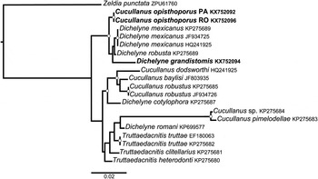
Partial sequence lengths of the 18S rRNA gene for C. opisthoporus n. sp. from C. melaniae and C. pinima were 1501 bp (KX752092) and 1679 bp (KX752096), respectively, and for D. grandistomis it was 1512 bp (KX752094). Sequence lengths of the 28S rRNA gene for C. opisthoporus n. sp. from C. melaniae and C. pinima were 773 bp (KX752091) and 795 bp (KX752095), respectively, and for D. grandistomis it was 749 bp (KX752093). Specimens of C. opisthoporus n. sp. from the two different hosts were highly similar, considering both 18S (sequence identity 99.5%) and 28S (sequence identity 99.0%). Sequence identities between C. opisthoporus and D. grandistomis were 97.7% for the 18S and 88.4% for the 28S. The topology of the trees generated using ML and BI inference were equal, in which samples of C. opisthoporus n. sp. from different hosts and localities clustered together with high support (fig. 6).
Bayesian tree from phylogenetic analyses of the sequences of the 18S rRNA gene from species of Cucullanidae, associated with their GenBank accession numbers. Nodes with high support are represented by white circles, i.e. Bayesian posterior probability >0.96 (4 × 10⁶ generations, sampling frequency = 4 × 10³, burn-in = 1 × 10⁶) and maximum likelihood bootstrap values > 96% (1000 replications). Specimens in bold are from the present study.

Phylogenetic reconstructions using the dataset of 18S from representatives of Cucullanidae showed that at least Cucullanus and Dichelyne are not monophyletic, and that Truttaedacnitis may be paraphyletic; however, some clades were weakly supported (fig. 6). Representatives of the genus Dichelyne appeared as a sister group of C. opisthoporus n. sp., including D. grandistomis (fig. 6).
Discussion
Systematic issues within Cucullanidae have been largely discussed from the morphological perspective (Petter, Reference Petter1974; Moravec et al., Reference Moravec, Sasal, Würtz and Taraschewski2005; Yooyen et al., Reference Yooyen, Moravec and Wongsawad2011; Li et al., Reference Li, Du, Xu, Guo, Wang and Zhang2014; Vieira et al., Reference Vieira, Pereira, Pantoja, Soares, Pereira, Timi, Scholz and Luque2015). Recently, Choudhury & Nadler (Reference Choudhury and Nadler2016) published an insight on the phylogeny of Cucullanidae. However, much more effort is needed to increase genetic and morphological data on these nematodes, in order to clarify their complex relationships.
The differentiation between C. opisthoporus n. sp. and C. tucunarensis, parasites collected from two different river basins (Amazonas and Tocantis-Araguaia, respectively), was interesting, since the morphological similarity of both species is considerable, except for the relative position of deirids and excretory pore. The relative position of such structures is believed to vary little within a species and, thus, to be reliable for specific diagnosis (Vieira et al., Reference Vieira, Pereira, Pantoja, Soares, Pereira, Timi, Scholz and Luque2015). Unfortunately, no genetic data for C. tucunarensis are available for comparison. Therefore, we preferred to retain C. opisthoporus n. sp. and C. tucunarensis as separate species until new data are available.
The molecular characterization and the phylogenetic analyses supported the conspecificity of C. opisthoporus n. sp. collected from C. melaniae in River Xingu and from C. pinima in River Jamarí (fig. 6). However, phylogenetic reconstructions based on 18S rRNA were not enough to resolve the relationships of the genera within Cucullanidae. Despite the apparent monophyly of Cucullanidae among Nematoda, the systematics of the superfamily Seuratoidea Hall, 1916 and all its taxa is still uncertain and poorly understood (Choudhury & Nadler, Reference Choudhury and Nadler2016). The classification of genera within Cucullanidae seems to be artificial based upon the present results and, in accordance with Choudhury & Nadler (Reference Choudhury and Nadler2016), we highly recommend taxonomists not to adopt the use of subgenera for these nematodes, since this practice complicates their fragile taxonomy and, at present, does not have phylogenetic support.
Considering the current panorama, the validity of at least Cucullanus, Dichelyne and Truttaedacnitis should be maintained until new data on different genes and specimens are available. In the present study, for example, the 28S rRNA gene exhibited fewer conserved regions than the 18S, being more effective for genetic differentiation (for details see sequences in GenBank under accessions numbers KX752091, KX752092, KX752093, KX752094, KX752095 and KX752096). Furthermore, the most representative genera of Cucullanidae, i.e. Cucullanus and Dichelyne, should still be separated, based upon the presence/absence of an intestinal caecum (or caeca), since this is the most consistent and most adopted diagnostic feature according to taxonomists. Thus, we suggest the transfer of C. grandistomis to Dichelyne.
Acknowledgements
The authors are thankful to Tomáš Scholz from the Institute of Parasitology, Biology Centre of the Czech Academy of Sciences (IPCAS) and the Staff of the Laboratory of Electron Microscopy from the same institute (IPCAS; RVO: 60077344). Thanks are also due to Marcelo Knoff, curator of the Helminthological Collection of the Instituto Oswaldo Cruz (CHIOC) for lending the type specimens of C. tucunarensis and D. grandistomis for observation, and Douglas McIntosh from the Laboratory of Molecular Biology of the Universidade Federal Rural do Rio de Janeiro (UFRRJ) for valuable support during molecular procedures. We also acknowledge Marcos Tavares-Dias, Philippe V. Alves, Fabiano Paschoal, Aldenice Pereira, Angélica Carvalho, Emil Hernández and Tomáš Scholz for support during the field trips.
Financial support
F.B.P. and J.L.L. were supported by a Postdoctoral fellowship from PNPD–CAPES (Programa Nacional de Pós-Doutorado–Coordenação de Aperfeiçoamento de Pessoal de Nível Superior-CAPES, Brazil) and a Research fellowship from CNPq (Conselho Nacional de Desenvolvimento Científico e Tecnológico do Brasil), respectively. The authors thank the Czech Science Foundation (project No. P505/12/G112) for support and for covering the costs of SEM observations at the IPCAS.
Conflict of interest
None.