Introduction
Recent studies have identified carbonates on Mars at Nili Fossae (Ehlmann et al. Reference Ehlmann, Mustard, Murchie, Poulet, Bishop, Brown, Calvin, Clark, Des Marais and Milliken2008; Brown et al. Reference Brown, Hook, Baldridge, Crowley, Bridges, Thomson, Marion, de Souza Filho and Bishop2010), at Leighton crater (Michalski & Niles Reference Michalski and Niles2010), at the Phoenix landing site (Boynton et al. Reference Boynton, Ming, Sutter, Arvidson, Hoffman, Niles and Smith2009; Smith Reference Smith2009), at Gusev crater (Morris et al. Reference Morris, Ruff, Gellert, Ming, Arvidson, Clark, Golden, Siebach, Klingelhöfer and Schröder2010) and in several crater pits including Huygens (Wray et al. Reference Wray, Murchie, Ehlmann, Milliken, Seelos, Noe Dobrea, Mustard and Squyres2011). Orbital detection of small carbonate outcrops on Mars has been possible recently using visible/near-infrared (VNIR) spectra acquired by the Compact Reconnaissance Imaging Spectrometer for Mars (CRISM) with spatial coverage at 18 m pixel−1 (Murchie et al. Reference Murchie, Mustard, Ehlmann, Milliken, Bishop, McKeown, Noe Dobrea, Seelos, Buczkowski and Wiseman2009a). These observations support previous detections of carbonates in the Martian dust (Pollack et al. Reference Pollack, Roush, Witteborn, Bregman, Wooden, Stoker, Toon, Rank, Dalton and Freedman1990; Calvin et al. Reference Calvin, King and Clark1994; Bandfield et al. Reference Bandfield, Glotch and Christensen2003; Christensen et al. Reference Christensen, Ruff, Fergason, Knudson, Anwar, Arvidson, Bandfield, Blaney, Budney and Calvin2004) and in meteorites (e.g. McKay et al. Reference McKay, Gibson, Thomas-Keprta, Vali, Romanek, Clemett, Chillier, Maechling and Zare1996; Bishop et al. Reference Bishop, Pieters, Hiroi and Mustard1998b; Brearley Reference Brearley2000). One challenge to remote spectral detection of any mineral is that the spectral signature is dominated by the surface. Hence, even thin surface coatings can greatly alter the spectral properties of a rock. Iron oxide-bearing coatings have been observed on most surface rocks on Mars measured by the Pathfinder rover (McSween et al. Reference McSween, Murchie, Crisp, Bridges, Anderson, Bell, Britt, Brückner, Dreibus and Economou1999) and the Mars Exploration Rovers (McSween et al. Reference McSween, Arvidson, Bell, Blaney, Cabrol, Christensen, Clark, Crisp, Crumpler and Des Marais2004; Bell et al. Reference Bell, Squyres, Arvidson, Arneson, Bass, Blaney, Cabrol, Calvin, Farmer and Farrand2004; Arvidson et al. Reference Arvidson, Squyres, Anderson, Bell, Blaney, Brückner, Cabrol, Calvin, Carr and Christensen2006). Another challenge to remote detection of carbonates on Mars is that the strongest reflectance bands near 3.4 and 4 μm are hindered by emissivity. We propose here that carbonate rocks in the Mojave Desert serve as potential analogues to the carbonates on Mars. Rocks collected from the Little Red Hill site contain iron oxide-bearing coatings that greatly suppress the carbonate features of the underlying material.
Background
The Mojave Desert once contained pedogenic soil rich in iron oxides, clays and carbonate (Wells et al. Reference Wells, McFadden and Dohrenwend1987). Much of these were eroded away, but remnants are still found, including a patch of soil preserved under a basaltic lava flow dated at ∼7–9 million years ago (Enzel et al. Reference Enzel, Brown, Anderson, McFadden and Wells1992; Harris et al. Reference Harris, Tuttle and Tuttle2004). Caliche and carbonate-bearing rocks are abundant in the Mojave Desert region as remnants of the former aqueous environment here (Schlesinger Reference Schlesinger1985). Varnishes containing clay, iron oxide and Mn oxide were formed on the surface of the rocks more than 2000 years ago (McFadden et al. Reference McFadden, Wells and Jercinovich1987, Reference McFadden, McDonald, Wells, Anderson, Quade and Forman1998; Quade Reference Quade2001). Reynolds et al. (Reference Reynolds, Reheis, Yount and Lamothe2006) noted concentrated abundances of magnetite and haematite in the dust across a wide expanse of the Mojave Desert that apparently resulted from alteration and erosion of other minerals and phases in the Cima volcanic ash. This is similar to the presence of magnetite and haematite in the Martian dust (e.g. Morris et al. Reference Morris, Klingelhöfer, Schröder, Rodionov, Yen, Ming, de Souza, Wdowiak, Fleischer and Gellert2006). Today the Mojave Desert is one of the most arid environments in North America with an average rainfall of ∼100–150 mm year−1 over the past 100 years (e.g. Beatley Reference Beatley1975; McFadden et al. Reference McFadden, Wells and Dohrenwend1986) and precipitation in the vicinity of the Little Red Hill site is 100 mm year−1 or less (http://cdo.ncdc.noaa.gov/climatenormals/clim81/CAnorm.txt).
Samples
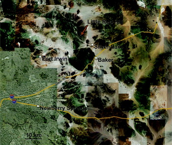
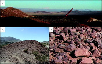
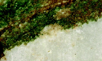
The Little Red Hill site is located northwest of Silver Lake near Baker, California at 35°23′N and 116°16′W at an elevation of about 514 m at the northeastern part of the Mojave Desert (Figs. 1 and 2). Several samples were collected that were representative of the area. Little Red Hill samples have a thin, exterior weathered rind (Figs. 2 and 3). This rock coating is reddish-brown in colour and typically on the order of 1 mm or less in thickness. The interior of these samples is white and semi-translucent. An HCl test indicated that these samples are dominantly carbonate. Some samples appear to have a planar foliation, but on the whole, most of the samples are massive. Many samples also exhibit green layers in the rock and/or green coatings on the underside of the rock. Rocks were cut with a saw to remove the exterior weathered surface. Samples were then broken and crushed using a shatterbox with an alumina ring and puck. This material was then crushed and passed through a 150 μm sieve to produce particulate material for analysis.
Map of the northern region of the Mojave Desert indicating the Little Red Hill site (red arrow) about 15 km northwest of Baker, CA.

(a) An aerial view of the Mojave Desert showing the Little Red Hill (marked with a red arrow) raised above its surroundings (image ∼0.5 km across in the foreground). (b) Red-toned rocks covering the Little Red Hill (image ∼5 m across). (c) Rocks at the Little Red Hill site (image ∼30 cm across).

Methods
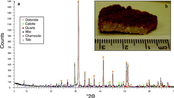
Mineralogy was determined from X-ray diffraction (XRD) analysis and is reported in Fig. 3. Powdered aliquots of white internal portions of the rocks were analysed in the Department of Earth and Planetary Sciences at the University of New Mexico, using a Scintag Pad V diffractometer with Data Scan 3.1 software (from MDI Inc.) for system automation and data collection. Ni-filtered, Cu-K-α radiation (40 kV, 35 mA) was used with a Bicron Scintillation detector (with curved graphite-crystal monochromator). Data were analysed with Jade 5 software (from MDI Inc.) using the ICDD PDF2 database (rev. 1999) for phase identification.
(a) XRD results for a Little Red Hill sample containing predominantly dolomite (CaMg(CO3)2), with small amounts of calcite (CaCO3), quartz (SiO2), chamosite ((Fe,Al,Mg)6(Si4−xAlx)O10(OH)8), illite (K(Al,Fe,Mg)2(Si4−x Alx)-O10(OH)8) and talc (Mg3Si4O10(OH)2) in order of reduced abundance. (b) Rock image showing the thin red coating.

Major and trace element composition of samples was determined using a Rigaku 4 KW RIX-2100 wavelength dispersive X-ray fluorescence spectrometer (XRF) with a rhodium target and an end-window. For major element analyses 9 g of lithium tetraborate (Li2B4O7) were mixed with 1 g of sample. Discs were then fused at 1000°C for 15 minutes with intermittent swirling. Fused discs were then cooled, tapped out of the mould and analysed by XRF. The major elements were determined at 50 kV and 50 mA. Trace elements were determined using pressed powders prepared in the ratio of 1.5–8.5 boric acid (H3BO3) to sample. Then 0.75 g of boric acid was combined with 4.25 g of sample (±0.001). Powders were pressed at ∼20 t in−2 using tungsten-carbide pellets polished with diamond paste. Trace elements were determined at 50 kV and 70 mA.
Visible and infrared (IR) reflectance spectra were measured of bulk powders and chips at the Brown University RELAB facility. Bidirectional spectra from 0.3 to 1.25 μm measured relative to Halon were spliced to biconical (on-axis) reflectance spectra from 1 to 25 μm measured relative to a rough gold surface using Fourier‑transform infrared (FTIR). A continuum was removed from the spectra across the ranges 2.20–2.65 and 3.175–4.25 μm in order to facilitate comparison of the band strengths using an algorithm implemented in the MR PRISM software suite (Brown & Storrie-Lombardi Reference Brown and Storrie-Lombardi2006).
Full resolution-targeted (FRT) CRISM images collect 544 wavelengths from 0.36 to 3.9 μm in ∼10 km swathes (Murchie et al. Reference Murchie, Seelos, Hash, Humm, Malaret, McGovern, Choo, Seelos, Buczkowski and Morgan2009b). The carbonate spectra presented in this study were retrieved from CRISM image FRT0000B072 in the Nili Fossae region west of Isidis Planitia. This image was processed for instrumental effects, converted to I/F and atmospheric gas absorptions were minimized using a ratio with a CRISM scene of Olympus Mons (Murchie et al. Reference Murchie, Seelos, Hash, Humm, Malaret, McGovern, Choo, Seelos, Buczkowski and Morgan2009b; McGuire et al. Reference McGuire, Bishop, Brown, Fraeman, Marzo, Morgan, Murchie, Mustard, Parente and Pelkey2009). Ratios to spectrally unremarkable pixels (e.g. flat and featureless spectra) in the same CRISM image column were used to minimize instrument contributions and help resolve spectral features.
Results: XRD
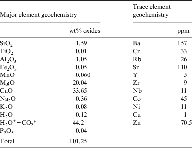
The XRD data are reported in Fig. 3 and indicate that the white internal portions of the Little Red Hill samples are primarily dolomite, with some calcite, and minor amounts of quartz, chamosite, illite and talc. The major and minor elements are given in Table 1 and are consistent with this mineralogy.
Major and trace element geochemistry of Little Red Hill samples
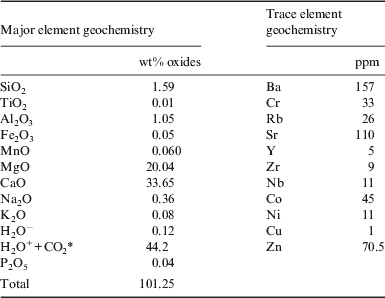
* H2O++CO2 determined by loss on ignition.
Results: VNIR spectra
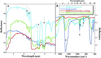
VNIR reflectance spectra of the Little Red Hill samples contain bands at 2.32 and 2.52 μm and a pair of doublets at 3.31 and 3.45 μm and 3.81 and 3.96 μm (Fig. 4) that are consistent with the carbonate mineral dolomite plus some magnesite (MgCO3) and aragonite (CaCO3) (Gaffey Reference Gaffey1987). In addition, a sharp band at 1.38 μm was observed in spectra of the Little Red Hill samples, which is characteristic of hydrous carbonates such as hydromagnesite (Mg5(CO3)4(OH)2⋅4H2O), also proposed to be present on Mars (Calvin et al. Reference Calvin, King and Clark1994) and very stable at moderate temperatures despite the presence of water (Zhang et al. Reference Zhang, Anderson, Kelly, Krumhansl and Papenguth2000). Alternatively, this could be due to the trace amount of talc identified by XRD (Fig. 3). The spectrum of the particulate (<125 μm) fraction of an internal portion of a rock from the Little Red Hill site exhibits stronger features at ∼2.3, 2.5, 3.4 and 4 μm than the spectrum of the fresh surface, as expected from previous studies showing that particulate spectra of carbonates have stronger VNIR features than coarse-grained size fractions or rocks (Gaffey Reference Gaffey1987).
(a) VNIR spectra of carbonate rocks from the Little Red Hill site, including a fresh rock surface, ground (<125 μm) rock interior and coated rocks. These spectra show carbonate bands near 2.32, 2.52, 3.46, 3.97 and 4.63 μm (black arrows). (b) Mid-IR reflectance spectra of the same samples include a strong carbonate reststrahlen band near 1550 cm−1 (6.45 μm), and weaker reflectance maxima at 875–900 and 720–740 cm−1 (11.1–11.4 and 13.5–13.9 μm) that flip to form reflectance minima for the fine-grained sample (black arrows).

The red coatings on the surface of the rocks include features at 0.86, 1.41, 1.91 and 2.2 μm that are attributed to a mixture of haematite and Al-bearing phyllosilicates such as montmorillonite. Red coatings on these rocks greatly suppress the carbonate features, producing much weaker spectral signatures than the non-coated material from interior portions of the rock. Green coatings on the subsurface of these rocks exhibit absorption features near 0.63 and 0.68 μm that are consistent with chlorophyll-type pigments.
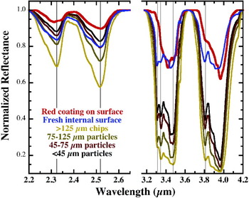
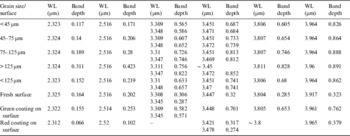
Continuum removed spectra are shown in Fig. 5 for several grain sizes in order to better illustrate the effects of grain size and coatings on the band depths of the key spectral features. Band centres and depths were determined for the features near 2.3, 2.5, 3.3, 3.4, 3.8 and 4 μm and are recorded in Table 2. These data show increasing band strength with decreasing particle size for the bands near 2.3, 2.5, 3.4 and 4 μm as expected. The bands are weaker for the fresh and coated rock surfaces. The shape of the 3.4 μm band in the spectrum of the red coating sample is also more consistent with CH vibrations than CO3 vibrations, suggesting the presence of organics in the coating as well.
Continuum removed spectra are shown here for the carbonate bands near 2.3, 2.5, 3.4 and 4 μm. These spectra show increasing band strength with increasing particle size for ground samples.

Band depths in continuum-removed spectra

Note: WL, wavelength; all data were continuum-removed across two regions (2.2–2.65 and 3.175–4.25 μm) using MR PRISM code (Brown & Storrie-Lombardi Reference Brown and Storrie-Lombardi2006).
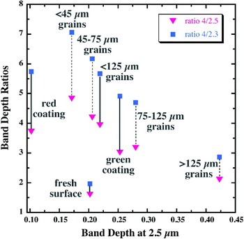
In order to compare the relative band strengths of these features, ratios were determined for the strength of the band at 4 μm compared with strengths of the bands near 2.3 and 2.5 μm. These results are displayed in Fig. 6 and show some variations in the relative band strengths with grain size. Although these trends may not be statistically significant, the finest size fractions tend to have a stronger 4-μm band relative to the bands near 2.3 and 2.5 μm than the coarser grains and the surfaces.
Band depth ratios are plotted against the band depth at 2.5 μm in order to illustrate the relative band strengths of the carbonate bands at 2.3, 2.5 and 4 μm for the different grain sizes and surface types in the samples.

Results: mid-IR spectra
Mid-IR spectra of the Little Red Hill samples are shown in Fig. 4b, where arrows indicate the primary carbonate vibrational bands near 1550 cm−1 (6.45 μm), 875–900 cm−1 (11.1–11.4 μm) and 720–740 cm−1 (13.5–13.9 μm). Additional bands are observed near 262 and 381 cm−1 (∼26 and 38 μm). These bands are consistent with dolomite plus small amounts of other carbonates (Lane & Christensen Reference Lane and Christensen1997). These features occur as scattering peaks in reflectance for the chip spectra, but are flipped for the bands near 727 and 900 cm−1 for the fine particulate spectra due to a larger contribution of volume scattering. Carbonates are particularly sensitive to the effects of volume and surface scattering, which determine whether the bands are concave up or concave down (Salisbury & Wald Reference Salisbury and Wald1992). Reflectance peaks (or emissivity lows) are observed for large grains where the vibrational absorptions occur as reststrahlen bands, while reflectance troughs (or emissivity highs) are observed in some cases for fine-grained particles. Spectra of our Mojave sample with the red surface coating exhibit severe masking of the characteristic carbonate features compared with the spectra of the fresh internal carbonate surface.
Discussion: spectral properties of carbonate mixtures
The spectral properties of mineral mixtures are not always linear representations of the mixture components. This has been described in detail for VNIR reflectance studies (e.g. Hapke Reference Hapke1981; Mustard & Pieters Reference Mustard and Pieters1989; Sunshine & Pieters Reference Sunshine and Pieters1993; Hiroi & Pieters Reference Hiroi and Pieters1994) and can be especially problematic if fine-grained materials are present. Recent mixtures of the <125 μm fractions of magnesite, nontronite and forsterite also showed non-linear effects on the spectral properties of the mixtures such that 10 wt% magnesite in forsterite was readily detectable at 2.3 and 2.5 μm, while a much greater abundance of magnesite was required for detection of these bands in mixtures with nontronite (Perry et al. Reference Perry, Bishop, Dyar, Blake, Peel and Brown2011). Brown et al. (Reference Brown, Walter and Cudahy2004) reported stromatolitic carbonate findings that were not detectable from airborne (5 km altitude) hyperspectral surveys (Brown Reference Brown2006). Dalton et al. (Reference Dalton, Bove, Mladinich and Rockwell2004) also observed non-linear effects on the spectra for mixtures of calcite, epidote and chlorite. Mineral mixtures of larger grain sizes typically produce linear relationships in the mid-IR spectral region that are dominated by fundamental vibrations (Ramsey & Christensen Reference Ramsey and Christensen1998).
Laboratory experiments of carbonates mixed in palagonitic soil and basalt showed that dolomite was easier to detect than siderite and that weak bands were observable for both sets of carbonate reflectance spectra down to 1 wt% carbonate abundance in the mixtures (Wagner & Schade Reference Wagner and Schade1996). This is similar to reflectance spectra of carbonates in sediments that are readily detectable using the 4 μm band down to a single percent abundance (Bishop et al. Reference Bishop, Koeberl, Kralik, Froeschl, Englert, Andersen, Pieters and Wharton1996, Reference Bishop, Lougear, Newton, Doran, Froeschl, Trautwein, Körner and Koeberl2001). A carbonate band was also observed at 4 μm in spectra of Martian meteorite ALH 84001 for tiny carbonate grains in the orthopyroxene matrix (Bishop et al. Reference Bishop, Mustard, Pieters and Hiroi1998a, Reference Bishop, Pieters, Hiroi and Mustardb).
Measurements of reflectance and emission spectra of carbonate mixtures showed that thermal emission contributes significantly towards reducing the spectral contrast of the carbonate bands near 3.4 and 4 μm at Martian surface temperatures (Wagner & Schade Reference Wagner and Schade1996). Grain size, absorptive properties of the matrix material and temperature all affect the emissivity over the range 3–4 μm in spectra of carbonate mixtures. Their modelling results show that the ∼3.4 and 4 μm bands are significantly weakened at 275–300 K, especially for larger grains, such that detection of these features in Martian spectra would be difficult (Wagner & Schade Reference Wagner and Schade1996).
Discussion: influence of coatings on spectral properties of rocks
VNIR spectral studies of rock coatings have shown that a thin layer of fine-grained material (Fischer & Pieters Reference Fischer and Pieters1993; Gilmore et al. Reference Gilmore, Merrill, Castaño, Bornstein and Greenwood2004) or a thin coating on rocks (Bishop et al. Reference Bishop, Minitti, Lane and Weitz2003; Minitti et al. Reference Minitti, Weitz, Lane and Bishop2007) can have a pronounced effect on the spectral character of the rock. For example, in the case of Hawaiian rock coatings, not only were the spectral features of the basaltic rocks masked, but a slope (decreasing reflectance towards longer wavelengths) was imparted on the spectra as well (Bishop et al. Reference Bishop, Minitti, Lane and Weitz2003; Minitti et al. Reference Minitti, Weitz, Lane and Bishop2007). A recent study of fine-grained iron-rich powders on phyllosilicate rocks (Parente et al. Reference Parente, Bishop and Cuadros2008) showed that a layer a few microns thick could alter the character of the visible region spectra of the rocks, while a thicker layer (hundreds of microns) was needed to significantly suppress the NIR clay bands. This suggests that if iron oxide-bearing coatings were masking spectral features of rocks near 2.3–2.5 μm, we would see evidence of Fe3+ features in the extended visible region. In fact, the OMEGA and CRISM spectra of Mars are characterized by this effect and it is presumed that ferric-bearing dust is dominating the spectral features near 0.5–1.0 μm such that differences in ferrous/ferric mineralogy in rocks is difficult to detect in many cases (Bibring et al. Reference Bibring, Langevin, Mustard, Poulet, Arvidson, Gendrin, Gondet, Mangold, Pinet and Forget2006; Murchie et al. Reference Murchie, Mustard, Ehlmann, Milliken, Bishop, McKeown, Noe Dobrea, Seelos, Buczkowski and Wiseman2009a). This coating effect would act to hide any surface below it and is not preferential to carbonates. Rock coatings have been investigated in detail at Gusev Crater by the instruments on Spirit, where substantial differences in chemistry and mineralogy were determined for the rocks and the coatings (Herkenhoff et al. Reference Herkenhoff, Squyres, Arvidson, Bass, Bell, Bertelsen, Cabrol, Gaddis, Hayes and Hviid2004; McSween et al. Reference McSween, Arvidson, Bell, Blaney, Cabrol, Christensen, Clark, Crisp, Crumpler and Des Marais2004).
Micro-organisms inhabiting the Mojave Desert rocks
Green coatings and green layers within the rock were also observed for carbonate-rich rocks at this site as shown for one sample in Fig. 7. The green layer is composed of dehydration-resistant cyanobacteria, predominantly Chroococcidiopsis (Schlesinger et al. Reference Schlesinger, Pippin, Wallenstein, Hofmockel, Klepeis and Hahall2003; Nienow Reference Nienow and Schaechter2009). These organisms live on the underside of rocks at the Mojave Desert site and can be revived after decades of storage in the laboratory. Only rocks that allow for some light (∼1%) to penetrate are colonized. This provides enough light for photosynthesis, while providing protection from extreme levels of sunlight and ultraviolet light (Nienow Reference Nienow and Schaechter2009). The rock also provides a small-scale environment in which moisture is preferentially retained (Nienow Reference Nienow and Schaechter2009). Microbes similar to Chroococcidiopsis could have existed in association with carbonate rocks in the past on Mars and could have been protected from solar radiation by the minerals. Other studies have shown how iron oxide coatings can provide a solar radiation screen and produce a niche for photosynthetic microbes to survive (Pierson et al. Reference Pierson, Oesterle and Murphy1987; Pierson & Parenteau Reference Pierson and Parenteau2000; Phoenix et al. Reference Phoenix, Konhauser, Adams and Bottrell2001; Bishop et al. Reference Bishop, Louris, Rogoff and Rothschild2006).
View of the green coating on the bottom side of a carbonate rock from the Little Red Hill at the Mojave Desert. Image is ∼5 mm across.

Applications to Mars
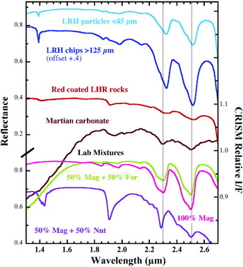
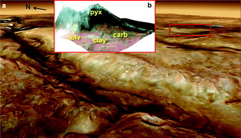
The carbonate rocks found at the Little Red Hill site in the Mojave Desert are mixtures of dolomite and other carbonates, with red coatings composed of haematite and Al-rich phyllosilicate. The carbonate signatures observed in CRISM images of the ancient rocks at Nili Fossae, Mars, found by Ehlmann et al. (Reference Ehlmann, Mustard, Murchie, Poulet, Bishop, Brown, Calvin, Clark, Des Marais and Milliken2008) occur at ∼2.30–2.31, 2.495–2.52, 3.45 and 3.9–4.0 μm in ratio spectra and are attributed to Mg-rich carbonate. These band centres are similar to those observed for the continuum-removed spectra of the Mojave Desert Little Red Hill rocks (Fig. 5). Shown in Fig. 8 are selected spectra of the Little Red Hill samples compared with an example Martian carbonate spectrum from Nili Fossae. The carbonate bands near 2.3 and 2.5 μm observed in spectra of the internal Little Red Hill samples occur at longer wavelengths than those observed in the Martian spectrum. This is not surprising as dolomite exhibits bands at slightly longer wavelengths than magnesite. However, the bands observed in spectra of the coated rocks are similar to those observed in the Martian carbonate spectra (Fig. 8). The band centres in the red-coated rock spectrum are broadened and shifted somewhat compared with the spectra of internal portions of the rock – either due to spatial mixing of the coating material with the carbonate or due to chemical alteration of the carbonate at the surface to make it more Mg-rich. Spectra are also shown in Fig. 8 of pure magnesite for comparison and mixtures of 50 wt% magnesite with 50 wt% nontronite or 50 wt% forsterite prepared for another study (Perry et al. Reference Perry, Bishop, Dyar, Blake, Peel and Brown2011). The magnesite/nontronite mixture spectrum shows that the bands near 2.3 and 2.5 μm shifted towards shorter wavelengths than observed for pure magnesite. Thus, the presence of Fe/Mg-clays mixed with carbonate (either spatially in the scene or together in the rocks) can influence the spectral signatures of the rocks. As Fe/Mg-clays are frequently observed near carbonate-rich rocks (Ehlmann et al. Reference Ehlmann, Mustard, Swayze, Clark, Bishop, Poulet, Marais, Roach, Milliken and Wray2009; Brown et al. Reference Brown, Hook, Baldridge, Crowley, Bridges, Thomson, Marion, de Souza Filho and Bishop2010) or even mixed with carbonates (Bishop et al. Reference Bishop, Makarewicz, Perry, McKeown, Parente, Tornabene, Swayze, Clark, Mustard and Murchie2010) on Mars, the effects of such mixtures are important for studies of Martian carbonates. A view of one carbonate-rich outcrop at Nili Fossae, Mars, is shown in Fig. 9 as an example. These carbonate rocks occur in a region 1–2 km wide near an outcrop of clay-rich rocks on top of olivine-bearing basalt and were studied in detail previously (Ehlmann et al. Reference Ehlmann, Mustard, Murchie, Poulet, Bishop, Brown, Calvin, Clark, Des Marais and Milliken2008, Reference Ehlmann, Mustard, Swayze, Clark, Bishop, Poulet, Marais, Roach, Milliken and Wray2009).
NIR reflectance spectra from 1.2 to 2.65 μm of Little Red Hill (LRH) samples, magnesite (Mag), mixtures of 50 wt% magnesite/50 wt% forsterite (For) and 50 wt% magnesite/50 wt% nontronite (Nnt) and a ratio spectrum of a Martian carbonate outcrop from CRISM image FRT0000B072. Vertical lines are drawn at 2.30 and 2.51 μm for comparison of the carbonate features in these spectra. Compared with the Martian spectrum, the bands near 2.3 and 2.5 μm are shifted shortwards in the mixture spectrum and longwards in the spectra of the internal LRH samples; however, the bands observed for the red-coated LRH rocks are similar to those observed in the Martian spectrum shown here and Martian carbonate spectra observed previously (Ehlmann et al. Reference Ehlmann, Mustard, Murchie, Poulet, Bishop, Brown, Calvin, Clark, Des Marais and Milliken2008).

(a) HRSC image of part of the Nili Fossae region enhanced ∼3× vertically. The red box marks the location of b. Image credits: ESA/DLR/FU Berlin (G. Neukum). (b) CRISM image draped over MOLA terrain. Pyroxene-bearing caprock overlays olivine-bearing basalt. Carbonate-rich outcrops are exposed as dark green rocks and clay-rich outcrops as light green rocks in this scene. The image is ∼10 km across and vertically enhanced 5×.

Spectral evidence for Mg- and Fe-bearing carbonates in ancient rocks on Mars is also present in the meteorite record where weak features were observed near 3.4, 3.9 and 6.4–7.1 μm due to a carbonate globule in Martian meteorite ALH 84001 (Bishop et al. Reference Bishop, Mustard, Pieters and Hiroi1998a). TEGA results from Phoenix show the presence of 1–5% Ca-carbonate, and possibly also Mg- or Fe-carbonate, in the soil near the lander (Boynton et al. Reference Boynton, Ming, Sutter, Arvidson, Hoffman, Niles and Smith2009). Coordinated analyses of chemistry, Mössbauer and Mini-TES spectra have identified the presence of Fe/Mg-rich carbonate in rocks at the Columbia Hills region in Gusev crater (Morris et al. Reference Morris, Ruff, Gellert, Ming, Arvidson, Clark, Golden, Siebach, Klingelhöfer and Schröder2010). Palomba et al. (Reference Palomba, Zinzi, Cloutis, D'Amore, Grassi and Maturilli2009) also detected a band in the 4 μm region in Mars Express Planetary Fourier Spectrometer (PFS) data at low abundances across many regions of the planet that is attributed to Mg-rich carbonate. TIR analyses of Martian dust have suggested the presence of 2–5 wt% Mg-carbonate (Bandfield et al. Reference Bandfield, Glotch and Christensen2003; Christensen et al. Reference Christensen, Ruff, Fergason, Knudson, Anwar, Arvidson, Bandfield, Blaney, Budney and Calvin2004). All these studies together suggest that carbonates may be present to a greater degree on Mars than detected to date. The ∼18 pixel−1 spatial resolution of CRISM could be a key factor in identifying carbonates in Martian rocks (Ehlmann et al. Reference Ehlmann, Mustard, Murchie, Poulet, Bishop, Brown, Calvin, Clark, Des Marais and Milliken2008). The iron oxide-coated carbonate rocks from the Mojave Desert provide an example of how a thin rock coating on Martian carbonates could be masking the spectral features used to identify carbonate minerals. Carbonates may be present in additional ancient rocks similar to those at Nili Fossae. A better spatial resolution NIR spectrometer in orbit or on a rover would greatly assist in detection of smaller outcrops of these carbonates.
The Little Red Hill rocks illustrate a terrestrial example of an iron oxide coating partially masking carbonate signatures that may be occurring on Mars as well. Thus, rock coatings could present a challenge for remote detection of carbonates. The Thermal Emission Spectrometer (TES) did not find carbonate rocks on the surface of Mars above the detection limits of the modelling (Christensen et al. Reference Christensen, Bandfield, Hamilton, Ruff, Kieffer, Titus, Malin, Morris, Lane and Clark2001). This is likely due to the small size of these outcrops (Ehlmann et al. Reference Ehlmann, Mustard, Murchie, Poulet, Bishop, Brown, Calvin, Clark, Des Marais and Milliken2008); if coatings are present on these carbonates that would make it even more difficult to detect them. However, TES did identify weak bands near 6.7 μm (∼1500 cm−1) due to Mg-rich carbonate in the dust (Bandfield et al. Reference Bandfield, Glotch and Christensen2003). Their spectral experiments suggested that 2–5 wt% fine-grained carbonate could be present in a silicate matrix on Mars (Bandfield et al. Reference Bandfield, Glotch and Christensen2003). Modelling of TES data generally results in a small carbonate component (at about the confidence level from the statistical errors) for many surfaces, indicating that carbonates could be contributing in a minor way to the Martian surface spectra (Christensen et al. Reference Christensen, Bandfield, Hamilton, Ruff, Kieffer, Titus, Malin, Morris, Lane and Clark2001; Bandfield et al. Reference Bandfield, Glotch and Christensen2003; Rogers & Christensen Reference Rogers and Christensen2007). Kirkland et al. (Reference Kirkland, Herr and Adams2003) described surface roughness and small particle size as factors contributing to weakening of the mid-IR carbonate features and challenges in detecting carbonates on Mars. Our study of iron-rich coatings on carbonate rocks from the Mojave Desert indicates that rock coatings contribute to the challenge of detecting carbonates from orbit as well.
Summary
Spectral analyses of rocks from the Little Red Hill site in the Mojave Desert resulted in NIR features at 2.32, 2.52, 3.31/3.45 and 3.81/3.96 μm and mid-IR features near 262, 381, 720–740, 875–900, and 1550 cm−1 (∼6.5, 11.2, 13.7, 26 and 38 μm) that are characteristic of dolomite with small amounts of other carbonates. The red rock surfaces include features at 0.86, 1.41, 1.91 and 2.2 μm that are attributed to a mixture of haematite and Al-phyllosilicates, while the subsurface green rock coatings exhibit absorption features near 0.63 and 0.68 μm that are consistent with chlorophyll-type pigments. The iron oxide-rich rinds on these rocks greatly suppress the NIR and mid-IR carbonate features, producing much weaker spectral signatures compared with spectra of interior portions of the rock. This indicates that carbonate rocks may be more abundant on Mars than observed to date due to the presence of rock coatings.
Spectral bands near 2.3, 2.5, 3.4 and 4 μm are used for detection of carbonates on Mars with CRISM. Due to emissivity contributions at longer wavelengths, the spectral features near 3.4 and 4 μm are very difficult to detect on Mars; thus the bands near 2.3 and 2.5 μm are primarily used for carbonate identification. This study illustrates the influence that iron oxide rock coatings and admixtures of Fe/Mg-clays can have on the carbonate bands near 2.3 and 2.5 μm. Further study of these effects are needed in order to better constrain the composition of carbonates on Mars. Contributions from emissivity and mineral mixing add challenges to detection of Martian carbonates. The observations of carbonates on Mars in several small (2 km or less) surface outcrops across the planet by CRISM, in Gusev crater by Spirit, at the Phoenix landing site, and in multiple Martian meteorites, together with the challenges for identification of carbonates presented here is consistent with a much greater abundance of carbonates on Mars than has been found to date.
The Mojave Desert provides an example of carbonate formation in an aqueous environment that subsequently became arid and formed varnishes on many exposed rock surfaces. A similar process may have occurred on Mars. One possible environment for microbes on Mars, if present, could have been carbonate-rich waters. Possibly, such microbes could have survived after the water was gone by living underneath the carbonate rocks as in the Mojave Desert. Carbonate rocks with iron oxide coatings could have provided a UV-protected niche. This study suggests that carbonate-bearing regions on Mars would be useful sites to search for biosignatures.
Acknowledgements
RELAB is operated as an open multi-user facility under NASA grant NNG06GJ31G. Funding is appreciated from NSF and the NAI for the SETI Institute REU program that supported KAP and from the ASTEP program for field work.