Introduction
The Colorado potato beetle (CPB) Leptinotarsa decemlineata belongs to the family Chrysomelidae, or leaf beetles, one of the seven largest families of Coleoptera. All members are phytophagous as larvae and as adults. CPB attacks mainly Solanaceae such as Solanum tuberosum (potato), and other economically important crops such as Solanum melongena (eggplant), Lycopersicon esculentum (tomato), and Nicotiana tabacum (tobacco). Potatoes are the preferred host for CPB, seriously attacking this crop, although it may also cause significant damage to tomatoes and eggplants (Alyokhin, Reference Alyokhin2009).
At present, the distribution of CPB covers more than 16 million km2 in Europe, North America, and Asia, and continues its expansion. The extensive distribution of CPB is due to its high fecundity (from several hundred to a few thousand eggs) and to its diversified and flexible life history. Diapause (Piiroinen et al., Reference Piiroinen, Lindström and Lyytinen2010, among others) allows a successful distribution of the offspring within and between different crops and years. Chemical insecticides are currently the main method used to control this beetle. However, CPB has a remarkable ability to develop resistance rapidly against insecticides. Specifically, the beetle has shown resistance to 51 active ingredients contained in the main types of insecticides (Whalon et al., Reference Whalon, Mota-Sanchez, Hollingworth and Duynslager2012). Another factor to take into consideration is the loss of natural enemies of the beetle as a result of insecticides. Consequently, control of this pest has become very difficult, requiring the development of new alternative strategies. The development of new biotechnology-based strategies requires a thorough knowledge of the CPB genome. Currently, numerous studies are being conducted to identify genes, especially genes encoding proteases, involved in the adaptation of CPB (Petek et al., Reference Petek, Turnšek, Gašparič, Novak, Gruden, Slapar, Popovič, Štrukelj, Gruden, Štrukelj and Jongsma2012), to produce transgenic plants with over-expression of proteinase inhibitors (Abdeen et al., Reference Abdeen, Virgós, Olivella, Villanueva, Avilés, Gabarra and Prat2005), and to obtain RNAi-based insecticides (Zhu et al., Reference Zhu, Xu, Palli, Ferguson and Palli2011).
Satellite DNA (stDNA), composed of long arrays of tandemly arranged repetition units, constitutes the major component of heterochromatin and is located mainly in the centromeric and telomeric chromosomal regions (Charlesworth et al., Reference Charlesworth, Sniegowski and Stephan1994). The tandem repeats can show a high diversity in terms of sequence, size, and number of repeats. However, stDNA may also be strain specific with all the repeat units of one stDNA family very similar or even identical to the individual or population level, as a result of concerted evolution. The divergence generally increases with distance between taxa (review in Palomeque & Lorite, Reference Palomeque and Lorite2008; Grechko, Reference Grechko2011). In general, there is no conservation of stDNA families at taxonomic levels higher than genus, although some exceptions have been reported. For instance, long-conserved stDNA has been found in several genera belonging to Acipenseridae (Acipenseriformes) and Tenebrionidae (Coleoptera) (Robles et al., Reference Robles, De la Herrán, Ludwig, Ruiz Rejón, Ruiz Rejón and Garrido-Ramos2004; Mravinac et al., Reference Mravinac, Plohl and Ugarković2005). This stDNA has high sequence conservation, ancestral mutations shared among monomers of different taxa, and no random pattern of variability of conserved and variable regions, which could indicate a functional constraint (Mravinac et al., Reference Mravinac, Plohl and Ugarković2005).
Thus far, stDNA has been studied only in some species of the family Chrysomelidae, i.e., in Chrysolina americana (Lorite et al., Reference Lorite, Palomeque, Garnería and Petitpierre2001), Chrysolina carnifex (Palomeque et al., Reference Palomeque, Muñoz-López, Carrillo and Lorite2005) and Xanthogaleruca luteola (Lorite et al., Reference Lorite, Carrillo, Garneria, Petitpierre and Palomeque2002). In contrast, the studies on the stDNA in darkling beetles (Tenebrionidae) are numerous (Mravinac & Plohl, Reference Mravinac and Plohl2010; Feliciello et al., Reference Feliciello, Chinali and Ugarković2011, among others). Transcription of stDNA from tenebrionid beetles from the genus Palorus has been reported and it has also been suggested that the DNA transcripts could play a structural role in the organization of pericentromeric heterochromatin (Pezer & Ugarković, Reference Pezer and Ugarković2009). In darkling beetles, an increase in stDNA variability has been linked to the colonization of new territories and to the origin of new taxa (Pons et al., Reference Pons, Bruvo, Petitpierre, Plohl, Ugarkovic and Juan2004; Grechko, Reference Grechko2011).
Historically, satellite and dispersed DNA repeats have been regarded as unnecessary for cell life and have even been described as junk or egoistic DNA, terms used in a recent paper (Faulkner & Carninci, Reference Faulkner and Carninci2009). A diametrically opposite view has been presented by other authors. Grechko (Reference Grechko2011) considered that ‘the regulation and plasticity of the genome is determined by the structure, plasticity, and evolution of genomic satellite and dispersed repetitive element, which, being tightly bound, with life phylogeny, result in somatic and inherited changes’. Similarly, Mravinac & Plohl (Reference Mravinac and Plohl2010) reported that ‘it becomes evident that comprehension of genomes in their entirety depends on research of repetitive DNAs’. These statements are based on the significant role that duplications or repeats of DNA fragments have played in forming and restructuring of the genome and their interaction with proteins at the level of chromatin, as many studies support (reviewed in Plohl et al., Reference Plohl, Luchetti, Meštrović and Mantovani2008; Hua-Van et al., Reference Hua-Van, Le Rouzic, Boutin, Filée and Capy2011; Kalitsis & Choo, Reference Kalitsis and Choo2012).
In the present paper, we analyze two different satellite-DNA families in the genome of L. decemlineata, studying their DNA features and chromosomal location, as a preliminary step in the understanding of repetitive DNA in this species.
Material and methods
Satellite-DNA isolation, cloning, and sequencing
A population of female and male adults of the beetle L. decemlineata (Coleoptera, Chrysomelidae) was collected at Canena (Jaén, Spain) in a potato cultivar. Genomic DNA was extracted from pools of 4–6 female adults, and genomic DNA was digested with restriction endonucleases using 4 units μg−1 DNA. A battery of restriction endonucleases was used for DNA digestion: AfaI, AluI, BglII, BseNI, Bsp119I, CfoI, DraI, EcoRI, HaeIII, PvuII, Sau3A, SfuI, SphI, and XbaI. The digested DNA was analyzed by electrophoresis on 2% agarose gels. Fragments of about 250 bp produced by digestion of genomic DNA with EcoRI were eluted from the agarose gel and inserted into the pUC18 vector EcoRI site. A portion of the eluted fragments was digoxigenin-labeled by random priming with the DIG system (Roche) and used as hybridization probes for plasmid screening. Recombinant plasmids yielding positive hybridization signals were directly sequenced on both strands by the dideoxy sequencing method.
stDNA sequence analysis
Multiple-sequence alignments were performed using CLUSTALW (Thompson et al., Reference Thompson, Higgins and Gibson1994). Estimates of evolutionary divergence between sequences were conducted using MEGA 5 (Tamura et al., Reference Tamura, Peterson, Peterson, Stecher, Nei and Kumar2011) using p-distance (Nei & Kumar, Reference Nei and Kumar2000). Putative gene-conversion events were estimated using DnaSP (Librado & Rozas, Reference Librado and Rozas2009). Short direct and inverted repeats were identified using Oligo Rep (http://wwwmgs.bionet.nsc.ru/mgs/programs/oligorep). The sequence data were analyzed and compared with the GenBank/NCBI DNA databases using the BLAST network service and the EMBL database (Altschul et al., Reference Altschul, Stephen, Madden, Schaffer, Zhang, Zhang, Miller and Lipman1997).
Southern analysis
Samples of 4 μg of genomic DNA were digested with several restriction endonucleases (EcoRI, EcoRV, Sau3A, SfuI, TaqI, AluI, and HaeIII) according to the supplier's manual. Agarose gel electrophoresis of the digested DNA samples and processing for transfer onto nitro-cellulose membranes were performed using standard procedures. Southern hybridization was performed at 60 °C using 20 ng of labeled probe/ml and a final wash in 2 × SSC (saline-sodium citrate buffer) at 60 °C. Since the mean G + C (guanine + cytosine) content for the two stDNA families is about 30%, these conditions allow hybridization between sequences sharing approximately 80% homology (McClean, Reference McClean1998). The probe was digoxigenin-labeled by random priming with the DIG system (Roche). Hybridization was detected with a DIG-detection kit (Roche).
Polymerase chain reaction (PCR) amplification and cloning of the PCR products
The primers, LEDE-I-1 and LEDE-I-2, were designed from conserved regions of the LEDE-I sequences: LEDE-I-1: 5′-ATTATGGGAAGAATTGAAG, and LEDE-I-2: 5′-CCCATAATTCATAAATCAG. PCRs were performed in 25 μl of reaction mixture containing 50 ng of genomic DNA, 20 pmol of each primer and 0.75 unit of Taq polymerase. The PCR program used was 1 min at 92 °C and 35 cycles: 30 s at 92 °C, 30 s at 50 °C, and 1 min at 72 °C, with a final extension of 5 min at 72 °C. The PCR-obtained bands were inserted into the pGEMT-Easy vector (Promega). Plasmids with inserted sequences were sequenced on both strands.
Analysis of DNA curvature
The satellite-DNA sequences were analyzed using a predictive model of sequence-dependent DNA bending. The magnitude of DNA curvature was calculated with the BEND server algorithm of Goodsell & Dickerson (Reference Goodsell and Dickerson1994) using the ICGEBnet server (http://hydra.icgeb.trieste.it/dna/bend_it.html; Vlahovicek et al., Reference Vlahovicek, Kajan and Pongor2003). The values of the curvature are presented as the deflection angle per 10.5 residue helical turn (1°/bp = 10.5/helical turn). The 3D reconstruction of DNA molecules was also performed using the Model.it program (Munteanu et al., Reference Munteanu, Vlahovicek, Parthasaraty, Simon and Pongor1998).
Retarded mobility was analyzed by electrophoresis on 6% non-denaturing polyacrylamide gels as described by Barceló et al. (Reference Barceló, Pons, Petitpierre, Barjau and Portugal1997). For this, inserts of LEDE-I and LEDE-II clones corresponding to monomeric and dimeric repeats were removed from the plasmid vectors by digestion with the appropriate restriction endonucleases and separated from the agarose gel. The gels were stained with AgNO3. The K value (migration calculated length/sequenced length) was determined with the 100-bp ladder fragments as a molecular-weight marker.
stDNA quantification
The amount of each stDNA family was estimated by dot-blot hybridization (Lorite et al. Reference Lorite, Palomeque, Garnería and Petitpierre2001). Hybridization experiments were performed at 60 °C using 20 ng of labeled probe/ml and a final wash in 0.5 × SSC at 60 °C. A series of dilutions of genomic DNA and plasmids containing the repetitive sequences were dot-blotted onto a nylon membrane and hybridized with a cloned stDNA labeled with digoxigenin dUTP (deoxyuridine triphosphate) by the random primer DNA labeling method. Genomic DNA from Drosophila melanogaster and pUC19 were used as negative controls.
Chromosome preparation, and in situ hybridization
Chromosome spreads were obtained from adult male gonads. In situ hybridization was carried out as described previously (Lorite et al., Reference Lorite, Carrillo, Garneria, Petitpierre and Palomeque2002; Palomeque et al., Reference Palomeque, Muñoz-López, Carrillo and Lorite2005). The stDNA probes (LEDE-I-B5 and LEDE-II-13) were labeled with biotin-16-dUTP by a biotin nick translation kit (Roche) or with digoxigenin-11-dUTP using a random primed labeling kit (Roche). Hybridization solutions were prepared to a final concentration of 2 ng probe μl−1, 50% formamide. Slides were denatured with the hybridization solution for 2–3 min at 80 °C and incubated in a moist chamber at 37 °C overnight. Fluorescence immunological detection was performed using the avidin–FICT/anti-avidin–biotin system with two rounds of amplification. The preparations were counterstained with propidium iodide. Hybridization signals with DIG-labeled probes were detected with an alkaline phosphatase DIG detection kit (Roche). The chromosomes were subsequently stained with Giemsa. In some metaphase plates and using an image analysis program, the hybridization images obtained with biotin- and DIG-labeled probes have been assembled to show both hybridizations in the same image.
Results and discussion
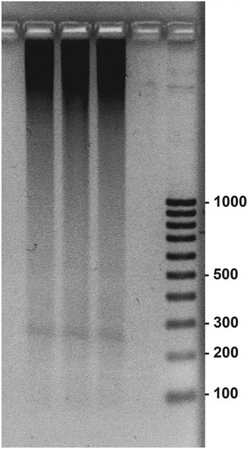
The tandem organization of stDNA gives rise to a characteristic ladder of bands after agarose gel electrophoresis of genomic DNA digested with restriction endonucleases. However, digestion with EcoRI generated exclusively a faint but discrete band of about 240 bp (fig. 1). Similar results were reported by Baus Lončar et al. (Reference Baus Lončar, Paić and Ugarković2005), who suggested that this fragment could be a dispersed repetitive DNA. We eluted and cloned the obtained band. Sequencing of the positive clones revealed the existence of two different repetitive DNA families, which we called LEDE-I and LEDE-II.
Electrophoretic separation on 2% agarose gels of L. decemlineata genomic DNA after digestion with EcoRI restriction enzyme showing a DNA band of about 240 bp. The numbers on the right indicate the size of DNA fragment in bp.

Study and characterization of LEDE-I and LEDE-II stDNA families
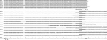
Thirteen of the 23 sequenced clones belong to the LEDE-I family (LEDE-I-2, 19, 27, 28, 29, 34, 37, 43, 44a, 44c, 49, 55, and 56) (EMBL accession nos. HE864283–HE864295). The CLUSTALW computer program was used to perform the alignment of sequences. The comparison of sequencing results is summarized in fig. 2, where the best alignment of sequences is presented.
Alignment of LEDE-I sequences. The first 13 LEDE-I (LEDE-2, 19, 27, 28, 29, 34, 37, 43, 44a, 44c, 49, 55, and 56) are sequences derived from L. decemlineata EcoRI digested DNA with 240 bp in length. The following LEDE-I sequences (LEDE-A1, A2, A4, A5, B33, C4, C5, D3, B4, B5, B13, and D1) were obtained after PCR. The primers used are shown. LEDE-I consensus sequence (295 bp) is also shown. The two EcoRI target sequences and the only one TaqI target sequence are also indicated.

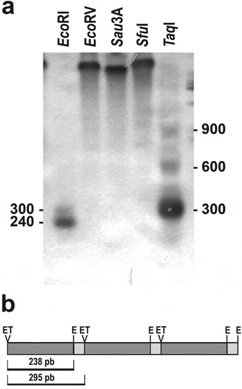
To determine whether the cloned repetitive DNA sequences were really dispersed or organized in tandem, we examined the genomic DNA by Southern-blot hybridization using the LEDE-I-37 fragments as a probe. Southern hybridization with EcoRI digested DNA caused the appearance of two bands, approximately 240 bp and 300 bp in length (fig. 3a).To determine the genetic organization of this repetitive DNA, we performed the PCR analysis using primers designed on the 240 bp EcoRI fragment. Primers were designed to amplify the DNA located between two of these fragments (fig. 2). The PCR generates bands of about 150, 300, 450, and 600 bp (see fig. 1 in supplementary material). These bands were cloned and sequenced (LEDE-I-A1, A2, A4, A5, B33, C4, C5, D3, B4, B5, B13, and D1) (EMBL accession nos. HE864297–HE864302). The alignment of all clones revealed that the 240-bp sequence, isolated by digestion with EcoRI, was part of a longer repeat of 295 bp (fig. 2). The 295-bp sequence contained two EcoRI targets which could generate the 240-bp band obtained in Southern hybridization (fig. 3a). The alignment of the sequences showed that the 300 (clones A1, −2, 5, B-3, C-4, −5) and 600-bp bands (clones B4, −5, −13) obtained by PCR represent the monomeric and dimeric repeats of the LEDE-I stDNA family. The bands of 150 (clones A4 and D3) and 450 bp (clone D1) correspond to monomeric and dimeric repeats with internal deletions of about 150 bp (fig. 2).
(a) Southern-blot hybridization of L. decemlineata genomic DNA digested with several restriction enzymes using LEDE-I-37 fragments as a probe. The numbers indicate the size of DNA fragment in bp. (b) Schematic representation of a repeat unit of the LEDE-I stDNA showing the origin of the 238 and 295 bp fragments generated by EcoRI digestion. E=EcoRI target sequences; T=TaqI target sequences.

These results could indicate that 295 bp is the size of the monomeric repeat in the LEDE-I stDNA family. The study of the consensus sequence of 295 bp showed only one target for the TaqI restriction enzyme (fig. 2). Thus, if this repetitive DNA is truly organized in tandem, digestion with this enzyme could result in a ladder pattern on a gel. Southern hybridization of TaqI digested DNA, using LEDE-I-37 sequence as a probe, originated a ladder with an approximately 300 bp unit, showing that LEDE-I is tandemly repeated DNA (fig. 3a, b). Some hybridization smears were also observed, possibly due to the existence of monomers and dimers with internal deletions.
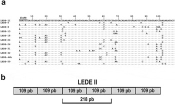
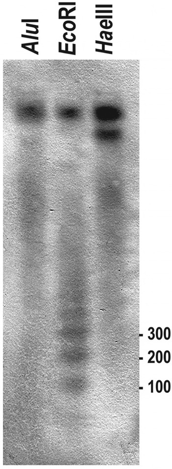
As mentioned above, sequencing of the positive clones revealed the existence of another repetitive DNA family in the 240-bp fragment obtained by digestion with EcoRI, called LEDE-II (EMBL accession nos. HE864308–HE864317). The cloned fragments were 218 bp in length and their analysis showed that they represent dimers of a repeat unit of 109 bp. Figure 4a shows the alignment of these sequences and the consensus sequence obtained with CLUSTALW. A schematic representation of a repeat unit of the LEDE-II stDNA is shown in fig. 4b. Southern-blot hybridization of genomic DNA digested with EcoRI demonstrated the tandem organization of the LEDE-II repeats (fig. 5).
(a) Alignment of LEDE-II stDNA sequences. The EcoRI sequence target is indicated. (b) Schematic representation of repeat unit of the LEDE-II stDNA showing the origin of the 218 bp fragments generated by EcoRI digestion.

Southern-blot hybridization of L. decemlineata genomic DNA digested with Alu I, EcoRI and HaeIII using the LEDE-II-5 repeat as a probe. The numbers on the right indicate the size of DNA fragments in bp.

In summary, we isolated two different and unrelated families of stDNA for which the consensus sequences were 295 bp (LEDE-I) and 109 bp (LEDE-II) in length. In both cases, searches in GenBank failed to find similarity of these stDNAs with other deposited sequences.
Dot-blot hybridization showed that LEDE-I and LEDE-II appear to comprise about 0.4 and 0.05% of the total genomic DNA, respectively. The estimated size of the L. decemlineata genome is 0.46 pg (Petitpierre et al., Reference Petitpierre, Segarra and Juan1993). Consequently, the estimated number of copies are 6.1 × 10⁴ and 2.06 × 10⁴, respectively, per haploid genome. This value is higher in C. americana, whose stDNA family constitutes about 10% of the total genomic DNA with an estimated 26.5 × 10⁴ copies (Lorite et al., Reference Lorite, Palomeque, Garnería and Petitpierre2001). A wide range of variability in relation to the quantity of stDNA has been observed in other species of insects (Palomeque & Lorite, Reference Palomeque and Lorite2008; Plohl et al., Reference Plohl, Meštrović, Mravinac and Garrido-Ramos2012). For example, the amount of stDNA in different Drosophila species varies from 2 to 24% of the genome (Bosco et al., Reference Bosco, Campbell, Leiva-Neto and Markow2007).
In LEDE-I stDNA, the estimate of average percent sequence divergence (p-distance) over all the sequence pairs was 0.176 ± 0.025. In LEDE-I fragments obtained by digestion with EcoRI, the p-distance was 0.109 ± 0.011 and in the monomers obtained by PCR was 0.207 ± 0.031. LEDE-II was less variable with an average p-distance over all monomer pairs of 0.098 ± 0.018.
Only one deletion was found in LEDE-II repeats, specifically a deletion of a nucleotide in the clone LEDE-I-24 (fig. 4a). In contrast, insertions and deletions were frequent in LEDE-I. Although there were short deletions of 1–4 nucleotides, long deletions over 150 bp were observed in some of the clones obtained by PCR. The sequence divergence in both stDNAs was caused by point mutations some of which were shared by several monomers. The existence of the same mutation shared by several monomers is likely to be a consequence of an individual mutation that spread to other repeats in the homogenization process. This pattern of gradual replacement of a nucleotide by another was analyzed by Strachan et al. (Reference Strachan, Webb and Dover1985), and has been observed in the stDNA evolution of several insect groups (Pons et al., Reference Pons, Bruvo, Petitpierre, Plohl, Ugarkovic and Juan2004) as well as in other organisms (Pérez-Gutiérrez et al., Reference Pérez-Gutiérrez, Suárez-Santiago, López-Flores, Romero and Garrido-Ramos2012). The new variants are spread by means of a variety of genomic-turnover mechanisms (unequal crossing over, gene conversion, slippage replication, transposition and RNA-mediated exchange) (Dover, Reference Dover2002). In LEDE-II sequences, and using the algorithm developed by Betrán et al. (Reference Betrán, Rozas, Navarro and Barbadilla1997) incorporated in the DnaSP program, we detected three putative gene conversion tracts. These were identified in the second monomer of the LEDE-8 and LEDE-26 clones between 97 and100 nucleotide sites and in the second monomer of the LEDE-44b clone between 57 and 68 nucleotide sites. These findings could suggest that gene conversion is involved in the homogenization processes that would spread these mutations to all monomeric repeats. Similar results have been found in the stDNA of another chrysomelid beetle, X. luteola (Lorite et al., Reference Lorite, Carrillo, Garneria, Petitpierre and Palomeque2002).
It has been proposed that the inverted and palindromic repeats could form secondary dyad structures or cruciform structures distributed throughout the stDNA of insects and could act as nucleosome-positioning signals (Palomeque & Lorite, Reference Palomeque and Lorite2008; Grechko, Reference Grechko2011). The LEDE-I consensus sequence showed four small inverted repeats, all short and in some cases overlapping (data not shown). Similar results have been found in the chrysomelid species C. americana (Lorite et al., Reference Lorite, Palomeque, Garnería and Petitpierre2001) and Xanthogaleruca luteola (Lorite et al., Reference Lorite, Carrillo, Garneria, Petitpierre and Palomeque2002). The LEDE-II consensus sequence does not include any inverted repeats. In contrast, long inverted repeats capable of forming stable dyad structures have been reported on stDNA from Tribolium brevicornis (Mravinac et al., Reference Mravinac, Plohl and Ugarković2005).
The stDNA curvature or its potential bendability has been related to heterochromatin organization as well as to specific protein binding (Lobov et al., Reference Lobov, Tsutsui, Mitchell and Podgornaya2001; Matyasek et al., Reference Matyasek, Fulnecek, Leitch and Kovarik2011). The bendability and/or curvature propensity, and the G + C content were plotted over the DNA sequence, and 3D reconstructions of DNA molecules were performed (see figs. 2, 4a, b in supplementary material). In addition, retarded electrophoretic mobility on a 6% non-denaturing polyacrylamide gel was also examined on both stDNAs to check DNA curvature. Generally, an inverse correlation between predicted curvature and G + C content was found. The AT richness and the presence of AT runs are a fundamental feature of DNA curvature (Lorite et al., Reference Lorite, Carrillo, Aguilar and Palomeque2004; Palomeque & Lorite, Reference Palomeque and Lorite2008; Escribá et al., Reference Escribá, Greciano, Méndez-Lago, de Pablos, Trifonov, Ferguson-Smith, Goday and Villasante2011). Recently, Matyasek et al. (Reference Matyasek, Fulnecek, Leitch and Kovarik2011) have reported a clear correlation between the existence of short and periodically phased T4–6 and stDNA curvature. LEDE-I and LEDE-II stDNA families were AT-rich (70.8% and 71.6%, respectively) and they present clusters of three or more consecutive T or A nucleotides along the monomeric repeat. This AT richness was higher than found in the stDNA of other chrysomelid species (Lorite et al., Reference Lorite, Palomeque, Garnería and Petitpierre2001, Reference Lorite, Carrillo, Garneria, Petitpierre and Palomeque2002; Palomeque et al., Reference Palomeque, Muñoz-López, Carrillo and Lorite2005), in which non-curved stDNA has been reported (Lorite et al., Reference Lorite, Carrillo, Garneria, Petitpierre and Palomeque2002). However, the AT richness from the stDNAs studied in this paper, is similar to those reported on stDNA from tenebrionid beetles (64–74%; Ugarkovic et al., Reference Ugarkovic, Petitpierre, Juan and Plohl1995; Mravinac & Plohl, Reference Mravinac and Plohl2010).
The estimated curvature and the K values (about 1.1) indicated that the monomer (LEDE-I-A1) and the smaller fragments (LEDE-I-D3 and A4) were only slightly curved (see figs. 2a,b,d and 3 in supplementary material). The LEDE-I-19 sequence had a somewhat higher value of K (about 1.3) in accordance with the predictive curvature model that showed the putative maximum peaks positioned in the middle of the repeat (see figs. 2c and 3a in supplementary material). The predictive models and the K values (about 2) strongly suggested that the dimeric sequences were curved (see figs. 2e, f, 3a, and b in supplementary material). The same studies carried out on the LEDE-II stDNA family (see fig. 4 in supplementary material) also showed that the dimeric repeats were also curved (K values between 1.4 and 1.7; see fig. 4c in supplementary material).
Chromosome location of the two stDNA families
The chromosome number in L. decemlineata is 2n = 34 + X0 in males and 2n = 34 + XX in females (Hsiao & Hsiao, Reference Hsiao and Hsiao1983). The karyotype is formed by metacentric to submetacentric chromosomes The X chromosome is a large metacentric chromosome. A polymorphism was detected in this species by the existence of a pericentromeric inversion in metacentric chromosome 2 that changes its morphology to acrocentric (Hsiao & Hsiao, Reference Hsiao and Hsiao1983). The material analyzed in this study belongs to this second karyomorph with an acrocentric chromosome (fig. 6i).
In situ hybridization using LEDE-I stDNA as a probe in mitotic (a) and meiotic (b) metaphases. The arrow indicates a chromosome with only one hybridization signal. In situ hybridization using LEDE-II stDNA as a probe in mitotic (c) and meiotic (d) metaphases. (e) A meiotic metaphase plate after in situ hybridization using as a probe the DIG-labeled LEDE-I stDNA repeat and the same meiotic metaphase (f) using as a probe the biotin-labeled LEDE-II stDNA repeat. (g) Selected bivalents of the previous images showing hybridization signals with both stDNA families, only with LEDE-I stDNA, only with LEDE-II stDNA or lacking hybridization signals with both repetitive DNAs, respectively. The X chromosome showing hybridization signals with both stDNA families is also shown. (h, l) Selected mitotic chromosomes hybridized with both stDNA families. In all figures the first and second chromosomes are obtained after in situ hybridization using as a probe the DIG-labeled LEDE-I stDNA repeat and the biotin-labeled LEDE-II stDNA repeat, respectively. In (h, i) the third chromosome is the result of the assembly of previous chromosomes. Scale bar = 5 μm.

FISH, using LEDE-I stDNA as a probe, showed positive hybridization signals at the pericentromeric region in at least 25 mitotic chromosomes, although in three of them the hybridization signals were very weak (fig. 6a). The odd number of signals in mitotic metaphases was probably due to the presence of this stDNA in the single X chromosome of the males. In meiosis, hybridization signals were visible in 12 bivalents (two hybridization signals in each one) and a chromosome with only one hybridization signal that probably corresponds to the X chromosome (fig. 6b). With LEDE-II stDNA as a probe, hybridization signals in 17 mitotic chromosomes were found (fig. 6c). Similar to LEDE-I stDNA, the LEDE-II stDNA was not present in all chromosomes. Nine meiotic chromosomes showed hybridization signals (fig. 6d). Also in this case, the odd number of signals in mitotic metaphases indicates that the X chromosome may carry this stDNA.
To test for chromosomal co-localization, in situ hybridizations were performed in the same meiotic and mitotic metaphases using both stDNA families as probes. The LEDE-I probe was labeled with digoxigenin, and chromosomes were stained with Giemsa. This stain allowed better observation of the chromosome morphology, enabling identification of the X chromosome. The analysis of the same metaphase plate in situ hybridized with both probes showed hybridization signals with both stDNA families on the X chromosome and seven bivalents. One bivalent contained only LEDE-II and five contained only LEDE-I. Finally, four bivalents lacked both repetitive DNAs (fig. 6e, f and g). Mitotic chromosomes were also observed carrying both stDNA families, only one, or neither. In addition, it was also possible to see that each stDNA family was located in a different chromosome arm in some chromosomes. This was evident when both hybridization signals were assembled on the same image (see figs 6h, i and 5 in supplementary material). The absence of LEDE-I or LEDE-II stDNA in some chromosomes could suggest the possible existence of other undetected stDNA families.
The results on the stDNA from L. decemlineata differ somewhat from those reported in other chrysomelid beetles although the knowledge of this type of DNA in leaf-beetle genomes is rather low. In the three chrysomelid species for which stDNA has been studied, only one stDNA family present in all or in the majority of the chromosomes has been detected (Lorite et al., Reference Lorite, Palomeque, Garnería and Petitpierre2001, Reference Lorite, Carrillo, Garneria, Petitpierre and Palomeque2002; Palomeque et al., Reference Palomeque, Muñoz-López, Carrillo and Lorite2005). The stDNA from C. carnifex is complex, showing monomers (211 bp), and higher-order repeats in the form of dimers or trimers, although probably with a common evolutionary origin (Palomeque et al., Reference Palomeque, Muñoz-López, Carrillo and Lorite2005). A major and single species-specific stDNA, constituting up to 60% of the genome, has been reported for the majority of darkling beetles (Tenebrionidae). However, other stDNA families were also present, although in the form of low-copy-number repeats (Pezer & Ugarković, Reference Pezer and Ugarković2009; Mravinac & Plohl, Reference Mravinac and Plohl2010 among others). This is the first molecular and cytogenetic study conducted on L. decemlineata repetitive DNA and specifically on stDNA which is one of the important constituents of the eukaryotic genomes. Certainly, more studies are needed using multiple CPB populations in order to establish the role of repetitive DNAs in the diverse and flexible life history of L. decemlineata.
The supplementary material for this article can be found at http://www.journals.cambridge.org/BER.
Acknowledgements
This work was supported by the Spanish Ministerio de Educación y Ciencia through project CGL2011-23841 (co-funded by the European Regional Development Fund) and by the Junta de Andalucía through the programs ‘Ayudas a Grupos de Investigación’, Group BIO220 and ‘Incentivos a proyectos de investigación de excelencia’, proyect CVI-6807 (co-funded by the European Regional Development Fund).