Schizophrenia is associated with a diverse array of clinical symptoms and cognitive deficits (Addington, Addington & Maticka-Tyndale, Reference Addington, Addington and Maticka-Tyndale1991; Andreasen et al., Reference Andreasen, Nopoulos, O'Leary, Miller, Wassink and Flaum1999; Johnstone, Owens, Frith, & Crow, Reference Johnstone, Owens, Frith and Crow1987). The symptoms include hallucinations, delusions, disorganized thought and speech, and negative symptoms such as avolition, alogia, and flattened affect (Andreasen & Olsen, Reference Andreasen and Olsen1982). In addition to the clinical symptoms, there are often cognitive deficits that span multiple neuropsychological domains (White, Ho, Ward, Leary, & Andreasen, Reference White, Ho, Ward, O'Leary and Andreasen2006). The diversity of these symptoms suggests perturbations that act on global scales of brain function. As the brain operates through an orchestration of intricately tuned neural networks, a disruption in the connectivity within or between these networks is one of the leading hypotheses underlying schizophrenia (Andreasen, Paradiso, & O'Leary, Reference Andreasen, Paradiso and O'Leary1998; Friston, Reference Friston1998; Konrad & Winterer, Reference Konrad and Winterer2008; Weinberger, Berman, Suddath, & Torrey, Reference Weinberger, Berman, Suddath and Torrey1992). Evidence supporting this theory has emerged through post mortem (Benes, Reference Benes1989; Colter et al., Reference Colter, Battal, Crow, Johnstone, Brown and Bruton1987; Davis et al., Reference Davis, Stewart, Friedman, Buchsbaum, Harvey and Hof2003; Heckers, Heinsen, Geiger, & Beckmann, Reference Heckers, Heinsen, Geiger and Beckmann1991), genetic (Hakak et al., Reference Hakak, Walker, Li, Wong, Davis and Buxbaum2001), and neuroimaging studies (Agartz, Andersson, & Skare, Reference Agartz, Andersson and Skare2001; Ardekani, Neirenberg, Hoptman, Javitt, & Lim, Reference Ardekani, Nierenberg, Hoptman, Javitt and Lim2003; Buchsbaum et al., Reference Buchsbaum, Tang, Peled, Gudbjartsson, Lu and Hazlett1998; Kendi et al., Reference Kendi, Kendi, Lehericy, Ducros, Lim and Ugurbil2008; Kubicki et al., Reference Kubicki, McCarley, Westin, Park, Maier and Kikinis2007; Kumra et al., Reference Kumra, Ashtari, McMeniman, Vogel, Augustin and Becker2004, Reference Kumra, Ashtari, Cervellione, Henderson, Kester and Roofeh2005; Lim et al., Reference Lim, Hedehus, Moseley, de Crespigny, Sullivan and Pfefferbaum1999; Rose et al., Reference Rose, Chalk, Janke, Strudwick, Windus and Hannah2006; Volkow et al., Reference Volkow, Wolf, Brodie, Cancro, Overall and Rhoades1988; White et al., Reference White, Kendi, Lehericy, Kendi, Karatekin and Guimaraes2007).
Theories of aberrant connectivity in distributed neural networks in schizophrenia have paralleled, and perhaps even driven, the exponential growth in techniques applied to measure brain connectivity. Measures of neural connectivity are divided into structural, functional, and effective connectivity, each which evaluates a different aspect of connectivity, although these aspects are interrelated. Structural connectivity refers to measures of the physical presence and pathways of the actual neural fibers. It is commonly established in animal models through anatomical tract tracing techniques (Köbbert et al., Reference Köbbert, Apps, Bechmann, Lanciego, Mey and Thanos2000), or less reliably in noninvasive human studies utilizing techniques such as diffusion tensor imaging (DTI; Shimony, Snyder, Conturo, & Corbetta, Reference Shimony, Snyder, Conturo and Corbetta2004). Although the structural connectivity of the human brain is not directly known, because of experimental limitations, more readily available noninvasive measures of functional connectivity, particularly for the “resting state,” have been taken as a proxy for human anatomical connections (Greicius, Supekar, Menon, & Dougherty, Reference Greicius, Supekar, Menon and Dougherty2009). Functional connectivity describes the functional association between brain regions that possess correlated temporal activation patterns (Friston, Reference Friston1994), as demonstrated by various functional imaging techniques. The previous two aspects of brain connectivity are linked through effective connectivity, which reflects the causal influence of the activity in one brain region on another. Thus, although functional connectivity is essentially model free, effective connectivity relies on a model that explains how functional connectivity is mediated by structural connectivity (Friston, Reference Friston1994).
The triad of structural (Konrad & Winterer, Reference Konrad and Winterer2008; Kubicki et al., Reference Kubicki, McCarley, Westin, Park, Maier and Kikinis2007), functional (Andreasen et al., Reference Andreasen, Nopoulos, O'Leary, Miller, Wassink and Flaum1999; Meyer-Lindenberg et al., Reference Meyer-Lindenberg, Poline, Kohn, Holt, Egan and Weinberger2001, Reference Meyer-Lindenberg, Olsen, Kohn, Brown, Egan and Weinberger2005; Micheloyannis et al., Reference Micheloyannis, Pachou, Stam, Breakspear, Bitsios and Vourkas2006), and effective (Schlosser et al., Reference Schlosser, Gesierich, Kaufmann, Vucurevic, Hunsche and Gawehn2003; Winterer, Coppola, Egan, Goldberg, & Weinberger, Reference Winterer, Coppola, Egan, Goldberg and Weinberger2003) connectivity has been shown to be aberrant in patients with schizophrenia. Early positron emission tomography studies demonstrated regional abnormalities (Volkow et al., Reference Volkow, Wolf, Brodie, Cancro, Overall and Rhoades1988), suggesting that the impairment is focused on specific neural subnetworks, such as hippocampal–prefrontal and cortical–cerebellar–thalamic–cortical connectivity (Andreasen et al., Reference Andreasen, Nopoulos, O'Leary, Miller, Wassink and Flaum1999). Aberrant connectivity in schizophrenia has been shown to be associated with the level of clinical (Lawrie et al., Reference Lawrie, Buechel, Whalley, Frith, Friston and Johnstone2002) and cognitive impairments (Meyer-Lindenberg et al., Reference Meyer-Lindenberg, Poline, Kohn, Holt, Egan and Weinberger2001), and can be altered with the administration of antipsychotic medication (Stephan et al., Reference Stephan, Magnotta, White, Arndt, Flaum and O'Leary2001).
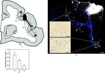
It has recently has been argued that structural connectivity also contributes to developmental processes that influence the shape of the folded cerebral cortex through axonal tension forces (Van Essen, Reference Van Essen1997). The overall interaction of these forces produces the characteristic convolutions of the human brain and leads to a compact layout that optimizes the transit of neuronal signals between brain regions (Hilgetag & Barbas, Reference Hilgetag and Barbas2005; Rakic, Reference Rakic1988; Van Essen, Reference Van Essen1997; Zilles, Armstrong, Schleicher, & Kretschmann, Reference Zilles, Armstrong, Schleicher and Kretschmann1988). Thus, the characteristic morphology of the brain, with its many gyri and sulci, may correspond to its efficient function (see Figure 1). Conversely, if form corresponds to function, and neural function is disrupted in schizophrenia, then is form disrupted as well? Is it possible that buried within the fissures and folds that make up the convoluted landscape of the cerebral cortex exists structural evidence of the disrupted connectivity reported in schizophrenia?
The trajectories and densities of projections support the axonal tension hypothesis of cortical folding. (a) Prediction of the axonal tension hypothesis: denser projections should be straighter in the adult brain because of the global balancing of tension forces; straight (st, thick line), intermediate (im, short dashed line), and curved (cu, long dashed line) projections link a tracer injection site (dark spot) with different areas. (b) An example of the densities and trajectories of straight, intermediate, and curved projections as labeled by neuronal tract tracing in the primate prefrontal cortex. Denser projections form straighter paths. (c) The average summed density of straight (st), intermediate (im), and curved (cu) projections linking cortical areas, demonstrating that straight connections dominate in the global tug of war. Adapted from “Role of Mechanical Factors in the Morphology of the Primate Cerebral Cortex,” by C. C. Hilgetag and H. Barbas, 2006, PLoS Computational Biology, 2(3), e22. Copyright 2006 International Society for Computational Biology. Adapted with permission. [A color version of this figure can be viewed online at journals.cambridge.org/dpp]

The goal of this paper is to provide a review of recent evidence for disruptions in the surface morphology or gyrification of the brain in schizophrenia. We start by reviewing the development and theories of the underlying mechanisms associated with gyrification. We then provide a review of studies of gyrification abnormalities in schizophrenia. Finally, we present evidence that abnormalities of gyrification may reflect abnormalities in the underlying neural connectivity. Because gyrification occurs during a relatively short window during prenatal development, specific changes seen in patients with schizophrenia may help elucidate the interplay between neurodevelopment and illness related changes. Recent theories of gyrification provide an important link for understanding the relationship between brain surface morphology and connectivity.
Theories of Gyrification
By the end of the second trimester (26 weeks gestational age) the developing brain is lissencephalic, with a generally smooth cortical surface (Welker, Reference Welker, Jones and Peters1990). Following the 26th week the brain surface morphology changes rapidly, and by term at 40 weeks the characteristic adult patterns of sulci and gyri are present. Thus, the major steps in the process of gyrification occur during the third trimester, which is a time of considerable brain growth (Neal, Takahashi, Silva, Tiao, Walsh, & Sheen, Reference Neal, Takahashi, Silva, Tiao, Walsh and Sheen2007). Genetic information forms the basis of the convolution patterns of the brain (Piao et al., Reference Piao, Hill, Bodell, Chang, Basel-Vanagaite and Straussberg2004). However, because even monozygotic twins show considerable differences in their folding patterns (Thompson, Cannon, et al., Reference Thompson, Cannon, Narr, van Erp, Poutanen and Huttunen2001; White, Andreasen, & Nopoulos, Reference White, Andreasen and Nopoulos2002), the characteristic convolutions are not specified directly from genetic information; they are probably specified via biological and physical self-organization, that is, through a multitude of interactions involving biological processes, such as regulatory metabolic cascades or anabolic growth, and physical forces, such as volume competition or biomechanical tissue tension (Hilgetag & Barbas, Reference Hilgetag and Barbas2005, Reference Hilgetag and Barbas2006; Richman, Stewart, Hutchinson, & Caviness, Reference Richman, Stewart, Hutchinson and Caviness1975; Toro & Burnod, Reference Toro and Burnod2005; Van Essen, Reference Van Essen1997; Welker, Reference Welker, Jones and Peters1990).
Early theories on brain gyrification hypothesized that differential growth rates in different brain regions could lead to competing volume demands and tension between these regions, resulting in local tissue deformation (His, Reference His1874). One school of morphogenetic concepts of the brain suggests that regions that are developing into gyri may grow at a faster rate than those destined to be become sulci (Kriegstein, Noctor, & Martinez-Cerdeno, Reference Kriegstein, Noctor and Martinez-Cerdeno2006). Thus, convolutions may be established by active, localized growth. This active growth could also occur in combination with friction between the different brain layers against each other, coupled with the friction with the surrounding skull (Le Gross Clark, Reference Le Gross Clark, LeGross and Medawar1945). This differential growth expansion of the individual cortical layers may lead to a buckling of the laminar cortical sheet (Hilgetag & Barbas, Reference Hilgetag and Barbas2005, Reference Hilgetag and Barbas2006; Richman et al., Reference Richman, Stewart, Hutchinson and Caviness1975; Toro & Burnod, Reference Toro and Burnod2005; Van Essen, Reference Van Essen1997; Welker, Reference Welker, Jones and Peters1990).
Theories of cortical morphogenesis should also consider the relationship between the development of brain convolutions and the pruning of neuronal and synaptic connections (Goldman-Rakic, Reference Goldman-Rakic1980; Goldman-Rakic & Rakic, Reference Goldman-Rakic, Rakic, Geschwind and Galaburda1984). It has been suggested that viscoelastic tension exerted by corticocortical fibers (Franze, Reichenbach, & Käs, Reference Franze, Reichenbach, Käs, Kamkin and Kiseleva2008) may contribute to shaping cortical convolutions (Van Essen, Reference Van Essen1997, Reference Van Essen2007). Specifically, this theory proposes that neuronal connectivity that develops with neuronal migration during the second trimester produces fiber tension that draws densely interconnected regions closer together, forming outward bulging gyri between them, whereas more sparsely connected regions drift apart and are separated by inward sulci. The tension, although very small for an individual axon (Heidemann, Lamoureux, & Buxbaum, Reference Heidemann, Lamoureux and Buxbaum1995), is summed by the very large population of neurons, thus creating differential forces that interact globally with the rapidly growing brain to form the gyri and sulci. Within this concept, the characteristic pattern of the convolutions is explained by the highly specific organization of the underlying connectivity (Sporns, Chialvo, Kaiser, & Hilgetag, Reference Sporns, Chialvo, Kaiser and Hilgetag2004).
If tension produced by neuronal connections is involved in the mechanisms of gyrification, then changes in the patterns of the gyri and sulci are an expected outcome of changes (intrinsic or experimental) that alter the connectivity between brain regions. Local and remote changes of gyrification have been observed after experimental white-matter lesions in the developing primate brain (Goldman-Rakic, Reference Goldman-Rakic1980; Goldman-Rakic & Rakic, Reference Goldman-Rakic, Rakic, Geschwind and Galaburda1984). The axonal tension concept has also been supported by recent experimental findings in the primate brain showing a strong correlation between the densities and trajectories of fiber projections, with denser fibers being straighter and most projections following straight or only mildly curved trajectories (Hilgetag & Barbas, Reference Hilgetag and Barbas2006; Toro & Burnod, Reference Toro and Burnod2005).
In summary, starting from genetically specified parameters, such as differential growth rates, various physical processes may interact to produce and organize the surface morphology of the cerebral cortex during brain development. In particular, experimental findings support the concept of a relationship between characteristic cortical convolutions and the specific layout of cortical fiber connections.
Measures of Gyrification
There have been a number of different techniques to quantify brain gyrification, and different approaches are used to quantify different aspects of gyrification. For example, sulcal depth may be considered as one characteristic feature; because sulci can branch in complex patterns, greater sulcal depth does not necessarily translate into a quantitative increase in other gyrification measures. Other features are sulcal frequency (Narr et al., Reference Narr, Thompson, Sharma, Moussai, Zoumalan, Rayman and Toga2001) or cortical complexity, which typically relates an estimate of brain surface area to volume, accounting for the two- (2-D) and three-dimensional (3-D) differences between surface area and volume measurements.
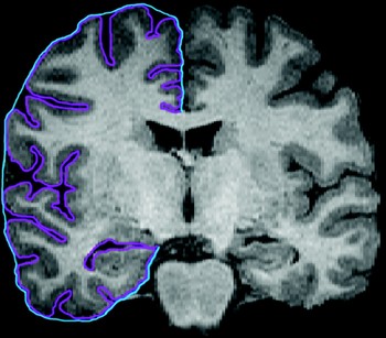
Zilles et al. (Reference Zilles, Armstrong, Schleicher and Kretschmann1988) utilized coronal sections of postmortem brains to measure the ratio of the length of the outer folded surface of the brain (including sulci) to the length of the outer surface excluding sulci. This measure, known as the gyrification index (GI), has been applied to study both the phylogeny (Armstrong et al., Reference Armstrong, Curtis, Buxhoeveden, Fregoe, Zilles and Casanova1991; Zilles et al., Reference Zilles, Armstrong, Schleicher and Kretschmann1988) and ontogeny (Armstrong, Schleicher, Omran, Curtis, & Zilles, Reference Armstrong, Schleicher, Omran, Curtis and Zilles1995) of cortical gyrification (see Figure 2). Brains that have a higher degree of cortical folding yield larger values of the GI. Anterior to posterior maps of human GI measures have shown greater gyrification in the prefrontal and temporal/parietal association regions of the brain (Zilles et al., Reference Zilles, Armstrong, Schleicher and Kretschmann1988). During development, the GI begins to increase prior to the third trimester and plateaus at birth. It is interesting that this plateau appears to remain constant across the life span, despite the rapid brain development and changes associated with aging (Armstrong et al., Reference Armstrong, Schleicher, Omran, Curtis and Zilles1995). However, because developmental studies typically require large samples, and because postmortem samples in younger individuals are difficult to obtain, it would be beneficial for the study of Armstrong et al. (Reference Armstrong, Schleicher, Omran, Curtis and Zilles1995) to be replicated with a larger sample size to confirm this finding. Age-related differences in gyrification measures using magnetic resonance imaging (MRI) techniques have been demonstrated (Magnotta et al., Reference Magnotta, Andreasen, Schultz, Harris, Cizadlo and Heckel1999; White, Su, Schmidt, Kao, & Sapiro et al., 2010), although factors such as cause of death, differences in the interval between fixation of the brains, brain shrinkage, or the generation of brain slices may complicate direct comparisons between MRI and postmortem GI measures.
The gyrification index (GI) is computed by dividing the length of the inner cortical contour (purple line online only) by the length of the outer contour (blue line online only). This yields a unitless measure that reflects a quantification of gyrification of the brain. [A color version of this figure can be viewed online at journals.cambridge.org/dpp]

Alternative methods for measuring the surface morphology of the brain involve regional measurements of curvature (i.e., convexity and concavity) of the cortical surface from MR images (Koenderink & van Doorn, Reference Koenderink and van Doorn1992; Luders et al., Reference Luders, Thompson, Narr, Toga, Jancke and Gaser2006; Magnotta et al., Reference Magnotta, Andreasen, Schultz, Harris, Cizadlo and Heckel1999). These methods first define a triangular isosurface covering a layer within the cortical rim. Once the isosurface is constructed, the angles between vectors normal to the triangular isosurfaces are used to calculate the regional curvature of the cortex. These regional measures can be utilized to study regional curvature between groups (Luders et al., Reference Luders, Narr, Thompson, Rex, Jancke and Steinmetz2004, Reference Luders, Thompson, Narr, Toga, Jancke and Gaser2006) or averaged to study global changes (Magnotta et al., Reference Magnotta, Andreasen, Schultz, Harris, Cizadlo and Heckel1999; Nopoulos, Flaum, O'Leary, Andreasen, Reference Nopoulos, Flaum, O'Leary and Andreasen2000; White et al., Reference White, Andreasen and Nopoulos2002).
Gyrification Abnormalities in Schizophrenia
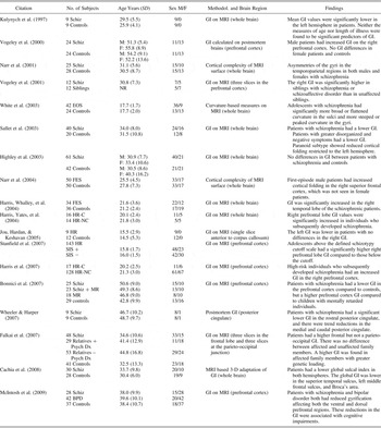
The ability to quantify various aspects of brain surface complexity has existed for decades (Thompson, Schwartz, Lin, Khan, & Toga, Reference Thompson, Schwartz, Lin, Khan and Toga1996; Zilles et al., Reference Zilles, Armstrong, Schleicher and Kretschmann1988), but there have been relatively few studies that applied these techniques to gyrification in schizophrenia (Table 1). The majority of these studies have employed the traditional 2-D GI measure to evaluate differences between patients and controls. These studies yielded mixed results showing GIs in patients that are higher (Harris, Yates, et al., Reference Harris, Yates, Miller, Best, Johnstone and Lawrie2004; Vogeley et al., Reference Vogeley, Schneider-Axmann, Pfeiffer, Tepest, Bayer and Bogerts2000, Reference Vogeley, Tepest, Pfeiffer, Schneider-Axmann, Maier and Honer2001), lower (Bonnici et al., Reference Bonnici, William, Moorhead, Stanfield, Harris and Owens2007; Kulynych, Luevano, Jones, & Weinberger, Reference Kulynych, Luevano, Jones and Weinberger1997; McIntosh et al., Reference McIntosh, Moorhead, McKirdy, Hall, Sussmann and Stanfield2009; Sallet et al., Reference Sallet, Elkis, Alves, Oliveira, Sassi and Campi de Castro2003; Wheeler & Harper, Reference Wheeler and Harper2007), or no different than controls (Highley et al., Reference Highley, DeLisi, Roberts, Webb, Relja and Razi2003).
Studies examining gyrification in schizophrenia spectrum disorders

Note: M, male; F, female; Schiz, chronic schizophrenia; GI, gyrification index; MRI, magnetic resonance imaging; NR, not reported; EOS, early-onset schizophrenia (children and adolescents with schizophrenia; FES, first-episode schizophrenia; HR-C, high risk who converted to schizophrenia; HR-NC, high risk who have not converted; HR, high risk; SIS, Structured Scale for Schizotypy (+ is above threshold/high score and – is below threshold/low score); MR, mental retardation; Dx, diagnosis; BPD, bipolar disorder.
The earliest study utilized coronal MRI slices from the left hemisphere to calculate the GI and reported an overall reduction in GI in both anterior and posterior regions in nine patients with chronic schizophrenia (Kulynych et al., Reference Kulynych, Luevano, Jones and Weinberger1997). Subsequent studies using MRI-based GI measures have replicated the finding of reduced GI in the frontal cortex (Bonnici et al., Reference Bonnici, William, Moorhead, Stanfield, Harris and Owens2007) and global decreases in the GI (Sallet et al., Reference Sallet, Elkis, Alves, Oliveira, Sassi and Campi de Castro2003). Using 50-μm coronal slices in a postmortem study of posterior cingulate cortex, Wheeler and Harper (Reference Wheeler and Harper2007) reported lower GI in the rostral cingulate, with a trend for a reduced GI in the medial and caudal regions. These studies demonstrating a decrease in the GI were all performed on patients with chronic schizophrenia.
There have been two postmortem studies of gyrification in patients with schizophrenia. The first study found an increase in frontal GI in a group of patients with chronic schizophrenia (Vogeley et al., Reference Vogeley, Schneider-Axmann, Pfeiffer, Tepest, Bayer and Bogerts2000). The second postmortem study focused only on the posterior cingulate and found a reduction in GI. Because typical whole-brain 2-D measures of the GI follow the entire contour of the brain, this finding of a reduced GI in the cingulate is difficult to compare to the more global GI measures.
The report of an increased GI in frontal regions, as shown in one of the postmortem studies, has also been reported in MRI studies of the frontal cortex in patients with chronic (Falkai et al., Reference Falkai, Honer, Kamer, Dustert, Vogeley and Schneider-Axmann2007; Vogeley et al., Reference Vogeley, Tepest, Pfeiffer, Schneider-Axmann, Maier and Honer2001) and first-episode schizophrenia (Harris, Yates, et al., Reference Harris, Yates, Miller, Best, Johnstone and Lawrie2004). Moreover, increased GI has been identified in temporal lobe structures (Harris, Yates, et al., Reference Harris, Yates, Miller, Best, Johnstone and Lawrie2004), but not occipitoparietal regions (Falkai et al., Reference Falkai, Honer, Kamer, Dustert, Vogeley and Schneider-Axmann2007; Harris, Yates, et al., Reference Harris, Yates, Miller, Best, Johnstone and Lawrie2004). However, findings of lower GI (Bonnici et al., Reference Bonnici, William, Moorhead, Stanfield, Harris and Owens2007; McIntosh et al., Reference McIntosh, Moorhead, McKirdy, Hall, Sussmann and Stanfield2009) have also been reported and include studies using the same techniques that reported an increase in GI (Harris et al., Reference Harris, Moorhead, Miller, McIntosh, Bonnici and Owens2007; Harris, Yates, et al. Reference Harris, Yates, Miller, Best, Johnstone and Lawrie2004). These apparent discrepant findings may point to the heterogeneity of gyrification or to the heterogeneity in the populations and the methodologies utilized to study GI.
Several approaches have studied whether abnormal surface folding is an endophenotypic marker (Gottesman & Gould, Reference Gottesman and Gould2003) for schizophrenia (Falkai et al., Reference Falkai, Honer, Kamer, Dustert, Vogeley and Schneider-Axmann2007; Vogeley et al., Reference Vogeley, Tepest, Pfeiffer, Schneider-Axmann, Maier and Honer2001). Evidence from studies of unaffected siblings and individuals at risk for developing schizophrenia support gyrification abnormalities a s being more severe in individuals who go on to develop the illness (Harris, Whalley, et al., Reference Harris, Whalley, Yates, Miller, Johnstone and Lawrie2004; Vogeley et al., Reference Vogeley, Tepest, Pfeiffer, Schneider-Axmann, Maier and Honer2001). Thus, it is possible that abnormalities of gyrification, which may surface early in cortical development (Armstrong et al., Reference Armstrong, Schleicher, Omran, Curtis and Zilles1995), are trait markers of the illness, which differentiate those at increased risk who eventually develop the illness (Harris et al., Reference Harris, Moorhead, Miller, McIntosh, Bonnici and Owens2007; Harris, Whalley, et al. Reference Harris, Whalley, Yates, Miller, Johnstone and Lawrie2004). An increase in the right frontal gyrification in individuals at a high risk for developing schizophrenia has been shown to be predictive for developing schizophrenia (Harris et al., Reference Harris, Moorhead, Miller, McIntosh, Bonnici and Owens2007; Harris, Whalley, et al., Reference Harris, Whalley, Yates, Miller, Johnstone and Lawrie2004; Stanfield et al., 2007).
Several studies have deviated from the traditional GI approach to measurements of gyrification and have utilized either 3-D GI measures of cortical surface complexity (Cachia et al., Reference Cachia, Paillere-Martinot, Galinowski, Januel, de Beaurepaire and Bellivier2008; White, Andreasen, Nopoulos, & Magnotta, Reference White, Andreasen, Nopoulos and Magnotta2003; Wiegand et al., Reference Wiegand, Warfield, Levitt, Hirayasu, Salisbury and Heckers2005) or 3-D curvature indices of the gryi and sulci (White et al., Reference White, Andreasen, Nopoulos and Magnotta2003). Two studies found no difference in surface complexity, one using a 3-D adaptation of the GI (Wiegand et al., Reference Wiegand, Warfield, Levitt, Hirayasu, Salisbury and Heckers2005) and the other using a comparison of surface area to volume, corrected to the two-thirds power (White et al., Reference White, Andreasen and Nopoulos2002). However, surface area measures are highly variable and patient–control differences may be more difficult to assess. Using a 3-D adaptation of the GI, Cachia et al. (Reference Cachia, Paillere-Martinot, Galinowski, Januel, de Beaurepaire and Bellivier2008) found that patients with chronic schizophrenia had lower sulcal complexity that was more pronounced in the temporal and left frontal lobes.
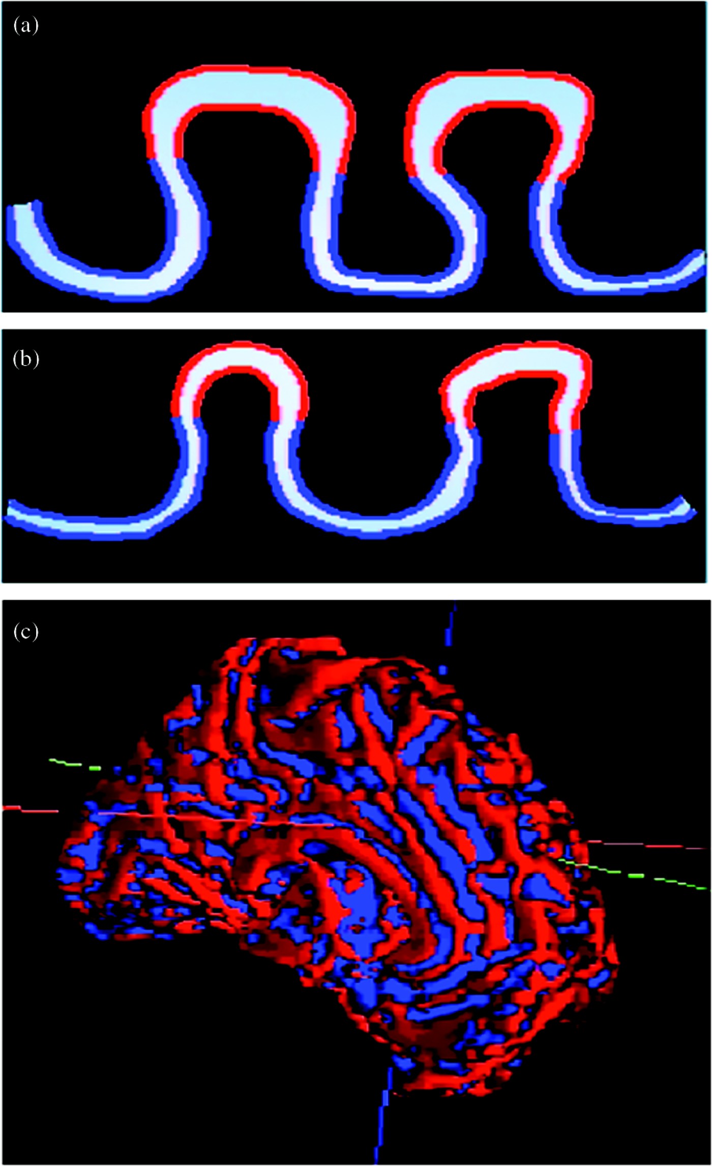
Direct comparisons between the methodologies that utilized surface curvature and those that implemented the GI are difficult, as the GI does not differentiate between gyral and sulcal regions. Thus, it is possible that sulcal widening may be cancelled by gyral or global brain changes (Kulynych et al., Reference Kulynych, Luevano, Jones and Weinberger1997).This is highlighted in children and adolescents with schizophrenia showing opposing changes in gyral and sulcal curvature in the frontal and temporal brain regions (White et al., Reference White, Andreasen, Nopoulos and Magnotta2003). These findings have been replicated and have been shown to be more pronounced in a younger sample of children and adolescent with schizophrenia (R. K. Lenroot, personal communication, 2006). In this sample, the sulci developed less curvature (i.e., became broader) but the gyri became more peaked, demonstrating an increased curvature (Figure 3). This alteration in morphology results in increased sulcal width, which has also been reported in adolescents and adults with schizophrenia (Allmann, Reference Allmann2000; Andreasen et al., Reference Andreasen, Flashman, Flaum, Arndt, Swayze V and O'Leary1994; Giedd et al., Reference Giedd, Jeffries, Blumenthal, Castellanos, Vaituzis and Fernandez1999; Pfefferbaum et al., Reference Pfefferbaum, Zipursky, Lim, Zatz, Stahl and Jernigan1988; Weinberger, Torrey, Neophytides, & Wyatt, Reference Weinberger, Torrey, Neophytides and Wyatt1979).
Children and adolescents with schizophrenia demonstrate differences in gyrification that include (a) a widening of the sulci and a more peaked appearance of gyri compared to (b) controls. These measures can be quantified regionally following the identification of (c) whole brain sulci and gyri. Adapted from “Gyrification Abnormalities in Childhood- and Adolescent-Onset Schizophrenia,” by T. White, N. C. Andreasen, P. Nopoulos, and V. Magnotta, 2003, Biological Psychiatry, 54, 418–426. Copyright 2003 by Elsevier. Adapted with permission. [A color version of this figure can be viewed online at journals.cambridge.org/dpp]

Neural Connectivity in Schizophrenia
New approaches and insights from the field of complex network studies (Strogatz, Reference Strogatz2001) are currently entering systems neuroscience. Applied to the global organization of neural networks, such approaches have revealed high modularity, complexity, and efficiency of the different types of anatomical and functional neural connectivity (Sporns et al., Reference Sporns, Chialvo, Kaiser and Hilgetag2004). Efficient connectivity, for instance, can be assessed by the average length of all shortest paths (ASP) across a network, where the shortest paths represent the smallest number of edges required to link two nodes. The ASP can be taken as an estimate for the minimal number of processing steps in a neural system, and it appears that neural networks are organized to maintain very short ASP (Kaiser & Hilgetag, Reference Kaiser and Hilgetag2006). Conversely, network alterations have been observed in schizophrenia, which lead to increased ASP and which may be associated with a loss of global network efficiency and functional impairments (see Figure 1; Liu et al., Reference Liu, Liang, Zhou, He, Hao and Song2008; Micheloyannis et al., Reference Micheloyannis, Pachou, Stam, Breakspear, Bitsios and Vourkas2006).
Network anomalies in schizophrenia can be identified in measures of functional of as well as structural connectivity (Karlsgodt, Sun, et al., Reference Karlsgodt, Sun, Jimenez, Lutkenhoff, Willhite and van Erp2008); however, a large number of recent studies have focused on white-matter integrity and structural connectivity as indicated by the noninvasive approach of DTI. These studies demonstrated mostly reductions of indicators of structural connectivity, such as fractional anisotropy, in particular for frontal and temporal white matter (Kyriakopoulos, Bargiotas, Barker, & Frangou, Reference Kyriakopoulos, Bargiotas, Barker and Frangou2008; White, Nelson, & Lim, Reference White, Nelson and Lim2008). The diminished integrity of structural connectivity may result from weaker connections, perhaps also due the loss of oligodendroglia (Konrad & Winterer, Reference Konrad and Winterer2008), or through the disconnection of network nodes and appears to precede the onset of the disease (Begre & Koenig, Reference Begre and Koenig2008). Nonetheless, there are also occasional reports of increases in functional (resting state) connectivity in schizophrenia, for instance, in paranoid schizophrenic subjects (Broyd et al., Reference Broyd, Demanuele, Debener, Helps, James and Sonuga-Barke2009).
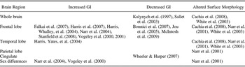
The approach of DTI has also indicated reductions in specific structural connection systems, such as the uncinate fasciculus, a fiber tract connecting the temporal pole near the amygdala and the orbitofrontal/ventrolateral prefrontal cortex, or connections of the cingulate gyrus (Karlsgodt, Sun, et al., Reference Karlsgodt, Sun, Jimenez, Lutkenhoff, Willhite and van Erp2008). Such weakened connections can be associated with specific functional aspects of schizophrenia. For example, the estimated strength of the left superior longitudinal fasciculus, which is decreased in schizophrenic patients, is correlated with performance in a verbal working memory task (Karlsgodt, van Erp, et al., Reference Karlsgodt, van Erp, Poldrack, Bearden, Nuechterlein and Cannon2008). However, the exact relationship between variations in projection systems and global or local aberrations of gyrification in schizophrenia is currently still unclear for two reasons. First, the results of the DTI investigations are themselves not consistent (Kyriakopoulos et al., Reference Kyriakopoulos, Bargiotas, Barker and Frangou2008; White et al., Reference White, Nelson and Lim2008). Investigated fiber systems varied across studies, and several studies investigating the same systems disagreed on whether they showed consistent changes in schizophrenic patients compared to healthy subjects (Kanaan et al., Reference Kanaan, Kim, Kaufmann, Pearlson, Barker and McGuire2005). Moreover, DTI findings, such as that of a weakened uncinate fasciculus in schizophrenic patients (Burns et al., Reference Burns, Job, Bastin, Whalley, Macgillivray and Johnstone2003), may be contradicted by corresponding studies of postmortem material (Highley, Walker, Esiri, Crow, & Harrison, Reference Highley, Walker, Esiri, Crow and Harrison2002). Second, changes of structural connectivity as well as gyrification in schizophrenia appear to be predominantly associated with the frontal lobes. However, although changes in connectivity mostly manifest as weakened projections, schizophrenia may go along with reduced as well as increased gyrification of the frontal gray matter (cf. Table 2). These issues still need to be investigated more systematically. For example, structural MR studies showed that the gray to white matter ratio in frontal and temporal lobes does not decrease over the life span in schizophrenic patients as it does in healthy subjects (Bartzokis et al., Reference Bartzokis, Nuechterlein, Lu, Gitlin, Rogers and Mintz2003).
Consolidated results of gyrification studies of schizophrenia and individuals at high risk

Note: GI, gyrification index. No change in the GI was noted by Highley et al. (Reference Highley, DeLisi, Roberts, Webb, Relja and Razi2003).
Discussion
Gyrification and brain efficiency
The phylogeny of the human nervous system has resulted in a highly complex brain with a high degree of cortical folding. Humans, dolphins, and porpoises stand apart from other species in having a disproportionately large cerebral cortex to body size ratio (Allmann, Reference Allmann2000). The cortical folding increases the surface area of the cortical gray matter and enhances the compactness of the brain.
Although increased gray matter could also be achieved by increasing the cortical thickness, the development of gyral and sulcal folds allows for an optimized compaction of neuronal fibers. For example, a slightly larger lisencephalic or smoothed surfaced human brain is theoretically possible by a threefold increase of the cortical thickness from approximately 5 to 15 mm. This increase in cortical thickness would preserve the total volume of cortical gray matter; however, computational models have shown that the necessary packing of connections within the cortex would preclude such an evolutionary change (Murre & Sturdy, Reference Murre and Sturdy1995; Ruppin, Schwartz, & Yeshurun, Reference Ruppin, Schwartz and Yeshurun1993). Because each neuron has more than a thousand connections with other neurons on average (Braitenberg & Schüz, Reference Braitenberg and Schüz1998), the volume of space required to allow for these connections grows with the power of the number of neurons (Changizi, Reference Changizi2001). The result would be highly inefficient neuronal pathways, with some neurons taking circuitous paths in order to reach their final destination (Chklovskii, Schikorski, & Stevens, Reference Chklovskii, Schikorski and Stevens2002).
The efficient pattern of cortical packing in the gyrified brain may account for the increase in surface area far exceeding the growth in cortical thickness (Welker, Reference Welker, Jones and Peters1990). The surface area of the human brain is 1,700 times greater than in shrews, yet the thickness of the cortex is only 6 times greater (Hofman, Reference Hofman1989). Furthermore, the surface area of the human brain is on average 10 times larger than that of the macaque monkey, yet only 2 times as thick (Rakic, Reference Rakic1995). Thus, the cortex expanded laterally rather than vertically (Chenn & Walsh, Reference Chenn and Walsh2002), resulting in a convoluted human cortical sheet that is about 3 times as large as the inner surface of the skull (Hilgetag & Barbas, Reference Hilgetag and Barbas2005, Reference Hilgetag and Barbas2006; Richman et al., Reference Richman, Stewart, Hutchinson and Caviness1975; Toro & Burnod, Reference Toro and Burnod2005; Van Essen, Reference Van Essen1997; Welker, Reference Welker, Jones and Peters1990). The parcellation of brain tissue into the computationally powerful cortical layers and efficient signal transmission through myelinated fibers, coupled with a compacted gyrification pattern, have resulted in an efficient wiring and volume arrangement for the very dense connectivity (Murre & Sturdy, Reference Murre and Sturdy1995; Ruppin et al., Reference Ruppin, Schwartz and Yeshurun1993; Wen & Chklovskii, Reference Wen and Chklovskii2005).
Such a “form-fitting function” design raises the question as to whether alterations in function also result in alterations of form, mediated by neural connectivity. There is a direct relationship between disorders of neuronal migration (i.e., lissencephaly) and aberrant neuronal connectivity (Stewart, Richman, & Caviness, Reference Stewart, Richman and Caviness1975). These disorders of neuronal migration have profound effects on the gyral and sulcal patterns in the brain and are associated with significant cognitive deficits. The timing of the pathology for these developmental disorders occurs before 24 weeks gestational age, a time when neuronal migration is laying the foundation for gyrification (Neal et al., Reference Neal, Takahashi, Silva, Tiao, Walsh and Sheen2007).
Gyrification in schizophrenia
There is an emerging literature describing differences in the brain surface morphology in patients with schizophrenia (Table 1). Studies in first episode and chronic schizophrenia in adults have primarily utilized the GI technique, which has an intrinsic property of controlling for differences in brain size. However, this approach provides more global measures of gyrification and is unable to differentiate the subtle changes that may occur separately within the sulci and gyri. Differences in methodologies applied and populations studied may account for some of the discrepancies in the literature to date. There does appear to be some convergence, however, toward increased gyrification in prefrontal brain regions in individuals who are early in the course of their illness (Table 2).
Differences in patient demographics and clinical variables (i.e., duration and type of medication use) between studies may contribute to the conflicting results. Most studies combine male and female subjects; however, differences are more likely present in male patients (Narr et al., Reference Narr, Bilder, Kim, Thompson, Szeszko and Robinson2004; Vogeley et al., Reference Vogeley, Schneider-Axmann, Pfeiffer, Tepest, Bayer and Bogerts2000). In addition, conflicting findings reported by the same site using similar approaches to measure GI (Bonnici et al., Reference Bonnici, William, Moorhead, Stanfield, Harris and Owens2007; Harris et al., Reference Harris, Moorhead, Miller, McIntosh, Bonnici and Owens2007; Harris, Whalley, et al., Reference Harris, Whalley, Yates, Miller, Johnstone and Lawrie2004; Harris, Yates, et al., Reference Harris, Yates, Miller, Best, Johnstone and Lawrie2004; McIntosh et al., Reference McIntosh, Moorhead, McKirdy, Hall, Sussmann and Stanfield2009; Stanfield et al., Reference Stanfield, Moorhead, Harris, Owens, Lawrie and Johnstone2008) also support differences in demographics or clinical variables contributing to the discrepant findings. High-risk (Harris et al., Reference Harris, Moorhead, Miller, McIntosh, Bonnici and Owens2007; Harris, Whalley, et al., Reference Harris, Whalley, Yates, Miller, Johnstone and Lawrie2004) and first-episode (Harris, Yates, et al., Reference Harris, Yates, Miller, Best, Johnstone and Lawrie2004) patients may be more likely to have increased GI, whereas chronic patients may have resultant changes in surface morphology, which result in lower GI measures (Bonnici et al., Reference Bonnici, William, Moorhead, Stanfield, Harris and Owens2007; McIntosh et al., Reference McIntosh, Moorhead, McKirdy, Hall, Sussmann and Stanfield2009).
Another possible explanation for discrepant findings between the studies is the differences in image processing approaches utilized to quantify the surface morphology. Two-dimensional approaches may be more prone to anatomic variability (White et al., Reference White, Su, Schmidt, Kao and Sapiro2010). For example, measures of GI can vary by as much as 6% to 8%, depending on the orientation of the slices (Zilles et al., Reference Zilles, Schleicher, Langemann, Amunts, Morosan and Palomero-Gallagher1997). However, this variability is likely significantly reduced by acquiring coronal sections and averaging over multiple slices, which is commonly done (Moorhead et al., Reference Moorhead, Harris, Stanfield, Job, Best and Johnstone2006). As an alternative to the standard 2-D approaches, 3-D approaches are not dependent on slice orientation and thus will likely provide more accurate measures of overall brain complexity (Schaer et al., Reference Schaer, Cuadra, Tamarit, Lazeyras, Eliez and Thiran2008; White et al., Reference White, Su, Schmidt, Kao and Sapiro2010). Studies utilizing 3-D approaches have also shown discrepant findings, showing increases in cortical complexity (Narr et al., Reference Narr, Bilder, Kim, Thompson, Szeszko and Robinson2004) and decreases in gyrification in schizophrenia (Cachia et al., Reference Cachia, Paillere-Martinot, Galinowski, Januel, de Beaurepaire and Bellivier2008; White et al., Reference White, Andreasen, Nopoulos and Magnotta2003). It is interesting that both increased gyrification (Goldman-Rakic, Reference Goldman-Rakic1980; Goldman-Rakic & Rakic, Reference Goldman-Rakic, Rakic, Geschwind and Galaburda1984) and decreased gyrification (Van Essen, Reference Van Essen1997) could be interpreted as a source of aberrant connectivity. The evidence to date, however, does support related alterations in both form and function, with “form” referring to gyrification patterns of the brain.
Heterogeneity in neurobiological findings is a common phenomenon in MRI studies of patients with schizophrenia. For example, there is considerable heterogeneity in the location of white matter differences in DTI studies in schizophrenia (Kyriakopoulos et al., Reference Kyriakopoulos, Bargiotas, Barker and Frangou2008; White et al., Reference White, Nelson and Lim2008). Furthermore, most of the DTI studies to date have been performed with the assumption that patients with schizophrenia have white matter deficits that are spatially homogeneous (White, Schmidt, & Karatekin, Reference White, Schmidt and Karatekin2009). For example, voxel-based approaches imply that white matter regions will spatially overlap when brains are coregistered into the same anatomical space; however, this may not be true (White et al., Reference White, Schmidt and Karatekin2009). Studies that combine DTI with measures of gyrification may result in findings that show similar patterns of abnormalities between white matter and gyrification.
Structural imaging studies of children and adolescents with schizophrenia have consistently shown a decrease in the volume of cortical gray matter and a resultant increase in the surface cerebrospinal fluid (Friedman et al., Reference Friedman, Findling, Kenny, Swales, Stuve and Jesberger1999; Giedd et al., Reference Giedd, Jeffries, Blumenthal, Castellanos, Vaituzis and Fernandez1999; Kumra et al., Reference Kumra, Giedd, Vaituzis, Jacobsen, McKenna and Bedwell2000; Rapoport et al., Reference Rapoport, Giedd, Blumenthal, Hamburger, Jeffries and Fernandez1999; Sowell, Toga, & Asarnow, Reference Sowell, Toga and Asarnow2000; Sporn et al., Reference Sporn, Greenstein, Gogtay, Jeffries, Lenane and Gochman2003; Thompson, Vidal, et al., Reference Thompson, Vidal, Giedd, Gochman, Blumenthal and Nicolson2001). Given these changes, it would be reasonable to expect that surface patterns show developmental changes in conjunction with the gray matter loss. However, as described above, postmortem studies of human brain gyrification appear to support a constant GI from birth to late adulthood in normal brains (Armstrong et al., Reference Armstrong, Schleicher, Omran, Curtis and Zilles1995; Zilles et al., Reference Zilles, Armstrong, Schleicher and Kretschmann1988). This would imply that postnatal developmental processes, such as synaptic pruning, dendritic arborization, and myelination (Benes, Reference Benes1998; Huttenlocher, Reference Huttenlocher1979, Reference Huttenlocher1990; Huttenlocher & Dabholkar, Reference Huttenlocher and Dabholkar1997; Huttenlocher, De Courten, Garey, & van der Loos, Reference Huttenlocher, De Courten, Garey and van der Loos1982; Yakovlev & Lecours, Reference Yakovlev, Lecours and Minkowski1967) alter the cortical gray matter in such a manner so as not to affect the GI. However, MRI measures of the GI have shown age-related differences in healthy subjects (Bonnici et al., Reference Bonnici, William, Moorhead, Stanfield, Harris and Owens2007; White et al., Reference White, Su, Schmidt, Kao and Sapiro2010); thus, there are discrepancies between these modalities.
Although the primary disorders of neuronal migration present early in life and cannot be directly compared to individuals with schizophrenia, they do provide evidence for a link between neuronal connectivity and changes in brain surface morphology. In addition, there is a temporal relationship between neuronal migration and patterns of gyrification, with disorders that affect migration early having more pronounced alterations in the pattern of gyrification. As an archeologist pieces together clues about past events based on current information, is it possible to “carbon date” the timing of specific developmental aberrations based on the present gyrification patterns and cortical thickness. Because developmental studies of gyrification suggest that the GI remains relatively stable following birth (Armstrong et al., Reference Armstrong, Schleicher, Omran, Curtis and Zilles1995) and gyrification measures have recently been shown to be associated to genetic factors (Gregorio et al., Reference Gregorio, Sallet, Do, Lin, Gattaz and Dias-Neto2009), patient–control differences may reflect changes that take place early in development (Kulynych et al., Reference Kulynych, Luevano, Jones and Weinberger1997). However, MRI studies of gyrification have now found a stable GI following birth (Bonnici et al., Reference Bonnici, William, Moorhead, Stanfield, Harris and Owens2007; Magnotta et al., Reference Magnotta, Andreasen, Schultz, Harris, Cizadlo and Heckel1999; White et al., Reference White, Su, Schmidt, Kao and Sapiro2010), and thus additional postmortem studies with larger numbers of subjects are necessary to confirm the stability in age-related trajectory of GI.
The pattern of gyrification abnormalities in children and adolescents with schizophrenia reflects a combination of cortical thinning and alterations in gyral and sulcal curvature (White et al., Reference White, Andreasen, Nopoulos and Magnotta2003). Studies with increased gyrification also are associated with increased cortical thickness (Thompson et al., Reference Thompson, Lee, Dutton, Geaga, Hayashi and Eckert2005), supporting an inverse relationship between the thickness of the cortex and gyrification measures. The sulci demonstrated more pronounced differences in schizophrenia, showing both a decrease in cortical thickness and a flattening of the convexity. In addition, the gyri developed greater concavity, although was also associated with greater gray matter thinning. Because the histology of cortical gyri and sulci shows differences in measures of global orientation (Welker, Reference Welker, Jones and Peters1990), it was postulated that these changes were consistent with aberrant pruning under the tension-based morphogenesis hypothesis of gyrification (Van Essen, Reference Van Essen1997).
Neural connectivity in schizophrenia
There is extensive evidence for altered functional and structural connectivity in schizophrenic subjects, for the whole brain as well as for specific regional connection systems. Although there are strong indications that connectivity is generally linked to gyrification (Hilgetag & Barbas, Reference Hilgetag and Barbas2006), likely via the shaping forces of axonal tension (Van Essen, Reference Van Essen1997), the detailed relation between specific projection systems and characteristic cortical landmarks is still unclear. A better understanding may perhaps be obtained from studying the mechanistic effects of fibers at the level of individual sulci and gyri. Neurons within sulci tend to orient horizontal to the cortical surface, due partially to fibers that traverse the sulci (Welker, Reference Welker, Jones and Peters1990). Thus, pruning, or a release of tension that occurs along these neuronal fibers, might create a broadening of the sulci. Moreover, the neurons within the gyri tend to be more numerous and run on average more tangential to the cortical surface (Hilgetag & Barbas, Reference Hilgetag and Barbas2005, Reference Hilgetag and Barbas2006; Richman et al., Reference Richman, Stewart, Hutchinson and Caviness1975; Toro & Burnod, Reference Toro and Burnod2005; Van Essen, Reference Van Essen1997; Welker, Reference Welker, Jones and Peters1990). Releasing tension in these fibers would cause the gyri to develop greater curvature or to become more peaked. Finally, the sulcal and gyral brain regions that showed the greatest differences in surface morphology also had the most pronounced decreases in cortical thickness (White et al., Reference White, Andreasen, Nopoulos and Magnotta2003), lending additional support to the link between neuronal connectivity and gyrification.
Toward improved models of gyrification in schizophrenia
In summary, there is emerging evidence for alterations in the gyrification of the brain in those who develop schizophrenia. Moreover, there are indications that high-risk individuals who later develop schizophrenia demonstrate greater gyrification abnormalities than those high-risk individuals who do not progress to the illness (Harris et al., Reference Harris, Moorhead, Miller, McIntosh, Bonnici and Owens2007; Harris, Whalley, et al., Reference Harris, Whalley, Yates, Miller, Johnstone and Lawrie2004). However, several discordant findings in the literature render the relationship between gyrification, brain connectivity, and schizophrenia still uncertain and a number of open questions remain. For example, does a loss of connectivity as observed in schizophrenia result in less or more overall cortical folding? Are there perhaps differences between the local and global impact of pathologically altered connectivity on gyrification? How exactly are connectivity and gyrification linked to other structural measures, such as the amount of white and gray matter? Specifically, because there appears to be an inverse relationship between sulcal cortical thickness and gyrification (Thompson et al., Reference Thompson, Lee, Dutton, Geaga, Hayashi and Eckert2005; White et al., Reference White, Andreasen, Nopoulos and Magnotta2003), it is important to determine the underlying neurobiologic mechanisms associated with this relationship. How does the developmental trajectory of gyrification differ between those who do and do not develop schizophrenia? Finally, do the different measures of GI remain constant over the life span, or do specific changes take place in morphology of the sulci and gyri over time. If age-related changes do occur, are different methodologies utilized to study gyrification more sensitive to different aspects of these changes.
Conclusion
There are a number of different directions for researchers to address these important questions. The field is still lacking a methodological study that evaluates the different quantitative techniques to measure gyrification. Optimally, this study would couple high-resolution postmortem and MRI techniques to evaluate differences in techniques and identify a gold standard. Ideally, this will also allow for an evaluation of the relationship between form (surface morphology) and function (connectivity) of the brain. Finally, longitudinal studies will be able to assess changes over time for the different measures of gyrification.