




No CrossRef data available.
Pheromones and other semiochemicals of ticks and their use in tick control
Published online by Cambridge University Press: 19 April 2005
Abstract
This review addresses the role of compounds secreted into the external environment that mediate important aspects of tick behaviour. Known as semiochemicals, these information-containing compounds include pheromones (used for conspecific communication), allomones (defence secretions) and kairomones (used for host identification and location). An impressive body of knowledge has accumulated concerning the identification of the compounds that comprise these semiochemicals. Pheromones are the best known and intensively studied, including arrestment (=assembly) pheromones, attraction–aggregation–attachment (AAA) pheromones and sex pheromones. Arrestment behaviour is mediated by contact with excreta from other ticks. In contrast, aggregating and sexual behaviours comprise a hierarchy of responses to different types of chemical compounds that must be recognized in a sequential order to achieve the end result. Ixodid ticks also produce an allomone that protects against certain insect predators. Finally, ticks use kairomones for host identification, e.g. volatiles such as CO2 and NH3 and various oils such as glandular secretions from deer. Knowledge of tick pheromones has been used to develop innovative new technologies for tick control. These products incorporate tick pheromones and small amounts of pesticide to attract and kill ticks on their hosts or in vegetation. The kairomones and the tick allomone also may be of interest for use in controlling ticks.
Information
- Type
- Research Article
- Information
- Copyright
- © 2004 Cambridge University Press
INTRODUCTION
In ticks, as in most animals, chemical mediators guide behaviour. These information-bearing compounds, known as semiochemicals, are secreted external to the animal body and, when recognized, direct a specific behavioural response such as food and mate location, escape and other behaviours. Chemical signaling between individuals is clearly one of the earliest types of information exchange to appear in the long history of life on earth, long before visual or auditory stimuli developed. Indeed, chemical communication via semiochemicals remains the dominant form of communication among many animals. Despite similarities with cell signaling (e.g. cytokines) among the cells of the metazoan animal, semiochemicals are fundamentally different in that they are secreted outside of the animal body, are recognized externally and modify the behaviour of the entire individual. With the advances in modern chemistry, biochemistry and molecular biology during the past several decades, a vast literature has accumulated concerning the variety of semiochemicals, their chemical composition, biosynthesis, secretion, perception and varying biological roles that these compounds regulate.
Collectively, the repertoire of chemical compounds used within a species or among competing species forms a simple chemical communication system, or chemical language. In many species, this chemical language consists of an ordered hierarchy of specific compounds that are secreted and perceived in a precise, sequential order leading to a desired end result. In others, a single compound (e.g. squalene) may be sufficient to accomplish a specific purpose such as defence against ant predators (Yoder, Pollack & Spielman, 1993b). Occasionally, two or more compounds, often mixed in a specific proportion may induce the maximal behavioural response, e.g. haematin, guanine and xanthine induces clustering in the black-legged tick, Ixodes scapularis (Sonenshine et al. in press).
Semiochemicals are defined by the type of behaviour they mediate, not the specific compound or compounds used to affect that behavior. Thus, a single compound such as cholesteryl oleate on the cuticular surfaces of female dog ticks, Dermacentor variabilis, enables males to recognize those females as suitable mates, whereas a specific mixture of cholesteryl–fatty acid esters is required to achieve the same response in different tick species such as the camel ticks, Hyalomma dromedarii (Sonenshine et al. 1991). The same behaviour, male mounting and probing for the genital pore, is accomplished in the two different species but with different compounds. In some cases, the same compound may function in a different role, depending upon the physiological state of the animal and its interaction with other species in its environment. Squalene, an abundant lipid on the surface of mammalian skin, is a potent attractant for hungry ticks (Yoder, Stevens & Crouch, 1999) but, as noted above, this same compound may be secreted by fed or feeding ticks when threatened by predatory ants and serve in a defensive role (Yoder et al. 1993b).
Semiochemicals are categorized into four major categories, namely: (1) pheromones; (2) allomones; (3) kairomones; and (4) synomones. These categories are based on the benefit that accrues to the animal that secretes them as well as to the animal that perceives them. Pheromones are secreted by individuals of a species that alter the behaviour of other individuals of that same species in a manner that benefits the species. Examples include sex pheromones and assembly pheromones. Often, pheromones are secreted only during certain life stages or periods of development, e.g. females feeding on a vertebrate host. In addition, the recipient animal that perceives the pheromone may only respond when it has reached a specific physiological and developmental stage, e.g. fed or feeding males but not unfed males. This high degree of specificity enhances the value of pheromonal communication, avoiding wasteful metabolic effort and limiting the synthesis, secretion and perception of the pheromone only to those individuals that will achieve the greatest benefit from its use. Allomones are information-bearing compounds or mixtures emitted by individuals of one species that affect the behaviour of individuals of a different species for the benefit of the emitter, e.g. hydrocarbon secretion by ticks to deter ant predators. Kairomones are information-bearing compounds or mixtures released by individuals of one species, detected by individuals of another species that benefit the recipient, e.g. host odours that enable a blood feeding ectoparasite to locate, recognize and feed on a suitable host. Often, kairomonal compounds are most effective when combined with visual cues, body heat and even sounds made by the vertebrate hosts. Synomones are semiochemicals produced by an individual of one species which, when they contact an individual of a different species, evoke a response favourable to both the emitter and the receiver (Evenden, Judd & Borden, 1999).
Improvements in instrumentation for detecting and identifying semiochemicals and electrophysiological methods for determining the tick's sensory responses to these compounds have provided a greater understanding of how these bioactive substances regulate the behaviour of ticks and other ectoparasites.
This chapter explores the characteristics of the various types of semiochemicals that are fundamental to the biology of ticks. In addition, a review of our knowledge of semiochemical biochemistry, including synthesis and secretion, and how semiochemicals are perceived is also provided. Finally, the practical applications of our accumulating knowledge of tick semiochemicals for development of new products to control ticks are addressed.
PHEROMONES
The need to recruit mates for sexual reproduction is critical to the success of any population of sexual animals. In insects and other invertebrates, a series of complex movements in time and space mediated by pheromones guides the process and minimizes wasteful non-specific encounters. An impressive variety of pheromones has been described in insects, covering the diverse behaviours found in this vast assemblage of species. Examples include varieties of pheromones, food finding (foraging or trail marking) pheromones, arrestment pheromones, alarm pheromones, nest-building pheromones and many others (Wheeler, 1976; Ayasse, Paxton & Tengo, 1995; Fauvergue, Hopper & Antolin, 1995; Roelofs, 1995; Stowe et al. 1995). An exhaustive list is beyond the scope of this review. In the case of ticks, a substantial body of new knowledge has accumulated since Berger's (1972) discovery of 2,6-dichlorophenol as a sex pheromone of the lone star tick Amblyomma americanum, the first pheromone discovered in these acarines. Since then, four different types of pheromones have been identified in ticks. These include: (1) arrestment (=assembly) pheromones; (2) attraction–aggregation–attachment pheromones; (3) sex pheromones; and (4) primer pheromones. Synomones, although well known in Lepidoptera and perhaps other arthropods (Evenden et al. 1999), have not been reported from ticks. Consequently, they will not be considered further in this review.
Arrestment (=assembly) is defined as the cessation of kinetic activity, a response that reduces the distance between individuals that perceive the stimulus in their environment (Cardé & Baker, 1984) and leads to clusters of individuals in their natural environment. These pheromones are widespread in ticks, having been found in both hard (Ixodidae) and soft ticks (Argasidae). The attraction–aggregation–attachment pheromones attract appetitive ticks (i.e. ticks attempting to feed) to a tick-infested host and induce them to cluster together at a single location before probing and attaching to the host skin. Sex pheromones comprise compounds or mixtures of compounds that mediate the various phases of mate recruitment, mate selection and, ultimately, insemination and fusion of gametes between the mating partners. Occasionally, sex pheromones and arrestment or aggregation–attachment pheromones have been confused. By definition, a sex pheromone is a semiochemical emitted by individuals of one sex that mediates the sexual behaviour of the opposite sex. Primer pheromones mediate physiological functions, e.g. reducing tick fecundity in response to overcrowding (Khalil, 1984; see also chapter by Kaufman in this Supplement). Little else is known about the existence of primer pheromones. Consequently, this review will be concerned only with the first three types of pheromones.
Arrestment (assembly pheromones)
Although arrestment pheromones are widespread in both argasid and ixodid ticks, they are best known in argasid ticks, where they lead to formation of tick clusters in caves, under ledges, cracks and crevices where the ticks hide in, or near, the nests of their hosts. These clusters are thought to enhance mating and host-finding success (Sonenshine, 1985). First described from Argas ticks by Leahy, Vandehay & Galun (1973), arrestment behaviour has since been reported from at least 14 species of soft ticks as well as several hard ticks including Ixodes ricinus (Graf, 1978), I. holocyclus and Aponnomca concolor (Treverrow, Stone & Cowie, 1977), Hyalomma dromedarii (Leahy, Hajkova & Bourchalova, 1981), Rhipicephalus appendiculatus and Amblyomma cohaerans (Otieno et al. 1985), R. evertsi (Gothe & Neitz, 1985) and I. scapularis (Allan & Sonenshine, 2002). As described by Leahy et al. (1975), clustering of soft ticks, Ornithodoros moubata (sensu latu) was initiated when individuals crawled over a substrate contaminated with excreta and body exudates from other ticks. Gradually, the individual ticks became akinetic, and remained in close contact with one another in tight clusters. Arrestment behaviour was so intense that an investigator could lift a large cluster with a pair of forceps without the individuals dispersing! A noteworthy feature of the arrestment response was its interspecific nature. O. moubata males formed clusters in response to ticks of the same genus, as well as to other species such as O. tholozani and even between genera, e.g. O. moubata with A. persicus and vice versa. Despite these interspecific interactions, these responses are still regarded as pheromone behaviour by most experts in the field (e.g. Dusbabek et al. 1991) although they could also be classified as synomones. Some differences were noted between the sexes; O. moubata females clustered more rapidly in response to O. moubata male extracts than female extracts. In I. scapularis, nymphs and adults also clustered in response to cast larval skins (Allan & Sonenshine, 2002). Clustering behaviour is believed to have survival value in that the individuals forming the mass accumulate in sites favourable for avoiding stressful environmental conditions, and where they are more likely to encounter hosts. In caves or crevices, this often leads to clusters forming in cool, sheltered niches near the entrances. In prostriate ticks, such as I. ricinus, clusters of host-seeking ticks are often found on vegetation and are believed to favour contact with passing hosts (Graf, 1974); up to 70% of females found in the vegetation are mated. Similar clusters have been found for the black-legged tick, I. scapularis, in the US.
Purines are the major component of the arrestment pheromone in most of the ticks examined. Purines are abundant in tick faeces. In I. ricinus, ticks respond to faeces from freshly moulted ticks of either sex and to faeces-contaminated filter papers (Grenacher et al. 2001). The most abundant component of the excreta in this tick is guanine. Other purines (e.g. xanthine and hypoxanthine) have been reported in the excreta from several other tick species. In Argas persicus, guanine comprised 89·8–98·6% of the purines in the tick's excreta, while the remainder comprised hypoxanthine (1·5%) and xanthine (up to 9·0%). Traces of uric acid and guanosine also were found in the faeces of some of the argasid ticks, but these compounds did not induce an arrestment response in the bioassays. Similar ratios of purines were found in some other argasid ticks, although hypoxanthine was proportionally more abundant in A. reflexus than in the other argasid ticks examined. Mixtures of xanthine and guanine (1[ratio ]25) or adenine[ratio ]xanthine[ratio ]guanine (1[ratio ]1[ratio ]25) most closely approximate the natural response (Dusbábek et al. 1991). Guanine was shown to be the primary stimulant for A. persicus and several other tick species and was found to be active at concentrations as low as 8×10⁻¹² moles/cm2 (Otieno et al. 1985). In I. ricinus, each of the faecal components guanine, xanthine, uric acid and 8-azaguanine (a bacterial breakdown product of guanine) presented individually caused arrestment responses by individual male ticks. However, the mixture of these compounds was 100 fold more effective in stimulating the arrestment response than any individual compound presented separately (an interesting finding since the authors failed to detect either xanthine or uric acid as natural constituents in the faeces of this tick) (Grenacher et al. 2001). In I. scapularis, ticks arrested in response to guanine, hypoxanthine, xanthine, inosine and haematin. The strongest response, however, was to a mixture of guanine, xanthine and adenine (in a ratio of 25[ratio ]1[ratio ]1) and was similar to the response to cast skins (Allan & Sonenshine, 2002). Further study of the chemical components of I. scapularis faeces and faecal contaminants showed the presence of haematin, guanine and xanthine, but no evidence of other purines. Haematin proved especially important as an arrestment stimulus for adults of this species (Sonenshine et al. in press). Clustering of I. ricinus and I. scapularis in vegetation facilitates contact between the sexes. As noted previously in the argasid ticks, arrestment behaviour is also believed to enhance host finding opportunities and possibly even protect the ticks against dessication (Yoder & Knapp, 1999; Kiszewski, Matuschka & Spielman, 2001). The use of this new knowledge for developing novel types of pheromone-assisted tick control products will be discussed below.
Volatiles also appear to contribute to clustering behaviour in the natural environment. Volatiles from female Argas walkerae stimulated engorged conspecific males to cluster on surfaces impregnated with volatile extracts from these females (Neitz & Gothe, 1984). Similarly, male and female Rhipicephalus evertsi were attracted to, and induced to, assemble in response to water soluble volatiles collected from unfed males (Gothe & Neitz, 1985). Grenacher et al. (2001) reported that I. ricinus responded to faeces-contaminated filter papers enclosed in a bronze mesh, but much slower (24 h) than when they were allowed contact with these surfaces. They found evidence of ammonia emanating from the tick faeces. Ammonia is a common by-product of the degradation of nitrogenous wastes and a potent attractant for ticks and many other haematophagous arthropods (Haggart & Davis, 1979, 1981a; Steullet & Guerin, 1994). Other volatiles, yet to be discovered, may also contribute to the tick arrestment responses.
Although widespread, arrestment pheromones may not be present in all ticks. Taylor et al. (1987) found no evidence of such behaviour in the American ticks Dermacentor variabilis or D. andersoni.
Tick clustering behaviour appears to involve a two step process: (1) attraction to a volatile source and (2) arrestment in response to various purines. Purines have very low vapour pressure and, consequently, are not attractants. Ammonia and perhaps other volatiles emanating from tick faeces gradually attract free living, unfed adults and even nymphs to the point source. Contact with the purines, especially guanine and xanthine, triggers the arrestment response and causes the ticks to cease activity, forming a cluster. Further studies are needed to determine the concentration and range over which the volatile attractant (e.g. ammonia) is effective and whether any other tick-originated volatiles (e.g. CO2) are also attractive.
Attraction–aggregation–attachment pheromones
So-named because they attract unfed males and females from grassy meadows, duff and sandy shelters of their natural environment, these pheromones also stimulate the attracted ticks to aggregate following contact with the vertebrate host and feed close together (Fig. 1). The attraction–aggregation–attachment (AAA) pheromone is a mixture of organic volatiles secreted solely by feeding males but attractive to both male and female ticks. Thus, although male originated, it is not a sex pheromone because it attracts both sexes. Males will attack cattle, buffalo and other large ungulates irrespective of the presence of females, but female ticks appear to require the AAA pheromone, without which they will not attach and feed. This pheromone occurs only in certain species of the genus Amblyomma in which the adults feed on large ungulates. In Africa, these include Amblyomma hebraeum, A. variegatum, A. lepidum, A. gemma, and A. marmoreum. A. variegatum also infests cattle and other livestock in islands in the Caribbean. In North America, the only species that has been reported to exhibit this type of pheromone is the Gulf Coast Tick, A. maculatum (Obenchain, 1984; Sonenshine, 1993).

Fig. 1. Photograph showing a aggregation of bont ticks, Amblyomma hebraeum, feeding on a cow in Zimbabwe. Photo credit Dr Suman Mahan, Harare, Zimbabwe; Heartwater Research Project, Department of Veterinary Pathology, University of Florida, Gainesville, Fl., USA.
Compared to the low emission rates of the volatile sex pheromone (2,6-dichlorophenol), the kinetics of AAA pheromone secretion are remarkable. The pheromonal components are secreted in large quantities, e.g. several micrograms of o-nitrophenol or methyl salicylate per tick per hour (Diehl et al. 1991; Pavis & Barré, 1993; Price et al. 1994) and are attractive for up to 3 m from a tick-infested host. However, CO2 emanating from the host is essential for maximum activity. CO2 is believed to act as a non-specific excitant, arousing ticks sheltering in the natural environment. Once excited, they respond to the pheromone which signals the presence of attached, feeding ticks and provides directional information that enables the host-seeking ticks to discriminate male-infested from uninfested hosts (Norval, Andrew & Yunker, 1989). In A. hebraeum and A. variegatum, males, females and even nymphal ticks are attracted. Females rarely attach to uninfested hosts (Norval, Andrew & Yunker, 1989) unless they encounter the male-originated pheromone. In some species, attached males move their hind legs vigorously when females, attracted by the pheromone, approach nearby and grasp the females if they make contact, thereby facilitating aggregation, attachment and subsequent conspecific mating (Obenchain, 1984). Although aggregations may form anywhere on the host body, A. hebraeum and A. variegatum usually persist mostly in the perianal region, groin, around the udders of cows or anteriorly on the head and neck or in the front axillary area where they unlikely to be removed by grooming.
The chemical composition of the AAA pheromone was first elucidated for the tropical bont tick, A. variegatum by Schöni et al. (1984) who showed that it consists of a mixture of three organic volatiles, o-nitrophenol (=2-nitropenol), methyl salicylate and nonanoic acid. They reported that the pheromone consists of a blend of these three compounds in a distinct ratio of 2[ratio ]1[ratio ]8, respectively. According to these authors, o-nitrophenol stimulates searching and aggregating behaviour, while methyl salicylate and n-nonanoic acid were believed to stimulate attachment. These same three compounds also are produced by the bont tick, A. hebraeum, a close relative in Zimbabwe and South Africa. However, except for parts of Zimbabwe where they are sympatric, their ranges do not overlap. Following attachment and feeding, o-nitrophenol and methyl salicylate were secreted at relatively high rates, from approximately 0·6–1·8 μg/h for o-nitrophenol and 0·02–0·6 μg/h for methyl salicylate. Although the ratios varied, the mean ratio for these two volatiles after 12 days of feeding was 4[ratio ]1 o-nitrophenol[ratio ]methyl salicylate. Emission rates for A. hebraeum were somewhat lower, from 0·23 to 0·28 μg/h for o-nitrophenol. Methyl salicylate was not detected in the emissions (Diehl et al. 1991). Subsequent studies revealed additional components, especially 2,6-dichlorophenol and benzaldehyde (Lusby et al. 1991; Price et al. 1994). Certain of these compounds, especially o-nitrophenol and methyl salicylate, are effective as long-range attractants, attracting ticks from as far as 4 m from the release site (Norval et al. 1991a). Methyl salicylate, o-nitrophenol and 2,6-dichlorophenol were strong stimulants for inducing the aggregation response by unfed female A. variegatum. Methyl salicylate and o-nitrophenol presented individually were as effective for inducing attraction as the crude extract. In contrast, none of the naturally occurring phenols or volatile acids was as effective as the natural extract in stimulating aggregations by unfed female A. hebraeum (Norval et al. 1992a). In addition, o-nitrophenol and methyl salicylate stimulated attachment by A. variegatum while o-nitrophenol and 2,6-dichlorophenol stimulated attachment by A. hebraeum, suggesting differences in the selective response by these two species. These findings indicate that the two tick species are capable of distinguishing differences in the composition of the natural pheromone, differences which may enable them to avoid interspecific mating. Substantial differences in the relative abundance and secretion rates of these compounds, as well as another volatile, benzaldehyde, are known to occur (Price et al. 1994) and these differences may facilitate the formation of species-specific aggregations in areas where ranges of the two species overlap. In both species, o-nitrophenol is secreted in large quantities and serves as a primary long-range attractant, attracting adults of both species more or less equally (Norval et al. 1991a). It also stimulates aggregation in A. variegatum, and aggregation and attachment in A. hebraeum. However, differences in the amounts of methyl salicylate, abundant in A. variegatum, but virtually absent in A. hebraeum, and benzaldehyde, abundant in A. hebraeum but virtually absent in A. variegatum, suggest differences in the AAA pheromone that may explain the formation of species-specific aggregations that have been observed in nature, even though many of the individual components are attractive to both species (Price et al. 1994). Other differences also occur. Apps, Viljoen & Pretorius (1988) reported the occurrence of heptanoic, octanoic and 2-methyl propanoic acids in effluents collected from feeding A. hebraeum males but their role as pheromones, if any, is unknown. Nothing is known about the AAA pheromone of the other African Amblyomma species or the North American Gulf Coast tick, A. maculatum.
Nymphal A. hebraeum and A. variegatum also attach to hosts in response to the AAA pheromone or to its individual components. Among the naturally occurring components presented individually, o-nitrophenol, methyl salicylate and 2-methyl propanoic acid induced attachment responses for A. hebraeum that were as strong or stronger than the naturally occurring mixture. For A. variegatum, only o-nitrophenol and methyl salicylate induced attachment responses similar to that induced by the natural mixture (Norval et al. 1991b). In both species, nymphs responded to a narrower range of volatile compounds than the adults (Norval et al. 1992b). Again, as with the adults, there is clear evidence that the two species discriminate among the naturally occurring volatiles.
A male-produced attachment pheromone also was found in A. cajennense, an important pest of bovines and other livestock in the New World. However, ticks attaching in response to this pheromone did not form clusters around the pre-attached, feeding males. The composition of the pheromone was not determined (Rechav, Goldberg & Fielden, 1997).
The AAA pheromone is produced and secreted by the large dermal glands (Type 2) located on the ventral surfaces of the males of these ticks. Both o-nitrophenol and methyl salicylate were identified by HPLC in extracts of these glands dissected from fed male ticks (Diehl et al. 1991) (Fig. 2). Similar large dermal glands also occur on the scutum of metastriate ticks where they secrete a hydrocarbon-rich fluid containing squalene and perhaps other compounds implicated in defensive reactions against predators (Yoder et al. 1993a; Yoder, Pollack & Spielman, 1993b). According to Rechav et al. (1977), A. hebraeum males commence pheromone secretion when attached at least 5 days and continue to maximum levels by 8–9 days of continuous feeding. In studies with A. variegatum, pheromone secretion did not begin until males were attached for at least 5 days. Subsequently, males that had been feeding for at least 10 days elicited attachment by unfed females. Thereafter, attractiveness of males for unfed females was greatest between 14 and 23 days of male engorgement. In bioassays performed on goats, females continued to be attracted to males attached as long as 50 days. A great deal of variation was observed in the day to day production of the individual pheromone components as well as differences between individual ticks (Pavis & Barré, 1993).
Sex pheromones of ixodid ticks
By definition, sex pheromones are compounds or mixtures of compounds secreted by individuals of one sex that are attractive to individuals of the opposite sex. Sex pheromones are important components of a complex process of courtship behaviour that leads to insemination of receptive females and, consequently, an essential element in the preservation of species integrity. Although kinetic activity, visual signals and other non-chemical cues may also contribute to the courtship process in many animals, most ticks depend upon a hierarchy of chemical messages to regulate their species-specific mating behaviour. Rather than a simple event comprising only one or two steps, mating is a complex sequence of discrete behavioural events arranged in a temporal sequence, each of which is mediated by different pheromones. This hierarchy of sexual selection procedures is the basis for the high degree of species-specific selection that maximizes conspecific mating.

Fig. 2. Light micrograph illustrating a histological section through the ventral region of a male Amblyomma variegatum showing the large dermal glands (Type II dermal glands). These glands were reported to be site of synthesis and secretion of the attraction–aggregation–attachment pheromone secreted during feeding (Diehl et al. 1991). Photo credit: Dr P. A. Diehl, University of Neuchatel, Neuchatel, Switzerland. The arrow indicates the cells of a large dermal gland; the asterisk indicates the pore of the dermal gland.
Most of our knowledge of tick mating behaviour comes from studies of metastriate ticks of the family Ixodidae (all Ixodidae except the genus Ixodes). In the metastriates, sexual activity occurs solely during blood feeding on hosts while in Ixodes, mating can occur off the host by unfed adults. Metastriate ticks are sexually immature when they emerge from the nymphal moult, and remain so until they attach to a host and begin feeding. Once feeding begins, spermatogenesis and oogenesis are initiated, and sex pheromone secretion soon follows. Three types of sex pheromones have been described: (1) an attractant sex pheromone (ASP); (2) a mounting sex pheromone (MSP); and (3) a genital sex pheromone (GSP), each of which mediates different aspects of the courtship process. Details of the process of pheromone-guided mating behaviour are summarized in the accompanying diagram (Fig. 3).

Fig. 3. Hypothetical model illustrating the sequence of behavioural events that occur during sexual courtship in a representative metastriate ixodid tick, Dermacentor variabilis. Phase 1, feeding female secretes volatile attractant sex pheromone, 2,6-dichlorophenol, exciting males feeding nearby. Phase 2, sexually excited male detaches and commences searching behaviour in response to sex attractant odour. Phases 3 and 4, sexually excited male orients to and walks toward the pheromone-emitting source and locates the pheromone-secreting female. These first four phases are mediated by 2,6-dichlorophenol. Phase 5, the male contacts the candidate female and detects the presence of cholesteryl oleate, the mounting sex pheromone. Phase 6, the male recognizes the female as a suitable mate and climbs onto the female's dorsal surface, turns posteriorly and crawls over the posterior end of the female's opisthosoma (tip-over behaviour) and onto the ventral surface. This behaviour constitutes the male mounting response. Phases 7–9, the male locates the female gonopore, positions itself under the female with legs intertwined, and inserts its chelicerae into the female's genital pore. Identification of the gonopore and genital probing with the chelicerae is mediated by the genital sex pheromone, comprising a mixture of long chain fatty acids and ecdysteroids. Following successful identification of the female as a conspecific mate, the spermatophore is formed and copulation ensues. Reproduced with permission from Annual Review of Entomology 30 (1985) and Modern Acarology. ASP=attractant sex pheromone; FA=fatty acids; GSP=genital sex pheromone; MSP=mounting sex pheromone.
Attractant sex pheromone (ASP)
2,6-dichlorophenol is the only proven ASP in metastriate ticks, although the possible role of other unknown volatiles is not excluded. This compound has been reported from 6 genera of ticks, including 15 different species. Fed or feeding males that recognize this compound become excited, detach from the host skin and crawl over the host in search of the pheromone-emitting females. Unfed males can detect the pheromone but do not respond to it (Haggart & Davis, 1981b). In contrast to the male-originated AAAP pheromone discussed above, all of the known tick sex attractant pheromones are produced by the females. In some species, e.g. D. variabilis, 2,6-dichlorophenol may also be produced by males and stored in their pheromone glands, but it is not secreted. In some other species, males produce and secrete this compound for other uses, e.g. as an attractant and attachment stimulus in A. variegatum and A. hebraeum (Norval et al. 1992a; Price et al. 1994) and even as an attractant stimulus for immature ticks (Yoder & Stevens, 2000). These findings should not alter our recognition of its primary role in sexual communication among adult ticks.
2,6-dichlorophenol is the pheromone that initiates the mate-finding process in most ixodid tick species. Females commence biosynthesis of the pheromone soon after emerging from the nymphal moult, whereupon it remains stored in oily droplets within the lobes of the foveal glands. These paired glands are located under the dorsal body cuticle where they are attached by ducts to the dorsal foveae. The dorsal foveae, located in the middle of the alloscutum just posterior to the scutum, contain clusters of slit-like pores (Fig. 4). Foveal glands occur in both males and females, although the glands in the females are larger and contain larger quantities of 2,6-dichlorophenol than those of the males. When female ticks attach and commence feeding, pheromone secretion is initiated. The vesicles within the cells of the foveal glands break down, and the pheromone-containing oils migrate into the ducts. The pheromone-containing oils pass out of the glands via these openings onto the cuticular surface, releasing the volatile 2,6-dichlorophenol into the atmosphere. This provides a simple type of controlled release of the pheromone from the tick's body surface.

Fig. 4. Diagrammatic reconstruction of the sex pheromone gland showing the secretory lobes, ductular zone and the foveal pores. In unfed ticks, the cells of the secretory lobes are filled with numerous tiny vesicles containing 2,6-dichlorophenol stored in lipid droplets. In feeding females, the individual vesicles rupture, releasing the oily droplets. The droplets migrate towards the ducts where they coalesce into large masses and pass through the ducts and out onto the external surface of the tick body. am=ampulla; cut=cuticle; d=duct; epd=epidermal cell; F=fovea; f=foveal nerve; FNZ=foveal neck zone; fp=foveal pore; nu=nucleus; rer=rough endoplasmic reticulum; sc=secretory cells; sd=secretory droplets; SL=secretory lobe; v=vesicle. From Sonenshine (1991) with permission from Oxford University Press, New York and the Journal of Parasitology.
The regulation of attractant sex pheromone secretion is discussed below (see Pheromone Glands). For a more detailed description of the ultrastructure and physiology of the foveal glands, see Sonenshine (1985).
A curious anomaly was the finding of 2,6-dichlorophenol in the cattle tick, Boophilus microplus in all of its different active life stages, but no evidence that it functions as a sex pheromone. Male ticks do not show the characteristic orientation and searching responses seen in males of other metastriate ticks during their courtship activities. Just how the males identify the females in the cattle ticks is unknown (de Bruyne & Guerin, 1998). This observation of 2,6-dichlorophenol in the various stages of this tick is consistent with Yoder et al.'s (2002) hypothesis linking chlorophenol production to water conservation.
Mounting sex pheromones (MSP)
These are contact sex pheromones produced by feeding females that enable males to identify the females as suitable mating partners. Even though attracted by ASP from a feeding female, the excited male will not mate with a female unless it recognizes this pheromone upon contact with the female body surface. Recognition of MSP mediates dorsal mounting, tip over (ventral turning) and genital searching behaviour (Fig. 3, phases 5 and 6). The critical importance of this pheromone was demonstrated in studies on D. variabilis by Hamilton & Sonenshine (1988), who showed that sexually excited males would not attempt mating with females that had been washed with a lipid solvent (hexane), even when 2,6-dichlorophenol was added. However, when lipid extracts made from fed females were applied, the mounting response was restored. Sexually excited males even attempted to mate with beads and other ‘dummy females’ coated with a mixture of the MSP extract and 2,6-dichlorophenol. Chemical studies showed that MSP of D. variabilis was cholesteryl oleate, a fatty acid ester of cholesterol. Comparison with other tick species showed a more complex picture, with a mixture of cholesteryl esters serving as the MSP instead of a single compound. Greatest differences were found between tick genera (Sonenshine et al. 1991). Male ticks showed only a limited ability to distinguish between the MSP extracts or artificial mixtures, i.e. the MSP enables the male to recognize the female as a possible mating partner but does not guarantee species specific identification. Subsequent studies showed that the same class of compounds functions as the mounting sex pheromone in the brown ear tick Rhipicephalus appendiculatus (Hamilton et al. 1994) as well as in the brown dog tick R. sanguineus and the camel tick Hyalomma dromedarii (Sobbhy et al. 1994). In the camel tick, chemical studies showed the presence of 4 relatively abundant cholesteryl esters, cholesteryl acetate, laurate, linoleate and oleate, as well as trace amounts of cholesteryl palmitate and cholesteryl stearate. Bioassays with sexually active males showed strong responses to hexane-washed, delipidized females treated, in addition to 2,6-dichlorophenol, with each of these four cholesterylesters. Cholesteryl acetate and cholesteryl oleate gave the strongest responses, not significantly different from the natural controls. However, when these compounds were incubated with esterases, the males no longer responded to the individual fatty acids or cholesterol released by the digestive enzymes. These findings provide support for the hypothesis that the males were responding to the cholesteryl esters, not to the fatty acids or cholesterol alone.
Genital sex pheromones (GSP)
This little-known pheromone occurs in at least a few species of closely related ticks, e.g. D. variabilis and D. andersoni. During the courtship process, identification of this pheromone by the sexually excited males stimulates synthesis and eversion of the spermatophore and subsequent insemination. Thus, this pheromone mediates the final stages of the courtship process, stages 7–9 in Fig. 3. Identification of this pheromone minimizes the occurrence of interspecific matings when individuals of both species infest the same host. Chemical studies showed that the pheromone occurs in the vestibular portion of the vagina. In D. variabilis, GSP consists of a mixture of long chain (C14–20), saturated fatty acids, and the steroid, 20-hydroxyecdysone (Allan et al. 1988; Taylor, Sonenshine & Phillips, 1991). Courting males crawling over the female's ventral body are guided to the genital pore when they detect these compounds with their chelicerae. Identification of the pheromone stimulates the males to insert their chelicerae into the vestibular vagina. In this location, the males soon receive further positive reinforcement as they detect even greater concentrations of the pheromone components. This in turn stimulates the males to synthesize and transfer the sperm-filled spermatophore into the female's vulva, using their chelicerae to complete the process. Examination of the fine structure of the cheliceral digits has revealed the presence of tiny pores innervated by sensory neurons. If the digits were ablated, the copulatory response was lost. The potency of the GSP is such that sexually excited males will even copulate with neutered females, i.e. females from which the vestibular vagina was excised, if GSP was placed in the residual gonopore (Allan et al. 1988). In D. andersoni, free fatty acids and ecdysteroids were much more abundant in the vestibular vagina and on the external genital surface than in D. variabilis. An interesting corollary was that D. andersoni males exhibited a much higher response threshold for the differing concentrations of the same compounds than do D. variabilis males. These differences in fatty acid and ecdysteroid concentrations and the corresponding differences in response thresholds are believed to explain the ability of the males of the two different species to discriminate their conspecific mates (Allan, Phillips & Sonenshine, 1989). Comparison with ticks of the genus Amblyomma showed evidence for a low level of GSP in A. americanum but not A. maculatum (Allan, Phillips & Sonenshine, 1991).
The sex pheromone(s) of prostriate ticks (genus Ixodes) has not been identified. Ticks of this genus lack foveal glands and no evidence of 2,6-dichlorophenol has been found in extracts. In a recent study of the lipophilic compounds extracted from the body surface of adult Ixodes persulcatus, no evidence of substituted phenols were found, although cholesterol, cholesterol derivatives, and other lipids were identified. However, whether any of these compounds act as sex pheromones for regulating I. persulcatus mating behaviour was not determined (Tkachev et al. 2000).
Sex pheromones of argasid ticks
Sex pheromone mediation of mating behaviour also occurs in argasid ticks. In ticks of the genus Ornithodoros, the sex pheromone is secreted in the coxal fluid of adult females several days after feeding. The females must also be moving in order to attract sexually active males. Males seeking mates recognize the ambulatory females, make contact and detect the pheromone. The pheromone also initiated courtship behaviour by males of different Ornithodoros species (Schlein & Gunders, 1981; Mohamed et al. 1990). Males did not respond to other males or nymphal ticks. In O. savignyi, unfed females were unattractive to males, although unfed males responded strongly to attractive females. In females, sex pheromone activity increased gradually to a maximum 6 days after feeding. Washing the female's body surface or sealing the coxal orifices eliminated sex attractant activity. Thus far, the chemical composition of the pheromone has not been identified.
ALLOMONES
When ticks such as D. variabilis are disturbed by grasping, especially the legs, large pores on the dorsolateral surfaces (large dermal glands=dermal glands type 2) secrete a hydrocarbon-based fluid that is rich in squalene (Yoder et al. 1993a; Yoder & Domingus, 2003). Previously termed sagittiform sensilla because of their presumed sensory functions (Dinnik & Zumpt, 1949), they are widespread on the bodies of metastriate ticks (Fig. 5) but absent in prostriate ticks (genus Ixodes) and argasid ticks. The amount of secretion is substantial, averaging approximately 2% of tick body weight. When attacked by fire ants (Solenopsis invicta), unfed adult ticks (D. variabilis or Amblyomma americanum) were observed to secrete waxy material that protected ticks from predation. Depletion of defensive waxes (repeated leg grasping) or immobility (e.g. fully engorged ticks) made ticks vulnerable to ant attack (Yoder et al. 1993b), and this is supported by field observations of ticks in ant-infested areas (Wilkinson, 1970; Harris & Burns, 1977; Barré, Garris & Lorvelec, 1997). Prostriate ticks, I. scapularis, or argasid ticks, Ornithodoros moubata,which lack large dermal glands, also were attacked immediately by foraging ants which dismembered them and ate their appendages. Beetles (Tenebrio molitor), however, attacked and ate any tick that they encountered. Ants also ate beetles. Therefore, when ants were presented with both ticks and beetles, ant predation on beetles protected or rescued many ticks that would otherwise have been eaten by beetles. This was the first report of an allomone in ticks. Ticks apparently lack the ability to synthesize squalene from precursor compounds and instead acquire it from their hosts during blood feeding. Subsequently, it appears that the ticks sequester large quantities of this compound in the large dermal glands, where it constitutes up to 25% of glandular secretions. Other hydrocarbons identified in the tick defence secretion (namely C20, C24 and a methyl branched C25 alkane) are as effective as squalene in protecting ticks against attack by ants (Jay Yoder, personal communication), implying that this allomone functions because it is hydrocarbon-based, not necessarily because it is squalene. Other chemicals may also play a role as defensive chemicals in ticks. Yoder, Wittenberg & Blomquist (1997) noted that larval ticks, which lack squalene, avoid predation by ants, suggesting squalene is not the only component of the tick allomone. However, to date no other defensive compound has been identified in ticks. The tick defence secretion does not have an obnoxious repellant odour; rather its role in predator avoidance is subtle and serves to temporarily neutralize predator aggressiveness. Interestingly, features of the tick allomone are comparable to the majority of allomones in insects (Blum, 1985).

Fig. 5. Apertures of large wax glands on the margin of the scutum of an adult female Dermacentor variabilis. The gland associated with these pores secrete a squalene-rich exudate when the tick legs are pressure-stimulated. Bar=100 μm. Photo credit Dr Jay Yoder. From Yoder et al. (1993a) Journal of Insect Physiology 39, 291–296 with permission of the editor.
KAIROMONES
In order to obtain blood for their nutritional needs, ticks must be able to detect the presence of hosts and recognize locations on the host body suitable for feeding. Kairomones are compounds emanating from prospective hosts, e.g. CO2 in animal breath, compounds on host skin such as NH3, lactic acid, or compounds deposited on vegetation by passing animals, that stimulate tick appetence behaviour.
Perhaps the best known kairomones that attract host-seeking ticks are CO2 and NH3, compounds typically found in animal breath. Tropical bont ticks (A. variegatum), for example, are aroused from their resting state by increases in the CO2 concentration above ambient levels (Steullet & Guerin, 1992a). For these ticks and many other species, CO2 is a non-specific, general excitant. Once stimulated, the ticks respond to differences in the concentration gradient that leads them to the CO2 source. Ammonia is also known to attract ticks, e.g. R. sanguineus (Haggart & Davis, 1981a).
Upon contact with the host body, several compounds on the host skin are attractive and apparently stimulate appetent behavior. In addition to CO2 and NH3 noted previously, the lipid squalene is a major component of the compounds found on human skin (Yoder, Atwood & Stevens, 1998) as well as urea, butyric acid and lactic acid. Human sweat is rich in aliphatic carboxylic acids, in addition to some of the other compounds noted above. One class of carboxylic acids, the C4–C6, 2-oxocarboxylic acids are highly attractive to mosquitoes (Anopheles gambiae) (Healy et al. 2002). Whether such sweat compounds also attract ticks is unknown.
A blend of different host-derived odourants was found to be highly attractive to both I. ricinus and Boophilus microplus. However, a phenolic fraction extracted from bovine odors stimulated B. microplus but not I. ricinus. No single odourant was as attractive to the ticks as a blend simulating the natural host odours. Two compounds in particular, 1-octen-3-ol and o-nitrophenol, compounds characteristically found in bovine odours, evoked a significantly stronger response from B. microplus than I. ricinus. Such differences in tick sensitivity to different host odour profiles may contribute to an understanding of how ticks recognize their preferred hosts and proceed to feed on them (Osterkamp et al. 1999). Prostriate ticks also recognize and respond to substances deposited by their hosts on vegetation. Black-legged ticks, I. scapularis, both males and females, exhibited an arrestment response when they contacted objects contaminated with substances rubbed from the pelage of the tarsal and interdigital glands of white tailed deer, Odocoileus virginianus. Substances collected from the preorbital glands of deer elicited a strong arrestment response among I. scapularis females. Extracts from other deer body regions had little or no effect (Carroll, Mills & Schmidtmann, 1996). Similar responses were observed with Ixodes neitzi that encounter vegetation contaminated by substances from klipspringer antelope, a favoured host for these ticks in southern Africa. The ticks cluster on vegetation marked with emanations of the animal's preorbital glands, which enhances the opportunities for future encounters with these hosts (Rechav et al. 1978). In both cases, these deer substances are believed to act as kairomones that, together with carbon dioxide, account for the accumulation of host-seeking ticks along animal trails.
PHEROMONE GLANDS
Glands associated with pheromone production have been identified in metastriate ixodid ticks but not in prostriate ixodids. The coxal glands are believed to serve as the source of the sex pheromone in argasid ticks (Schlein & Gunders, 1981). 2,6-dichlorophenol is produced in the foveal glands, small paired glands located under the foveae dorsales on the dorsal alloscutum (i.e. region immediately posterior to the scutum) (Figs. 4, 6). Evidence for the role of these glands in the production, storage and secretion of 2,6-dichlorophenol was obtained by experimental methods and electron microscopy, as summarized by Sonenshine (1991). In unfed females, the dominant feature of the cells that make up the lobes of the gland is the large number of vesicles scattered throughout the cytoplasm, each containing an oil droplet. The phenol is sequestered in these oil droplets. When examined by X-ray microanalysis, the oil droplets but not other parts of these cells were found to contain large amounts of chlorine bound in organic form (Sonenshine et al. 1983). Feeding initiates the disruption of storage vesicles, allowing the 2,6-dichlorophenol-containing oil droplets to migrate to the ducts near the apices of the glandular cells. From here, masses of oil droplets accumulate in the ducts of foveal glands and disperse out of the tiny slit-like pores of each fovea onto the external body surface. The foveal glands are innervated by small nerves. The finding of abundant neurosecretory granules in the axons of the foveal nerves as well as the inhibition of 2,6-dichlorophenol secretion following treatment with catecholamine antagonists suggests that pheromone secretory activity is mediated, at least in part, by the neurosecretory pathway. Other studies summarized by Sonenshine (1991) also indicated a stimulatory role for the steroid, 20-hydroxyecdysone.

Fig. 6. Scanning electron micrograph of one of the paired foveae dorsales from an adult female Dermacentor variabilis. This image illustrates the numerous slit-like pores in each fovea. Oily droplets containing the sex attractant pheromone, 2,6-dichlorophenol ooze from these pores, releasing the volatile pheromone to the atmosphere. From Sonenshine (1991) with permission from Oxford University Press, New York and the Entomological Society of America.
In contrast to the foveal glands, virtually nothing is known about the biosynthesis or regulation of secretory activity by the ventrally-located Type II dermal glands in Amblyomma species, the glands responsible for the secretion of the AAA pheromone. The source of the mounting sex pheromone is unknown, but presumed to be the ubiquitous Type I dermal glands scattered over the body surface. The source of the genital sex pheromone also is unknown. Two likely candidates are (1) the lobular accessory gland, which surrounds the cuticle-lined vestibular region of the vagina or (2) the tubular accessory glands at the junction of the vestibular and cervical regions of the vagina.
PERCEPTION OF SEMIOCHEMICALS BY TICKS
Ticks, like other animals, bear numerous sensory organs that enable them to sense chemicals in their environment as well as neural networks for interpreting and responding to the information received. In addition to the numerous setiform sensilla (=hair-like setae) on the legs and most of the tick's body that function as mechanosensilla, ticks have clusters of specialized chemosensory sensilla that detect chemical compounds in their environment. Two major types of chemosensilla occur: olfactory sensilla and gustatory sensilla.
Ticks extend their forelegs like antennae when actively exploring their environment or responding to stimuli from a stationary perch. This behaviour enables olfactory sensilla located on the foreleg tarsi to detect odours. Olfactory sensilla are located in the Haller's organ, a prominent sensory apparatus on the dorsal surface of the tarsus, while others are located anterior and posterior to this structure (Fig. 7). The single-wall (SW) olfactory sensilla are easily recognized by the innumerable tiny pores all over the surface when examined by high magnification scanning electron microscopy (SEM) (Fig. 8). The pores open directly into the lymph cavity within the sensillum, allowing compounds absorbed from the atmosphere on the surface to migrate into the interior and stimulate the dendrites to produce nerve impulses. Most metastriate ticks that have been studied have 6 or 7 setiform sensilla in the anterior capsule, one or two of which are the large, SW multiporose olfactory sensilla. In D. variabilis, this is represented by the large setiform sensillum, Ap1 shown in Fig. 7 and Fig. 8. Anterior to the Haller's organ are other prominent setiform sensilla, at least one of which is an olfactory sensillum. These multiporose olfactory sensilla detect the sex pheromone, 2,6-dichlorophenol and other substituted phenols. Moreover, different neurons among the large number of sensory neurons that innervate any particular sensilla are responsive to different compounds, e.g. NH3 as well as substituted phenols (Haggart & Davis, 1981a). Several multiporose sensilla also occur in the Haller's organ capsule. In argasid ticks, the number of anterior pit sensilla among the different species is more variable (from 5–11) but again there are one or two SW multiporose olfactory sensilla.

Fig. 7. Scanning electron micrograph of the tarsus of leg I in Dermacentor variabilis showing the Haller's organ. Arrows show the location olfactory sensilla. Ap=Anterior pit; Ap1=multiporose olfactory sensilla; Pc=Posterior capsule. Bar=50 μm.

Fig. 8. Scanning electron micrograph illustrating the large olfactory sensillum, Ap1 in the anterior pit of Haller's organ surrounded by other sensilla. Bar=5 μm. Ap=anterior pit; Ap1=olfactory sensillum.
In addition to its impressive morphological complexity, the Haller's organ presents an amazing complex of functionally diverse neurons that innervate the few visible setiform sensilla. For example, NH3 is detected by two sensilla on the tarsus of the brown dog tick, Rhipicephalus sanguineus, one located in the anterior pit, the other in the cluster of 4 sensilla distal to the Haller's organ (Holscher, Gearhart & Barker, 1980). In the lone star tick, A. americanum, 2,6-dichlorophenol is detected by the large multiporose olfactory sensilla (AP1). Other neurons detect CO2 (see below). In A. variegatum, Haller's organ sensilla also detect H2S (Steullet & Guerin, 1992b). In view of the multiple sensory neuron innervation of the AP1 sensilla, it is likely that it detects other odourants as well (Haggart & Davis, 1981a). The response characteristics of the typical olfactory sensilla such as the pheromone detecting AP1 are phasic-tonic, i.e. the neuronal spike activity accelerates rapidly when exposed to the odourant at concentrations slightly above threshold, and then decays gradually. Such neurons are sensitive to extremely low concentrations of products, e.g. parts per billion. Once maximum spike activity has occurred, exposure to higher concentrations of the odourant has no effect. This type of sensory response is exceptionally well suited for detection of distant odours. In the case of CO2, differences in odour concentration can be determined by two types of sensilla. A. variegatum has two CO2-sensitive receptors, one excited by and the other inhibited by CO2. Both exhibit phasic-tonic responses. The CO2-excitable receptor responds to a relatively wide range of concentrations above 0·1% (atmospheric concentrations are approximately 0·04%). In contrast, the CO2-inhibitable receptor is extremely sensitive to a much narrower range of concentrations slightly above ambient, and small increases in the range of 10–20 ppm above ambient are sufficient to inhibit this receptor. This combination of receptor responses is thought to enable the tick to identify small changes in CO2 concentrations above ambient and recognize the increasingly higher concentrations it encounters as it approaches a vertebrate host (Steullet & Guerin, 1992a; Perritt, Couger & Barker, 1993).
Gustatory sensilla also occur in the Haller's organ on tarsus I as well as elsewhere on the leg I tarsus and on the terminal segments of the palps. Gustatory sensilla are recognized by presence of a single pore at the tip end of the shaft and smooth walls. Studies by Phillips & Sonenshine (1993) showed (by ablation or surface coating) that the tip pore sensilla at the end of the leg I tarsus (claw sensilla), located at the base of the claw apotele, are used by the male ticks to detect cholesteryl esters (mounting sex pheromone) during mating behaviour (Fig. 9). Three pairs of claw sensilla occur, arranged in a semicircle around the claw apotele. The dorsal and middle pairs of sensilla are mechanogustatory, while the ventral pair is solely mechanosensory. Small sensilla located on the terminal segments of the palps form a sensory field, serving as gustatory and/or mechanogustatory sensilla to detect compounds on the skin.

Fig. 9. Scanning electron micrograph illustrating the three pairs of gustatory sensilla at the base of the claw apotele on the leg I tarsus of a Dermacentor variabilis male tick. Ventral view. AT=apotele; DP=dorsal pair of claw sensilla; MP=middle (lateral) pair of claw sensilla; VP=ventral pair of claw sensilla. Bar=25 μm. From Phillips & Sonenshine (1993) with permission from Kluwer Academic Publishers, Van Godewijckstraat 30, P.O. Box 17, 3300 AA Dordrecht, The Netherlands.
The chelicerae also bear sensilla on the cutting edges of the digits. In addition to a papilla (B. microplus) or conical depression (D. variabilis) that is a mechanoreceptor, pores occur that are innervated by sensory neurons. The cheliceral digits respond to physiological concentrations of NaCl, ATP and glutathione, compounds normally present in host blood. In addition to detecting these phagostimulants, the cheliceral digits of sexually active D. variabilis males also respond to extracts of the conspecific female vulva and to physiological concentrations of ecdysone and 20-hydroxyecdysone. In contrast, sexually active D. andersoni males responded to 20-hydroxyecdysone but not ecdysone (Taylor et al. 1991). When the cheliceral digits were excised, sexually active males attempted to mate with normal, unaltered females. They inserted their mouthparts in the female's genital pore, but were unable to form spermatophores and failed to copulate (Sonenshine et al. 1984). These findings suggest that the cheliceral digits play an important role in mating, as well as feeding behaviour.
APPLICATIONS TO TICK CONTROL
Control of ticks on livestock, domestic fowl and pets has depended almost entirely on the large-scale administration of toxic substances onto the external body surfaces of these hosts or by the administration of systemics (see chapter by George, Pound & Davey in this Supplement). Used as acaricides, these toxicants also have been dispersed in large quantities in the natural environment to kill ticks clinging to vegetation along roadsides, trails or surrounding homes and gardens where they might attack humans and their companion animals. The discovery of insecticidal compounds with low mammalian toxicity such as the pyrethroids or avermectins and improvements in the methods of delivery has enhanced their efficacy for tick control but at greatly increased cost. Moreover, the rapid development of insecticidal resistance to each new compound, the increased cost of new product registration with regulatory agencies and opposition from environmental activists has limited their usefulness for tick control. As a consequence, efforts to develop tick control strategies using tick semiochemicals in combination with acaricides have attracted considerable interest.
The earliest recorded attempt to use tick pheromones to assist in tick control was by Gladney et al. (1974). These workers combined an extract of the aggregation–attachment pheromone from male Gulf Coast ticks, A. maculatum, with an acaricide (isobenzan) and deposited it onto a bovine. Female ticks were lured to the treated site, attached nearby and were killed. Similar results were obtained in South Africa when an extract of fed male bont ticks, A. hebraeum was combined with an acaricide (toxaphene) and applied to cattle. The resulting combination lured nymphal and adult ticks to the treated areas where they attached and died (Rechav & Whitehead, 1978). In both cases, however, efficacy was short-lived and soon became apparent that, to be effective, the pheromone must be delivered continuously by means of a slow-release device. In addition, the pheromone-acaricide delivery system must be optimized to exploit those characteristics of the target species, e.g. host location behaviour or mating behaviour where its use would be most effective. This review will examine three specific types of pheromone-assisted tick control devices that have been developed in recent years that exploit different kinds of tick behaviour, namely, arrestments, confusants (=disrupt mating behaviour) and ‘attract and kill’ devices.
Arrestment pheromone-impregnated device for Ixodes ticks
A patented device (Allan, Sonenshine & Burridge, 2002) incorporating purines from the faecal wastes of the prostriate tick I. scapularisinto oily droplets released from a pump sprayer was prepared for delivery to vegetation. The oily droplets (Last CallTM, IPM Technologies, Portland, OR) adhere to vegetation where I. scapularisquest for hosts. The incorporation of the arrestment pheromone components guanine and xanthine along with an acaricide (Permethrin) causes ticks that encounter the droplets to cling to the contaminated surfaces where they acquire a lethal dose of the acaricide. Laboratory studies showed that the incorporation of the arrestment pheromone increased the mortality from 70% for the device with acaricide alone to 95% for pheromone-acaricide mixtures (S. A. Allan, personal communication).
Confusants (mating disruption)
One of the reasons that ticks are difficult to kill is that they remain fixed (attached) in specific locations on the animal body. Large amounts of highly toxic acaricides must be spread over the host's body to reach the stationary ticks. Male ticks, however, are more susceptible when they become excited by the female sex attractant, 2,6-dichlorophenol, detach and crawl over the surface searching for females. A confusant exploits this mate-searching behaviour by creating a ubiquitous background of 2,6-dichlorophenol, exciting the males to search but minimizing their ability to locate females as the emitting source. In one experiment, a water emulsion of gelatin-microencapsulated 2,6-dichlorophenol was combined with an acaricide (Propoxur) and applied to tick-infested dogs. The gelatin microcapsules adhered to the hairs, providing a slow release mechanism that persisted for many days. Significantly more males than females were killed by the treatment, and most of the surviving females died without laying eggs. Oviposition was reduced to less than 10% of the amount observed with the controls (Sonenshine, Taylor & Corrigan, 1985).
‘Attract and kill’ devices
Tick decoy
This device was developed by Hamilton and Sonenshine (Hamilton & Sonenshine, 1989) to attract D. variabilis mate-seeking males to bead-shaped plastic spherules containing small quantities of toxicant and kill them. The plastic spherules made of common plastic, polyvinyl chloride (PVC), served as female mimics, i.e. decoys. These devices were impregnated with the sex attractant pheromone, 2,6-dichlorophenol and the organophosphorus acaricide, Propoxur. Following manufacture, the plastic devices were coated with the D. variabilis mounting sex pheromone cholesteryl oleate. When completed, the PVC decoys were attached with cement to the hair coat of a tick-infested rabbit at a rate of 10 decoys per naturally attached female tick. Sexually excited males were released onto the rabbit and allowed to search for mates, either decoys or live females. Within 30 minutes, 89% of the males were found in the mating posture on the decoys; the remainder were attached to the host skin adjacent to these devices. All of the males were dead, presumably as a result of the lengthy exposure to the acaricide-emitting decoys. In contrast, when the experiment was repeated with decoys with 2,6-dichlorophenol but no cholesteryl oleate, only 20% of the males attempted to mate with the decoys and only 36% were dead after 30 minutes. Clearly, the presence of the mounting sex pheromone (cholesteryl oleate) was essential to increase the contact time with the acaricide emitting source if the ticks were to acquire a lethal dose (Table 1). The advantage of this tick control strategy is expressed in its ability to disrupt tick reproduction. Although targeted against mate seeking males, surviving females failed to engorge to repletion. Most females died and the few that dropped off failed to lay eggs. Thus, no F1 generation capable of infesting new hosts resulted from this treatment. When used in a confined environment, e.g. kennel, barn, etc., this strategy may be expected to lead to total eradication of the tick infestation with only a few consecutive applications (assuming no new introductions). When the same procedure was applied to tick-infested cows in a barn, the results were similar. Most of the sexually active males were lured to the decoys instead of the naturally occurring females; 92·3% died within 1 hour and all were dead within 1·5 hours (Sonenshine, Hamilton & Lusby, 1992). In Egypt, this treatment method was used to control infestations of Hyalomma dromedarii, the vector of tropical theileriosis, on camels. Application of decoys impregnated with pheromone+cyfluthrin showed an efficacy of 85·3% vs. the controls. Sexually active males migrated rapidly to the decoys and some even mounted these devices for attempted copulation (Adbel-Rahman, Fahmy & Aggour, 1998).

Bont tick decoy
A modified version of the decoy strategy was adapted for use on cattle and other livestock attacked by the African bont ticks, A. variegatumand A. hebraeum, the major vectors of the heartwater-causing Anaplasma (=Cowdria) ruminantium. In this application, a mixture of the known AAA pheromonal components o-nitrophenol, methyl salicylate and 2,6-dichlorophenol as well as the proven artificial attractant phenylacetaldehyde (Norval et al. 1992a) were impregnated into plastic tags attached to the tails of cattle (Fig. 10). Also included was a pyrethroid, either Cyfluthrin or Flumethrin (Bayer). The pheromone components emerging from the plastic tags attracted bont ticks from the surrounding vegetation. Attracted to the treated animals, the ticks formed aggregations on the animal body adjacent to, or near, the decoys, acquired lethal doses of the acaricides and died. In a large-scale trial in Zimbabwe with hundreds of animals, including a control group, tick control for cattle treated with cyfluthrin-impregnated tags was 94·9% for the first 3-month trial and 99·3% for a second 3-month trial. Efficacy using flumethrin-impregnated tags was slightly lower, 87·5% and 95·1%, respectively. Tags impregnated with α-cypermethrin (FMC) were much less effective (Norval et al. 1996) (Table 2).

Fig. 10. Photograph showing a bont tick decoy attached to the tail of a cow in Zimbabwe. These pheromone-acaricide-impregnated tags proved highly effective in controlling infestations of bont tick, Amblyomma hebraeum, for as long as three months. Photo credit Dr Suman Mahan, Harare, Zimbabwe; Heartwater Research Project, Department of Veterinary Pathology, University of Florida, Gainesville, Fl., USA.

The efficacy of the bont tick decoy was evaluated against tropical bont ticks, A. variegatum on cattle in the island of Guadeloupe in the Caribbean. Pheromone- or pheromone-acaricide-impregnated tags were applied to both the neck and tails of cattle in this study. The efficacy, expressed as tick mortality on treated vs. untreated cattle, was similar for the pheromone plus acaricides-impregnated tags and the tags with acaricides alone. However, cattle with pheromone-impregnated tags had significantly greater proportions of ticks on the hind quarters and front regions as compared to untreated cattle, indicating that ticks aggregated in response to the attractants. Chemical analysis showed detectable levels of acaricides on all body regions of the animals throughout the trial. Emission rates for the pheromone components from the tags were most rapid during the first 4 weeks after the tags were installed. On average, the concentrations of the pheromone components in the tags were 78% lower at week 4 than at the time of manufacture. Thereafter, emission rates were slower, averaging 51·5% for all pheromone components during the remaining 9 weeks. Small quantities of pheromone components, from 1·4–2·6%, remained in the plastic tags when the trials were terminated at 13 weeks (Allan et al. 1998).
FUTURE INVESTIGATIONS
Interest in the use of tick semiochemicals to aid in tick control has increased as a result of the several successful demonstrations, described above. In addition, continued opposition of environmental activists to the large-scale use of acaricides and insecticides in general has stimulated interest in alternative approaches to tick control. Although none of the patented technologies have been commercialized to date, negotiations are in progress with different companies to license and market the tick decoy for use on livestock and the arrestment pheromone-impregnated droplets (Last CallTM, IPM Technologies, Inc., Portland, OR) for treatment of vegetation to control ticks transmitting Borrelia burgdorferi sensu stricto, the agent of Lyme disease. New research is being directed to enhancing the effectiveness of the latter device by the incorporation of NH3 and CO2 emitting substances to broaden its attraction for I. scapularis.
In addition to the need to continue research on the efficacy of the existing pheromone-based technologies described above, other avenues also exist that can be exploited for possible use in tick control. One concept that has already received some attention is to combine extracts of the tarsal glands of deer with an acaricide in a suitable matrix (e.g. Last CallTM) for delivery to tick infested vegetation. This concept exploits the use of kairomones that attract black-legged ticks, I. scapularis.Preliminary investigations currently in progress suggest that this strategy may be useful for controlling I. scapularis in their natural habitat. Another avenue involves the development of vaccines to target the receptor proteins that bind and transfer pheromone compounds to the dendrites in the olfactory and gustatory receptors that detect the pheromones. Finally, molecular methods may prove useful for determining the enzymatic steps in the biosynthetic pathway responsible for pheromone biosynthesis. Knowledge of pheromone biosynthesis offers unique opportunities for disrupting pheromone secretion.
ACKNOWLEDGEMENTS
I express my gratitude to Dr Jay Yoder, Wittenberg University, Springfield, OH for his assistance in reviewing and editing this manuscript.
References
REFERENCES
ABDEL-RAHMAN, M. S., FAHMY, M. M. &
AGGOUR, M. G. (
1998).
Trials for control of ixodid ticks using pheromone-acaricide tick decoys.
Journal of the Egyptian Society of Parasitology
28, 551–557.Google Scholar
ALLAN, S. A., BARRÉ, N., SONENSHINE, D. E. &
BURRIDGE, M. J. (
1998).
Efficacy of tags impregnated with pheromone and acaricides for control of Amblyomma variegatum.
Medical and Veterinary Acarology
12, 141–150.CrossRefGoogle Scholar
ALLAN, S. A., PHILLIPS, J. S. &
SONENSHINE, D. E. (
1989).
Species recognition elicited by differences in composition of the genital sex pheromone in Dermacentor variablis and Dermacentor andersoni (Acari: Ixodidae).
Journal of Medical Entomology
26, 539–546.CrossRefGoogle Scholar
ALLAN, S. A., PHILLIPS, J. S. &
SONENSHINE, D. E. (
1991).
Amblyomma americanum and Amblyomma maculatum (Acari: Ixodidae). Role of genital sex pheromones.
Experimental and Applied Acarology
11, 9–21.CrossRefGoogle Scholar
ALLAN, S. A., PHILLIPS, J. S., TAYLOR, D. E. &
SONENSHINE, D. E. (
1988).
Genital sex pheromones of ixodid ticks: evidence for the role of fatty acids from the anterior reproductive tract in mating of Dermacentor variabilis and Dermacentor andersoni.
Journal of Insect Physiology
34, 315–323.CrossRefGoogle Scholar
ALLAN, S. A. &
SONENSHINE, D. E. (
2002).
Evidence of an assembly pheromone in the black-legged deer tick, Ixodes scapularis.
Journal of Chemical Ecology
28, 15–27.CrossRefGoogle Scholar
ALLAN, S. A., SONENSHINE, D. E. &
BURRIDGE, M. J. (
2002).
Ticks pheromones and uses thereof.
United States Patent Office, patent no. 6,331,297.
APPS, P. J., VILJOEN, H. W. &
PRETORIUS, V. (
1988).
Aggregation pheromones of the bont tick Amblyomma hebraeum: identification of candidates for bioassay.
Onderstepoort Journal of Veterinary Research
55, 135–137.Google Scholar
AYASSE, M., PAXTON, R. J. &
TENGO, J. (
1995).
Mating behavior and chemical communication in the order Hymenoptera.
Annual Review of Entomology
46, 31–78.Google Scholar
BARRÉ, N., GARRIS, G. I. &
LORVELEC, O. (
1997).
Field sampling of the tick Amblyomma variegatum (Acari: Ixodidae) on pastures in Guadeloupe; attraction of CO2 and/or tick pheromones and conditions of use.
Experimental and Applied Acarology
21, 95–108.CrossRefGoogle Scholar
BERGER, R. S. (
1972).
2,6-dichlorophenol, sex pheromone of the lone star tick.
Science
177, 704–705.CrossRefGoogle Scholar
BLUM, M. S. (
1985).
Fundamentals of Insect Physiology.
New York, John Wiley & Sons.
598 pp.
CARDÉ, R. T. &
BAKER, T. C. (
1984).
Sexual communication with pheromones. In
Chemical Ecology of Insects
(ed. Bell, W. C. & Cardé, R. T.), pp.
355–383.
Sunderland, MA, Sinauer Associates Inc.CrossRef
CARROLL, J. F., MILLS, G. D. &
SCHMIDTMANN, E. T. (
1996).
Field and laboratory responses of adult Ixodes scapularis (Acari: Ixodidae) to kairomones produced by white-tailed deer.
Journal of Medical Entomology
33, 640–644.CrossRefGoogle Scholar
DE BRUYNE, M. &
GUERIN, P. M. (
1998).
Contact chemostimuli in the mating behaviour of the cattle tick, Boophilus microplus. Archives of Insect Biochemistry and Physiology
39, 65–80.3.0.CO;2-W>CrossRefGoogle Scholar
DIEHL, P. A., GUERIN, P. M., VLIMANT, M. M. &
STEULLET, P. (
1991).
Biosynthesis, production site and emission rates of the aggregation–attachment pheromone in males of two Amblyomma ticks.
Journal of Chemical Ecology
17, 833–847.CrossRefGoogle Scholar
DINNIK, J. &
ZUMPT, F. (
1949).
The integumentary sense organs of the larvae of the Rhipicephalinae (Acarina).
Psyche
56, 1–17.Google Scholar
DUSBÁBEK, F., SIMEK, P., JEGOROV, A. &
TROSKA, J. (
1991).
Identification of xanthine and hypoxanthine as components of assembly pheromone in excreta of argasid ticks.
Experimental and Applied Acarology
11, 307–316.CrossRefGoogle Scholar
EVENDEN, M. L., JUDD, J. R. &
BORDEN, J. H. (
1999).
A synomone imparting distinct sex pheromone communication channels for Choristonuera rosaceana (Harris) and Pandemis limitata (Robinson) (Lepidoptera: Tortricidae).
Chemoecology
9, 73–80.CrossRefGoogle Scholar
FAUVERGUE, X., HOPPER, K. R. &
ANTOLIN, M. F. (
1995).
Mate finding via a trial sex pheromone by a parasitoid wasp.
Proceedings of the National Academy of Sciences, USA
92, 900–904.CrossRefGoogle Scholar
GLADNEY, W. J., GABBE, R. R., ERNST, S. E. &
OEHLER, D. D. (
1974).
The Gulf Coast Tick: evidence of a pheromone produced by males.
Journal of Medical Entomology
11, 303–306.CrossRefGoogle Scholar
GOTHE, R. &
NEITZ, A. W. H. (
1985).
Investigation into the participation of male pheromones of Rhipicephalus evertsi evertsi during infestation.
Onderstepoort Journal of Veterinary Research
52, 6–70.Google Scholar
GRAF, J. F. (
1974).
Ecologie and ethology d'Ixodes ricinus L. en Suisse (Ixodoidea: Ixodidae). Troisième note: Copulation, nutrition et ponte.
Acarologia
16, 636–642.Google Scholar
GRAF, J. F. (
1978).
Ecologie and ethology d'Ixodes ricinus L. en Suisse (Ixodoidea: Ixodidae). 2 e partie.
Bulletin de la Société Entomologuiqe Suisse
51, 241–253.Google Scholar
GRENACHER, S., KRÖBER, T., GUERIN, P. M. &
VLIMANT, M. (
2001).
Behavioral and chemoreceptor cell responses of the tick, Ixodes ricinus to its own faeces and faecal constituents.
Experimental and Applied Acarology
25, 641–660.CrossRefGoogle Scholar
HAGGART, D. A. &
DAVIS, E. E. (
1979).
Electrophysiological responses of two types of ammonia-sensitive receptors on the first tarsi of ticks. In
Recent Advances in Acarology
(ed. Rodriguez, J. G.), pp.
421–425.
New York, Academic Press.CrossRef
HAGGART, D. A. &
DAVIS, E. E. (
1981 a).
Ammonia-sensitive neurons on the first tarsi of the tick, Rhipicephalus sanguineus.
Journal of Insect Physiology
26, 51–523.Google Scholar
HAGGART, D. A. &
DAVIS, E. E. (
1981 b).
Neurons sensitive to 2,6-dichlorophenol on the tarsi of the tick, Amblyomma americanum (Acari: Ixodidae).
Journal of Medical Entomology
18, 187–193.Google Scholar
HAMILTON, J. G. C., PAPADOPOULOS, E., HARRISON, S. J., LLOYD, C. M. &
WALKER, A. R. (
1994).
Evidence for a mounting sex pheromone in the brown ear tick Rhipicephalus appendiculatus, Neuman 1901 (Acari: Ixodidae).
Experimental and Applied Acarology
18, 331–338.CrossRefGoogle Scholar
HAMILTON, J. G. C. &
SONENSHINE, D. E. (
1988).
Evidence for the occurrence of mounting sex pheromone on the body surface of female Dermacentor variabilis (Say) (Acari: Ixodidae).
Journal of Chemical Ecology
14, 401–410.Google Scholar
HAMILTON, J. G. C. &
SONENSHINE, D. E. (
1989).
Methods and apparatus for controlling arthropod populations. United States Patent Office, patent no. 4,884,361.
HARRIS, W. G. &
BURNS, E. C. (
1977).
Predation of the lone star tick by the imported fire ant.
Environmental Entomology
1, 362–365.Google Scholar
HEALY, T. P., COPLAND, M. J., CORK, A., PRZVBOROWSKA, A. &
HALKET, J. (
2002).
Landing responses of Anopheles gambiae elicited by oxocarboxylic acids.
Medical and Veterinary Entomology
16, 126–132.CrossRefGoogle Scholar
HOLSCHER, K. H., GEARHART, H. L. &
BARKER, R. W. (
1980).
Electrophysiological responses of three tick species to carbon dioxide in the laboratory and field.
Annals of the Entomological Society of America
73, 288–292.CrossRefGoogle Scholar
KHALIL, G. M. (
1984).
Fecundity-reducing pheromone in Argas (Persicargas) arboreus (Ixodoidea: Argasidae).
Parasitology
88, 395–402.CrossRefGoogle Scholar
KISZEWSKI, A. E., MATUSCHKA, F. R. &
SPIELMAN, A. (
2001).
Mating strategies and spermiogenesis in ixodid ticks.
Annual Review of Entomology
46, 167–182.CrossRefGoogle Scholar
LEAHY, M. G., HAJKOVA, Z. &
BOURCHALOVA, J. (
1981).
Two females pheromones in the metastriate ticks, Hyalomma dromedarii (Acarina: Ixodidae).
Acta Entomologica Bohemoslavia
78, 224–230.Google Scholar
LEAHY, M. G., KARUHIZE, G., MANGO, C. C. &
GALUN, R. (
1975).
An assembly pheromone and its perception in the tick, Ornithodoros moubata (Murray) (Acari: Argasidae).
Journal of Medical Entomology
12, 284–287.CrossRefGoogle Scholar
LEAHY, M. G., VANDEHAY, R. &
GALUN, R. (
1973).
Assembly pheromones in the soft tick, Argas persicus (Olsen).
Nature (London)
246, 515–517.CrossRefGoogle Scholar
LINTHICUM, K. J. &
BAILEY, C. L. (
1994).
Ecology of Crimean-Congo Hemorrhagic Fever. In
Ecological Dynamics of Tick-borne Zoonoses
(ed. Sonenshine, D. E. & Mather, T. N.), pp.
392–437.
New York, Oxford University Press.
LUSBY, W. R., SONENSHINE, D. E., YUNKER, C. E., NORVAL, R. A. I. &
BURRIDGE, M. J. (
1991).
Comparison of known and suspected pheromonal constituents in males of the African ticks Amblyomma hebraeum Koch and Amblyomma variegatum (Fabricius).
Experimental and Applied Acarology
13, 143–152.CrossRefGoogle Scholar
MOHAMED, F. S. A., KHALIL, G. M., MARZOUK, A. S. &
ROSHDY, M. A. (
1990).
Sex pheromone recognition of mating behavior in the tick Ornithodoros (Ornithodoros) savignyi (Audouin) (Acari: Argasidae).
Journal of Medical Entomology
27, 288–294.CrossRefGoogle Scholar
NEITZ, A. W. H. &
GOTHE, R. (
1984).
Investigations into the volatility of female pheromones and the aggregation-inducing property of guanine in Argas (Persicargas) walkerae.
Onderstepoort Journal of Veterinary Research
54, 197–201.Google Scholar
NORVAL, R. A. I., ANDREW, H. R. &
YUNKER, C. E. (
1989).
Pheromone mediation of host selection in bont ticks (Amblyomma hebraeum Koch).
Science
243, 364–365.CrossRefGoogle Scholar
NORVAL, R. A. I., PETER, T., MELTZER, M. I., SONENSHINE, D. E. &
BURRIDGE, M. J. (
1992 b).
Response of the ticks Amblyomma hebraeum and A. variegatum to known or potential components of the aggregation–attachment pheromone. IV. Attachment stimulation of nymphs.
Experimental and Applied Acarology
16, 247–253.Google Scholar
NORVAL, R. A. I., PETER, T., YUNKER, C. E., SONENSHINE, D. E. &
BURRIDGE, M. J. (
1991 a).
Response of the ticks Amblyomma hebraeum and A. variegatum to known or potential components of the aggregation–attachment pheromone. I. Long range attraction.
Experimental and Applied Acarology
13, 11–18.Google Scholar
NORVAL, R. A. I., PETER, T., YUNKER, C. E., SONENSHINE, D. E. &
BURRIDGE, M. J. (
1991 b).
Response of the ticks Amblyomma hebraeum and A. variegatum to known or potential components of the aggregation–attachment pheromone. II. Attachment stimulation.
Experimental and Applied Acarology
13, 19–26.Google Scholar
NORVAL, R. A. I., PETER, T., YUNKER, C. E., SONENSHINE, D. E. &
BURRIDGE, M. J. (
1992 a).
Response of the ticks Amblyomma hebraeum and A. variegatum to known or potential components of the aggregation–attachment pheromone. III. Aggregation.
Experimental and Applied Acarology
16, 237–245.Google Scholar
NORVAL, R. A. I., SONENSHINE, D. E., ALLAN, S. A. &
BURRIDGE, M. J. (
1996).
Efficacy of pheromone-acaricide impregnated tail-tag decoys for control of bont ticks, Amblyomma hebraeum, on cattle in Zimbabwe.
Experimental and Applied Acarology
20, 31–46.Google Scholar
OBENCHAIN, F. D. (
1984).
Behavioral interactions between the sexes and aspects of species specificity pheromone mediated aggregation and attachment in Amblyomma. In
Acarology Vol. 1
(ed. Griffith, D. A. & Bowman, C. E.), pp.
387–392.
Chichester, Ellis Horwood.
OSTERKAMP, J., WAHL, U., SCHMALFUSS, G. &
HAAS, W. (
1999).
Host-odour recognition in two tick species is coded in a blend of vertebrate volatiles.
Journal of Comparative Physiology A
185, 59–67.CrossRefGoogle Scholar
OTIENO, D. A., HASSANALI, A., OBENCHAIN, F. D., STEINBERG, A. &
GALUN, R. (
1985).
Identification of guanine as an assembly pheromones of ticks.
Insect Science Applications
6, 667–670.CrossRefGoogle Scholar
PAVIS, C. &
BARRÉ, N. (
1993).
Kinetics of male pheromone production by Amblyomma variegatum (Acari: Ixodidae).
Journal of Medical Entomology
30, 961–965.CrossRefGoogle Scholar
PERRITT, D. W., COUGER, G. &
BARKER, R. W. (
1993).
Computer-controlled olfactometer system for studying behavioral responses of ticks to carbon dioxide.
Journal of Medical Entomology
30, 571–578.CrossRefGoogle Scholar
PHILLIPS, J. S. &
SONENSHINE, D. E. (
1993).
Role of the male claw sensilla in perception of female mounting sex pheromone in Dermacentor variabilis, Dermacentor andersoni and Amblyomma americanum.
Experimental and Applied Acarology
17, 631–653.CrossRefGoogle Scholar
PRICE, T. L. Jr., SONENSHINE, D. E., NORVAL, R. A. I., YUNKER, C. E. &
BURRIDGE, M. J. (
1994).
Pheromonal composition of two species of African Amblyomma ticks: similarities, differences and possible species specific components.
Experimental and Applied Acarology
18, 37–50.CrossRefGoogle Scholar
RECHAV, Y., GOLDBERG, M. &
FIELDEN, L. J. (
1997).
Evidence for attachment pheromone in the Cayenne tick (Acari: Ixodidae).
Journal of Medical Entomology
34, 234–237.CrossRefGoogle Scholar
RECHAV, Y., NORVAL, R. A. I., TANNOCK, J. &
COLBORNE, J. (
1978).
Attraction of the tick Ixodes neitzi to twigs marked by the klipspringer antelope.
Nature (London)
275, 310–311.CrossRefGoogle Scholar
RECHAV, Y., PAROLIS, H., WHITEHEAD, G. B. &
KNIGHT, M. M. (
1977).
Evidence of an assembly pheromone(s) produced by males of the bont tick Amblyomma hebraeum (Acarina: Ixodidae).
Journal of Medical Entomology
14, 71–78.CrossRefGoogle Scholar
RECHAV, Y. &
WHITEHEAD, G. B. (
1978).
Field trials with pheromone-acaricide mixtures for control of Amblyomma hebraeum.
Journal of Economic Entomology
71, 149–151.CrossRefGoogle Scholar
ROELOFS, W. L. (
1995).
Chemistry of sex attraction.
Proceedings of the National Academy of Sciences, USA
92, 44–49.CrossRefGoogle Scholar
SCHLEIN, Y. &
GUNDERS, A. E. (
1981).
Pheromone of Ornithodoros spp. (Argasidae) in the coxal fluid of female ticks.
Parasitology
83, 467–471.Google Scholar
SCHÖNI, R., HESS, E., BLUM, W. &
RAMSTEIN, K. (
1984).
The aggregation–attachment pheromone of the tropical bont tick Amblyomma variegatum Fabricius (Acari: Ixodidae): isolation, identification and action of its components.
Journal of Insect Physiology
30, 613–618.CrossRefGoogle Scholar
SOBBHY, H., AGGOUR, M. G., SONENSHINE, D. E. &
BURRIDGE, M. J. (
1994).
Cholesteryl esters on the body surface of the camel tick Hyalomma dromedarii (Koch, 1844) and the brown dog tick, Rhipicephalus sanguineus (Latreille, 1806).
Experimental and Applied Acarology
18, 265–280.CrossRefGoogle Scholar
SONENSHINE, D. E. (
1985).
Pheromones and other semiochemicals of the Acari.
Annual Reveiw of Entomology
30, 1–28.CrossRefGoogle Scholar
SONENSHINE, D. E. (
1991).
Biology of Ticks. Vol. 1.
New York, New York, Oxford University Press.
447 pp.
SONENSHINE, D. E. (
1993).
Biology of Ticks. Vol. 2.
New York, New York, Oxford University Press.
465 pp.
SONENSHINE, D. E., ADAMS, T., SALLAN, S. A., McLAUGHLIN, J. R. &
WEBSTER, F. X.
Chemical composition of some components of the arrestment pheromone of the black-legged tick, Ixodes scapularis (Acari: Ixodidae) and their use in tick control.
Journal of Medical Entomology
40, 849–859.
SONENSHINE, D. E., HAMILTON, J. G. C. &
LUSBY, W. R. (
1992).
Use of cholesteryl esters as mounting sex pheromone in combination with 2,6-dichlorophenol and pesticides to control populations of hard ticks. United States Patent Office, No. 5,149,526.
SONENSHINE, D. E., HAMILTON, J. G. C., PHILLIPS, J. S. &
LUSBY, W. R. (
1991).
Mounting sex pheromone: its role in regulation of mate recognition in the Ixodidae. In
Modern Acarology
(ed. Dusbabek, F. & Bukva, V.), vol. 1,
pp. 69–78.
Academia, Prague, The Hague, SPB Academic Publishing bv.
SONENSHINE, D. E., HOMSHER, P. J., DEES, W. H., CARSON, K. A. &
WANG, V. B. (
1984).
Evidence of the role of the cheliceral digits in the perception of genital sex pheromones during mating in the American dog tick, Dermacentor variabilis (Say).
Journal of Medical Entomology
21, 296–306.CrossRefGoogle Scholar
SONENSHINE, D. E., KHALIL, G. M., HOMSHER, P. J., DEES, W. H., CARSON, K. A. &
WANG, V. B. (
1983).
Development, ultrastructure and activity of the foveal glands and foveae dorsales of the camel tick, Hyalomma dromedarii (Acari: Ixodidae). I.
Journal of Medical Entomology
20, 424–439.CrossRefGoogle Scholar
SONENSHINE, D. E., TAYLOR, D. &
CORRIGAN, G. (
1985).
Studies to evaluate the effectiveness of sex pheromone impregnated formulations for control of populations of the American dog tick Dermacentor variabilis (Say) (Acari: Ixodidae).
Experimental and Applied Acarology
1, 23–34.CrossRefGoogle Scholar
STEULLET, P. &
GUERIN, P. M. (
1992 a).
Perception of breath components by the tropical bont tick Amblyomma variegatum Fabricius (Ixodidae). I. CO2-excited and CO2-inhibited receptors.
Journal of Comparative Physiology A
170, 665–676.Google Scholar
STEULLET, P. &
GUERIN, P. M. (
1992 b).
Perception of breath components by the tropical bont tick Amblyomma variegatum Fabricius (Ixodidae). II. Sulfide receptors.
Journal of Comparative Physiology A
170, 677–685.Google Scholar
STEULLET, P. &
GUERIN, P. M. (
1994).
Identification of vertebrate volatiles stimulating olfactory receptors on tarsus I of the tick Amblyomma variegatum Fabricius (Ixodidae). II. Receptors outside the Haller's organ capsule.
Journal of Comparative Physiology A. Sensory, Neural and Behavioral Physiology
174, 39–47.CrossRefGoogle Scholar
STOWE, M. K., TURLINGS, T. C., LOUGHRIN, J. H., LEWIS, W. J. &
TUMLINSON, J. H. (
1995).
The chemistry of eavesdropping, alarm and deceit.
Proceedings of the National Academy of Sciences, USA
92, 23–28.CrossRefGoogle Scholar
TAYLOR, D., PHILLIPS, J. S., ALLAN, S. A. &
SONENSHINE, D. E. (
1987).
Absence of assembly pheromones in the hard ticks, Dermacentor variabilis and Dermacentor andersoni (Acari: Ixodidae).
Journal of Medical Entomology
24, 628–632.CrossRefGoogle Scholar
TAYLOR, D., SONENSHINE, D. E. &
PHILLIPS, J. S. (
1991).
Ecdysteroids as a component of the genital sex pheromone in two species of hard ticks, Dermacentor variabilis (Say) and Dermacentor andersoni Stiles (Acari: Ixodidae).
Experimental and Applied Acarology
12, 275–296.CrossRefGoogle Scholar
TKACHEV, A. V., DOBROTVORSKY, A. K., VJALKOV, A. I. &
MOROZOV, S. V. (
2000).
Chemical composition of lipophylic compounds from the body surface of adult Ixodes persulcatus ticks (Acari: Ixodidae).
Experimental and Applied Acarology
24, 145–158.CrossRefGoogle Scholar
TREVORROW, N. L., STONE, B. F. &
COWIE, M. (
1977).
Aggregation pheromone in two Australian hard ticks, Ixodes holocyclus and Aponomma concolor.
Experientia
33, 680–682.CrossRefGoogle Scholar
WILKINSON, P. R. (
1970).
Factors affecting the distribution and abundance of the cattle tick in Australia: Observations and hypotheses.
Acarologia
3, 492–508.Google Scholar
YODER, J. A., ATWOOD, A. D. &
STEVENS, B. W. (
1998).
Attraction to squalene by ticks (Acari: Ixodidae): First demonstration of a host-derived attractant.
International Journal of Acarology
24, 143–147.CrossRefGoogle Scholar
YODER, J. A. &
DOMINGUS, J. L. (
2003).
Identification of hydrocarbons that protect ticks (Acari: Ixodidae) against fire ants (Hymenoptera: Formicidae) but not lizards (Squamata: Polychrotidae), in allomonal defense secretion.
International Journal of Acarology
29, 87–91.CrossRefGoogle Scholar
YODER, J. A., HANSON, P. E., PIZZULI, J. L., SANDERS, C. I. &
DOMINGUS, J. L. (
2002).
Sex pheromone production and its relationship to water conservation: Studies on a trichlorophenol in the American dog tick, Dermacentor variabilis (Acari: Ixodidae).
International Journal of Acarology
28, 379–385.CrossRefGoogle Scholar
YODER, J. A. &
KNAPP, D. C. (
1999).
Cluster-promoted water conservation by larvae of the American dog tick, Dermacentor variabilis (Acari: Ixodidae).
International Journal of Acarology
25, 5–57.CrossRefGoogle Scholar
YODER, J. A., POLLACK, R. J. &
SPIELMAN, A. (
1993 b).
An ant-diversionary secretion of ticks: first demonstration of an acarine allomone.
Journal of Insect Physiology
39, 42–435.Google Scholar
YODER, J. A., POLLACK, R. J., SPIELMAN, A., SONENSHINE, D. E. &
JOHNSTON, D. E. (
1993 a).
Secretion of squalene by ticks.
Journal of Insect Physiology
39, 291–296.Google Scholar
YODER, J. A. &
STEVENS, B. W. (
2000).
Attraction of immature stages of the American dog tick (Dermacentor variabilis) to 2,6-dichlorophenol.
Experimental and Applied Acarology
24, 159–164.CrossRefGoogle Scholar
YODER, J. A., STEVENS, B. W. &
CROUCH, K. C. (
1999).
Squalene: A naturally abundant mammalian skin secretion and long distance tick-attractant.
Journal of Medical Entomology
36, 526–529.CrossRefGoogle Scholar
YODER, J. A., WITTENBERG, T. L. &
BLOMQUIST, G. J. (
1997).
Dietary contribution to the defense secretion of ixodid ticks. In
Acarology IX. Proceedings (
ed. Mitchell, R., Horn, D. J., Needham, G. R. & Welbourn, W. C.), pp.
713–714.
Columbus, Ohio Biological Survey.
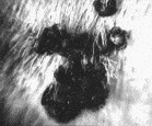
Fig. 1. Photograph showing a aggregation of bont ticks, Amblyomma hebraeum, feeding on a cow in Zimbabwe. Photo credit Dr Suman Mahan, Harare, Zimbabwe; Heartwater Research Project, Department of Veterinary Pathology, University of Florida, Gainesville, Fl., USA.
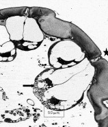
Fig. 2. Light micrograph illustrating a histological section through the ventral region of a male Amblyomma variegatum showing the large dermal glands (Type II dermal glands). These glands were reported to be site of synthesis and secretion of the attraction–aggregation–attachment pheromone secreted during feeding (Diehl et al. 1991). Photo credit: Dr P. A. Diehl, University of Neuchatel, Neuchatel, Switzerland. The arrow indicates the cells of a large dermal gland; the asterisk indicates the pore of the dermal gland.
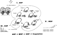
Fig. 3. Hypothetical model illustrating the sequence of behavioural events that occur during sexual courtship in a representative metastriate ixodid tick, Dermacentor variabilis. Phase 1, feeding female secretes volatile attractant sex pheromone, 2,6-dichlorophenol, exciting males feeding nearby. Phase 2, sexually excited male detaches and commences searching behaviour in response to sex attractant odour. Phases 3 and 4, sexually excited male orients to and walks toward the pheromone-emitting source and locates the pheromone-secreting female. These first four phases are mediated by 2,6-dichlorophenol. Phase 5, the male contacts the candidate female and detects the presence of cholesteryl oleate, the mounting sex pheromone. Phase 6, the male recognizes the female as a suitable mate and climbs onto the female's dorsal surface, turns posteriorly and crawls over the posterior end of the female's opisthosoma (tip-over behaviour) and onto the ventral surface. This behaviour constitutes the male mounting response. Phases 7–9, the male locates the female gonopore, positions itself under the female with legs intertwined, and inserts its chelicerae into the female's genital pore. Identification of the gonopore and genital probing with the chelicerae is mediated by the genital sex pheromone, comprising a mixture of long chain fatty acids and ecdysteroids. Following successful identification of the female as a conspecific mate, the spermatophore is formed and copulation ensues. Reproduced with permission from Annual Review of Entomology 30 (1985) and Modern Acarology. ASP=attractant sex pheromone; FA=fatty acids; GSP=genital sex pheromone; MSP=mounting sex pheromone.
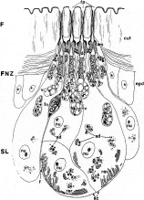
Fig. 4. Diagrammatic reconstruction of the sex pheromone gland showing the secretory lobes, ductular zone and the foveal pores. In unfed ticks, the cells of the secretory lobes are filled with numerous tiny vesicles containing 2,6-dichlorophenol stored in lipid droplets. In feeding females, the individual vesicles rupture, releasing the oily droplets. The droplets migrate towards the ducts where they coalesce into large masses and pass through the ducts and out onto the external surface of the tick body. am=ampulla; cut=cuticle; d=duct; epd=epidermal cell; F=fovea; f=foveal nerve; FNZ=foveal neck zone; fp=foveal pore; nu=nucleus; rer=rough endoplasmic reticulum; sc=secretory cells; sd=secretory droplets; SL=secretory lobe; v=vesicle. From Sonenshine (1991) with permission from Oxford University Press, New York and the Journal of Parasitology.
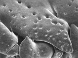
Fig. 5. Apertures of large wax glands on the margin of the scutum of an adult female Dermacentor variabilis. The gland associated with these pores secrete a squalene-rich exudate when the tick legs are pressure-stimulated. Bar=100 μm. Photo credit Dr Jay Yoder. From Yoder et al. (1993a) Journal of Insect Physiology 39, 291–296 with permission of the editor.
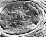
Fig. 6. Scanning electron micrograph of one of the paired foveae dorsales from an adult female Dermacentor variabilis. This image illustrates the numerous slit-like pores in each fovea. Oily droplets containing the sex attractant pheromone, 2,6-dichlorophenol ooze from these pores, releasing the volatile pheromone to the atmosphere. From Sonenshine (1991) with permission from Oxford University Press, New York and the Entomological Society of America.
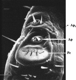
Fig. 7. Scanning electron micrograph of the tarsus of leg I in Dermacentor variabilis showing the Haller's organ. Arrows show the location olfactory sensilla. Ap=Anterior pit; Ap1=multiporose olfactory sensilla; Pc=Posterior capsule. Bar=50 μm.
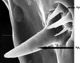
Fig. 8. Scanning electron micrograph illustrating the large olfactory sensillum, Ap1 in the anterior pit of Haller's organ surrounded by other sensilla. Bar=5 μm. Ap=anterior pit; Ap1=olfactory sensillum.
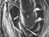
Fig. 9. Scanning electron micrograph illustrating the three pairs of gustatory sensilla at the base of the claw apotele on the leg I tarsus of a Dermacentor variabilis male tick. Ventral view. AT=apotele; DP=dorsal pair of claw sensilla; MP=middle (lateral) pair of claw sensilla; VP=ventral pair of claw sensilla. Bar=25 μm. From Phillips & Sonenshine (1993) with permission from Kluwer Academic Publishers, Van Godewijckstraat 30, P.O. Box 17, 3300 AA Dordrecht, The Netherlands.
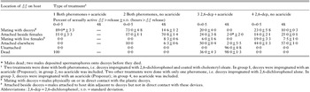
Table 1. Pheromone-pesticide decoys kill male ticks (D. variabilis) and prevents mating when administered at a ratio of 10[ratio ]1 decoys: live females ticks on rabbits
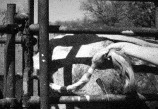
Fig. 10. Photograph showing a bont tick decoy attached to the tail of a cow in Zimbabwe. These pheromone-acaricide-impregnated tags proved highly effective in controlling infestations of bont tick, Amblyomma hebraeum, for as long as three months. Photo credit Dr Suman Mahan, Harare, Zimbabwe; Heartwater Research Project, Department of Veterinary Pathology, University of Florida, Gainesville, Fl., USA.
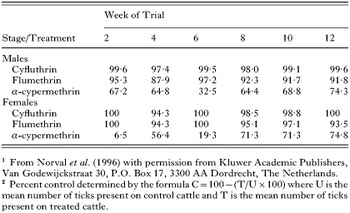
Table 2. Control of A. hebraeum ticks on cattle using tail tags impregnated with AAA pheromone and cyfluthrin, flumethrin or α-cypermethrin during field trials in Zimbabwe1,2
 You have
Access
- 98
- Cited by