Introduction
Conductive hearing loss may be secondary to eustachian tube dysfunction. Anatomically, the middle ear is a box linked by the eustachian tube to the nasopharynx. Blockage of the eustachian tube prevents drainage and pressure equalisation of the middle ear. When the eustachian tube is blocked, negative pressure may develop within the middle ear as the nitrogen and oxygen contained within it is absorbed. The negative pressure causes transudate to collect from the surrounding mucosa, creating an effusion. This may cause symptoms of conductive hearing loss, and potentially otorrhoea, should secondary infection and perforation of the tympanic membrane occur.
The eustachian tube may become blocked from within its lumen, within its wall, or due to extrinsic pressure from outside. The commonest causes in adults are mucosal inflammation associated with infection, smoking, reflux, paranasal sinus disease and barotrauma.Reference Finkelstein, Ophir, Talmi, Shabtai, Strauss and Zohar1 Though relatively rare, nasopharyngeal malignancy must be excluded as a cause of eustachian tube pathology in adults.Reference Mair, Schroder and Kearney2 Recommended investigations in adults with eustachian tube dysfunction include nasendoscopy, audiometry, computed tomography (CT) of the temporal bone and magnetic resonance imaging, if intratemporal or intracranial complications are suspected.Reference Finkelstein, Ophir, Talmi, Shabtai, Strauss and Zohar1
A rare cause of eustachian tube compression is an aneurysm of the petrous or cavernous segment of the extradural internal carotid artery. Such aneurysms may present with otological manifestations such as hearing loss and tinnitus, or with devastating haemorrhage from the nose or ear.Reference Moonis, Hwang, Ahmed, Weigele and Hurst3, Reference Davies, Dale and Renowden4 Congenital, mycotic, iatrogenic and traumatic causes have all been implicated in aneurysm formation.Reference Lau and Chow5, Reference Mori, Murayama, Irie, Takao, Ebara and Ishibashi6 Congenital carotid aneurysms may be associated with underlying connective tissue disorders,Reference Finney, Roberts and Anderson7 and possibly also aneurysms elsewhere, such as in the abdominal aorta, if atherosclerosis is the causative factor.Reference Johnsen, Forsdahl, Solberg, Singh and Jacobsen8 Aneurysms of the petrous or cavernous segment of the internal carotid artery are difficult to access, so endovascular embolisation may be used as a treatment strategy,Reference Abe, Takemoto, Higashi and Inoue9 as may proximal surgical ligation.Reference Singh, Thomas, Hoe and Sethi10
Case report
A 37-year-old man presented to a tertiary teaching hospital in the UK with one year's history of right-sided deafness and recent serous otorrhoea. He had no history of trauma to the ear, no symptoms of tinnitus or dizziness, and no otalgia. He had been unsuccessfully treated for unilateral chronic otitis media with effusion by his general practitioner, using oral antibiotics and ear drops. He was otherwise healthy, with no recent weight loss, pyrexia, or past medical or relevant family history. He was a non-smoker, took no regular medications and had no allergies. He incidentally complained of low back pain for the last two years, which did not respond to analgesia and for which no cause had been identified on blood tests or plain lumbar X-ray.
On examination, the patient appeared comfortable. He had a retracted tympanic membrane on the right side, described as Sadé grade two (i.e. in contact with the incus or stapes).Reference Sadé11 An effusion was evident in the middle ear and there was no tympanic membrane perforation. He had no mastoid tenderness or skin changes around the ear, and was afebrile. His cranial nerves were grossly intact. The rest of his examination, with nasendoscopy, was unremarkable.
Pure tone audiometry demonstrated a right-sided conductive deafness, with an air–bone gap of 40 dB. On the left side, he displayed normal thresholds.
The patient's CT scan demonstrated a large, expansile mass, most likely a giant aneurysm of the petrocavernous segment of the right internal carotid artery.
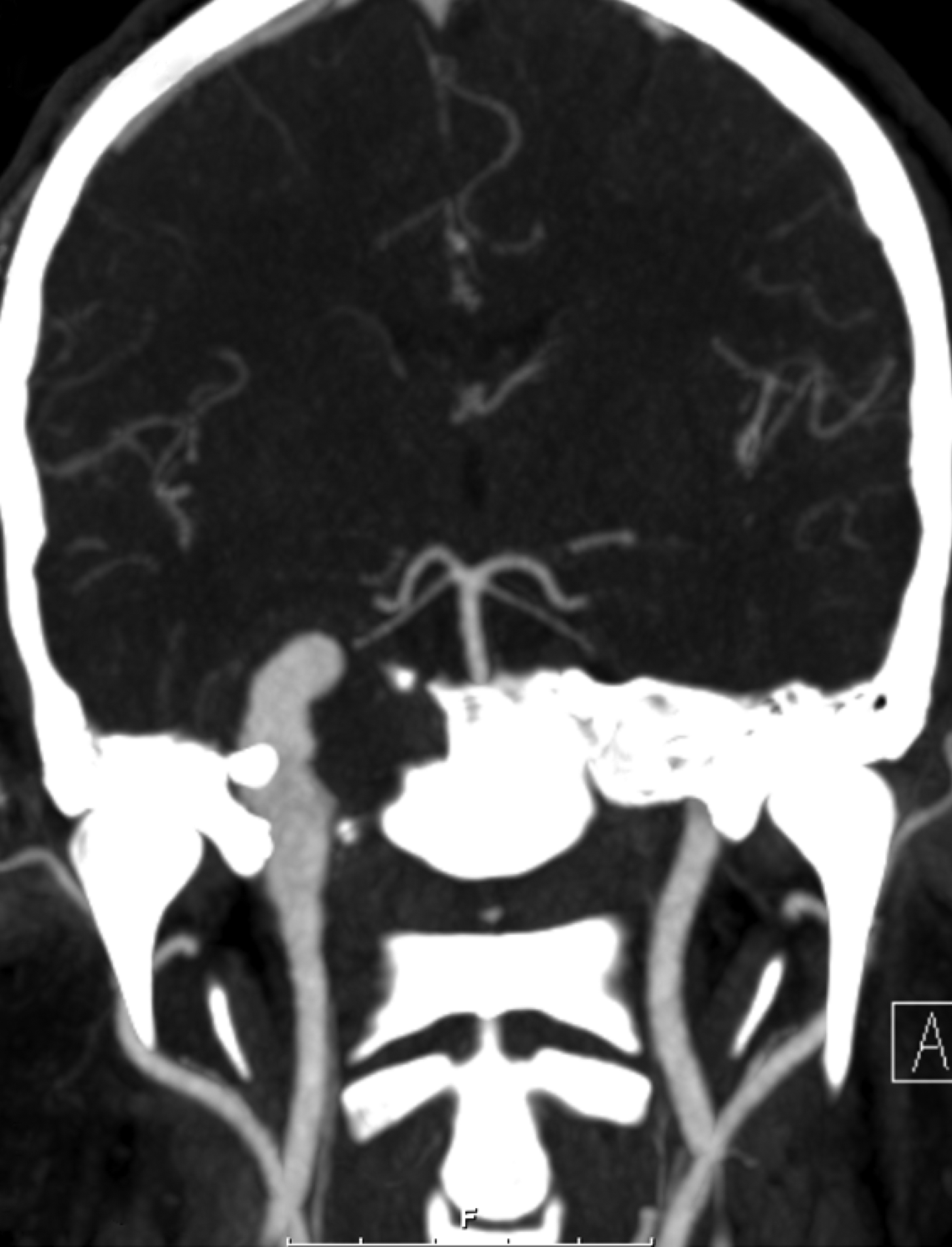
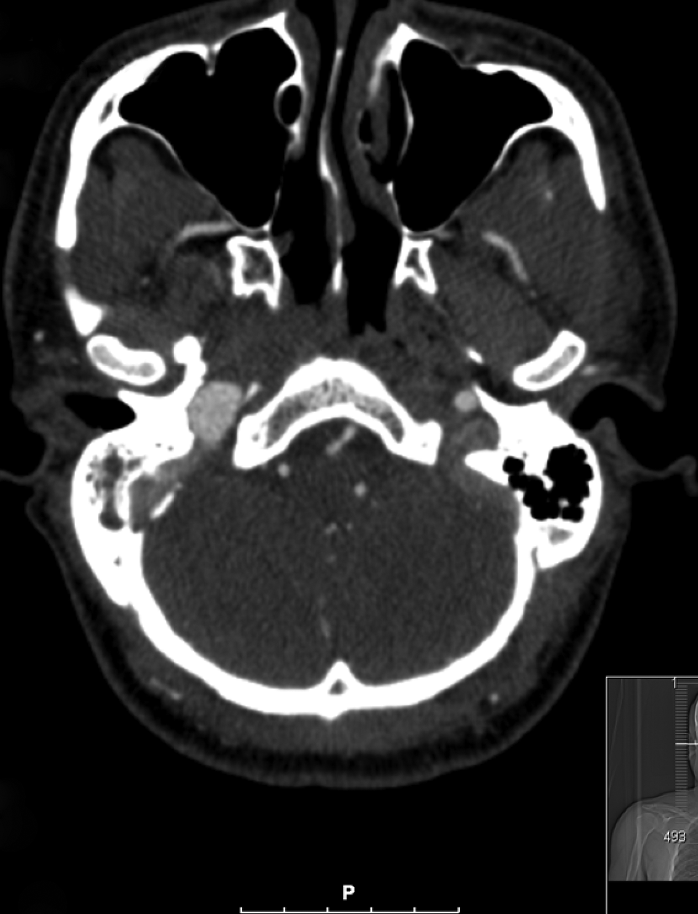
When further investigated with an arterial contrast study (see Figures 1 and 2), an aneurysm was seen, described as follows.
There is a marked, predominantly fusiform aneurysmal dilatation from the level of just below the skull base and entry point into the carotid canal up to the carotid siphon. However, the segment of the right internal carotid artery related to the carotid canal exhibits frank fusiform aneurysmal change with non-enhancing peripheral thrombus related to the aneurysmal ‘sac’. As a result of the expansile nature of the aneurysm related to the carotid canal segment of the internal carotid artery, there is compromise of the eustachian tube with secondary opacification of the right middle-ear cavity and mastoid air cells. Medially, as a result of the expansile nature of the aneurysm related to the right carotid canal, there is pressure erosion of the lateral aspect of the right side of the osseous basisphenoid. The petrocavernous junction of the aneurysm is indenting the posterolateral wall of the sphenoid sinus with thinning and pressure erosion of this part of the sphenoid sinus and radiologically, this is a concerning potential site of rupture into the sphenoid sinus for the aneurysm.
Coronal computed tomography angiogram showing the intracranial internal carotid artery aneurysm. F=feet; A=anterior.

Axial computed tomography angiogram showing the intracranial internal carotid artery aneurysm. P=posterior.
The patient had a successful balloon occlusion test and proceeded to interventional radiological parent vessel occlusion in the upper cervical segment. Several days post-operatively, his hearing had not yet been restored; however, the procedure would be expected to alleviate the risk of a life-threatening haemorrhage caused by the carotid eroding through the sphenoid sinus. Abdominal ultrasonography showed no evidence of an abdominal aortic aneurysm.
• Intracranial internal carotid artery aneurysms may present with symptoms of compression of local structures, e.g. the eustachian tube, causing otitis media with effusion
• Patients should be screened for underlying causes of or pre-dispositions to aneurysms
• Management of giant intracerebral aneurysms depends on collateral cerebral blood supply, and may be interventional or surgical
Discussion
This patient presented with a rare complication of what was probably a congenital intracranial internal carotid artery aneurysm. Whilst this is a rare differential for the common condition of otitis media with effusion, it is an important one to bear in mind. Each of the causes of aneurysm may have systemic associations; for example, a patient with a congenital or acquired predisposition to aneurysms may have aneurysms elsewhere, as may a patient with aneurysms due to mycotic disease. It is also important to exclude aneurysms as underlying causes of symptoms in other systems.