Introduction
A lichen thallus is mainly composed of one fungal species (mycobiont), one or more photosynthetic micro-organisms (photobionts), and other micro-organisms such as basidiomycete yeasts (Spribille et al. Reference Spribille, Tuovinen, Resl, Vanderpool, Wolinski, Aime, Schneider, Stabentheiner, Toome-Heller and Thor2016). In the lichen thallus, green algal photobionts release specific acyclic polyols (i.e. erythritol, ribitol or sorbitol) while cyanobacterial photobionts release glucose (Richardson et al. Reference Richardson, Hill and Smith1968; Smith Reference Smith1968). The mycobionts convert the photosynthetic products into arabitol and mannitol (Drew & Smith Reference Drew and Smith1967; Richardson & Smith Reference Richardson and Smith1968). Polyols have been confirmed as the major soluble carbohydrate compounds in previously examined lichen species (Lewis & Smith Reference Lewis and Smith1967; Roser et al. Reference Roser, Melick, Ling and Seppelt1992; da Silva et al. Reference da Silva, Iacomini, Jablonski and Gorin1993; Gaio-Oliveira et al. Reference Gaio-Oliveira, Dahlman, Palmqvist, Martins-Loução and Máguas2005; Henskens et al. Reference Henskens, Green and Wilkins2012). Chapman et al. (Reference Chapman, Roser and Seppelt1994) reported that six lichen species contained 11–65 mg g−1 dry mass of polyol (consisting of arabitol, mannitol and ribitol), and 0.7–10 mg g−1 dry mass of sugars (glucose, fructose and sucrose), whereas neither mannitol nor ribitol was stored by free-living algae and mosses in their study.
Meanwhile, non-lichenized fungal communities known as ‘lichen-inhabiting fungi’ (Muggia & Grube Reference Muggia and Grube2018) have been shown to occur in lichen thalli using isolation techniques (Li et al. Reference Li, Zhou, Guo and Guo2007; Arnold et al. Reference Arnold, Miadlikowska, Higgins, Sarvate, Gugger, Way, Hofstetter, Kauff and Lutzoni2009; U'Ren et al. Reference U'Ren, Lutzoni, Miadlikowska and Arnold2010; Suryanarayanan et al. Reference Suryanarayanan, Govindarajulu, Rajamani, Tripathi and Joshi2017; Lagarde et al. Reference Lagarde, Jargeat, Roy, Girardot, Imbert, Millot and Mambu2018), next generation sequencing (U'Ren et al. Reference U'Ren, Riddle, Monacell, Carbone, Miadlikowska and Arnold2014; Zhang et al. Reference Zhang, Wei, Zhang, Liu and Yu2015; Wang et al. Reference Wang, Zheng, Wang, Wei and Wei2016; Fernández-Mendoza et al. Reference Fernández-Mendoza, Fleischhacker, Kopun, Grube and Muggia2017) and microscopic visualization methods (Honegger et al. Reference Honegger, Axe and Edwards2013; Spribille et al. Reference Spribille, Tuovinen, Resl, Vanderpool, Wolinski, Aime, Schneider, Stabentheiner, Toome-Heller and Thor2016; Tuovinen et al. Reference Tuovinen, Ekman, Thor, Vanderpool, Spribille and Johannesson2019). Honegger (Reference Honegger and Hock2012) called them ‘endolichenic fungi’ and defined them as symptomless endophytes living within lichen thalli. Although many studies have shown the presence of lichen-inhabiting fungi, their lifestyle in the lichen thallus is poorly understood. For example, Xylariaceae (Sordariomycetes), one of the major families of endophytes from lichens (Suryanarayanan et al. Reference Suryanarayanan, Govindarajulu, Rajamani, Tripathi and Joshi2017; Lagarde et al. Reference Lagarde, Jargeat, Roy, Girardot, Imbert, Millot and Mambu2018) and land plants (Davis et al. Reference Davis, Franklin, Shaw and Vilgalys2003; Osono et al. Reference Osono, Tateno and Masuya2013), has not been investigated to clarify its functions and trophic mode in these hosts. Fungal metabolic information, such as the available carbon source, is essential for understanding its trophic mode and life history. However, the use of polyols has not yet been examined in these lichen-inhabiting fungi, although polyols are major potential carbon sources in lichen thalli.
Ramalina spp. and Flavoparmelia caperata have a green algal photobiont, Trebouxia (e.g. Beck et al. Reference Beck, Friedel and Rambold1998), which provides ribitol to the mycobionts. Komiya & Shibata (Reference Komiya and Shibata1971) reported that Ramalina crassa and R. subbreviuscula contained three polyols: ribitol in the photobiont, and arabitol and mannitol in the mycobionts. Unlike these lichens, many species of Peltigera are associated with a cyanobacterial photobiont, Nostoc, which provides glucose to the mycobionts. Mannitol is reported as the predominant storage compound in Peltigera spp. associated with Nostoc (Smith Reference Smith1961, Reference Smith1963, Reference Smith1968; Tysiaczny & Kershaw Reference Tysiaczny and Kershaw1979; Eisenreich et al. Reference Eisenreich, Knispel and Beck2011) such as P. polydactyla (current name P. polydactylon; note, material from southern England used by Smith and co-workers was P. hymenina), P. horizontalis and P. canina var. praetextata (current name P. praetextata). It is possible that lichen-inhabiting fungi show different polyol-assimilation capacities between green algal lichens (phycolichens) and cyanolichens. Therefore, we isolated lichen-inhabiting fungi from the surface-sterilized Ramalina spp., Flavoparmelia caperata and Peltigera degenii, and assessed the polyol-assimilation capacities of the fungal isolates.
Materials and Methods
Sampling and identification of host lichens
Three symptomless, mature Ramalina spp. (Supplementary Material Fig. S1, available online) were collected from a riverside forest near the Oguro River (Nagano, Japan, 35°49′N, 137°52′E) on 26 May 2018. The Ramalina specimens were placed in paper bags and transported to the laboratory where the external morphology was examined and the chemistry analysed by standard thin-layer chromatographic methods (Culberson & Kristinsson Reference Culberson and Kristinsson1970; Culberson Reference Culberson1972; Culberson & Johnson Reference Culberson and Johnson1976, Reference Culberson and Johnson1982), using two solvent systems (A and B′). Specimens were subsequently identified by referring to the current taxonomy of Japanese Ramalina (Sakata et al. Reference Sakata, Matsuzaki, Yoshikawa and Harada2019).
Isolation of lichen-inhabiting fungi
The Ramalina thalli were stored at 4 °C and processed within one week. Surface sterilization of lichen thalli and isolation of lichen-inhabiting fungi were performed following the methods described in U'Ren et al. (Reference U'Ren, Lutzoni, Miadlikowska, Laetsch and Arnold2012) with minor modifications. Visible extraneous material attached to the thalli was removed under a dissection microscope using tweezers, and each thallus was washed thoroughly under running tap water; healthy parts of the thalli were then cut into c. 5 mm fragments. The fragments were surface-sterilized by agitating sequentially in 95% ethanol for 30 s, 0.5% sodium hypochlorite (NaOCl) solution for 1 or 2 min, and 70% ethanol for 2 min. Surface sterilised fragments were placed on sterilized filter paper inside a Petri dish to dry overnight in a sterile lamina flow cabinet. The fragments were cut in half and placed on two media, 2% malt extract agar (MA) (Becton, Dickinson and Company) or 2% malt extract and 0.2% yeast extract agar (MYA) (Becton, Dickinson and Company), in plastic Petri dishes sealed with Parafilm, and incubated in the dark at 25 °C for up to 2 months. Mycelia growing from each fragment were sub-cultured and pure cultures were established. After isolation, the Ramalina specimens were dried and deposited in the herbarium of the Natural History Museum and Institute, Chiba, Japan (CBM) (Table 1).
Sample information for Ramalina species collected during this study, with GenBank Accession numbers for mycobionts and photobionts. All specimen ID numbers are preceded by K. Yoshino.

*Herbarium of the Natural History Museum and Institute, Chiba, Japan
DNA sequencing and molecular identification
Genomic DNA was extracted from harvested mycelia growing on media, or lichen thalli, using the methods of Izumitsu et al. (Reference Izumitsu, Hatoh, Sumita, Kitade, Morita, Gafur, Ohta, Kawai, Yamanaka and Neda2012). Several extractions were unsuccessful, therefore the standard hexadecyltrimethylammonium bromide (CTAB) method was also used for thalli of the Ramalina specimens and some cultures. The nuclear ribosomal internal transcribed spacer region (nrITS) was amplified and sequenced using primers ITS5 and ITS4 for fungi (White et al. Reference White, Bruns, Lee, Taylor, Innis, Gelfand, Sninsky and White1990) and ITS1T and ITS4T for lichen photobionts (Kroken & Taylor Reference Kroken and Taylor2000). PCR conditions, purification of PCR products, and sequencing followed previously described procedures (Yoshino et al. Reference Yoshino, Yamamoto, Hara, Sonoda, Yamamoto and Sakamoto2019a). The sequences were edited using BioEdit ver. 7.2.5 (Hall Reference Hall1999) and assembled using GENETYX ver. 12 (Genetyx Corporation, Tokyo, Japan). An operational taxonomic unit (OTU) of fungi was defined as >97% nrITS sequence similarity and each OTU set was used for the subsequent polyol-assimilation tests. Isolates were identified from the nrITS sequences using the GenBank database (mainly from type specimens, reliable vouchers and the strains described in published papers). The current scientific names were confirmed after consulting MycoBank (http://www.mycobank.org/) and Index Fungorum (http://www.indexfungorum.org/).
Polyol-assimilation tests
One isolate was selected from each OTU of the Ramalina spp.-inhabiting fungi for an assimilation test. Lichen-inhabiting fungi from phyco- and cyanolichens possibly have different assimilation capacities therefore a further six isolates were also used from Flavoparmelia caperata and Peltigera degenii (Masumoto & Degawa Reference Masumoto and Degawa2019). Isolates from these lichens were obtained using the methods outlined above with some modifications: thalli were collected at Sugadaira Research Station, Mountain Science Center, University of Tsukuba (Nagano, Japan) between 2015 and 2016, cut into c. 5-mm2 fragments and surface-sterilized in surfactant, 0.5% Aerosol OT (Wako Pure Chemical Industries, Osaka, Japan), for 1 min. Subsequently, they were surface sterilized using one of three different procedures: 70% ethanol for 2 min, 0.5% NaClO for 2 min or 20% hydrogen peroxide (H2O2) for 2 min. Once dried, all fragments were further cut into c. 2.5 mm2 pieces and incubated on 2% malt extract agar at room temperature (c. 25 °C) for up to 3 months.
Isolates were pre-cultured on MYA at room temperature for 17–38 days until sufficiently grown. An agar disc (6 mm diam.) was punched from the edge of the colony and transferred to the centre of a plate of slightly modified Lilly and Barnett's medium (Lilly & Barnett Reference Lilly and Barnett1951) covered with a cellulose filter. Three treatments of the medium were prepared which differed in carbon source content; 20 g of glucose (as a positive control), mannitol or ribitol as a sole carbon source, or no additional carbon source as the control. The other components were 2.273 g of L-asparagine monohydrate, 1 g of KH2PO4, 0.5 g of MgSO4⋅7H2O, 0.2 mg of Fe (NO3)3⋅9H2O, 0.2 mg of ZnSO4⋅7H2O, 0.1 mg of MnSO4⋅4H2O, 0.1 mg of thiamine and 5 μg of biotin, with up to 1 l of distilled water. Lilly and Barnett's medium has been confirmed as a suitable synthetic culture medium for the growth of the mycobiont of Ramalina conduplicans (Yoshino et al. Reference Yoshino, Kawakami and Sakamoto2019b). The plates were incubated in the dark at 25 °C for 2–8 weeks according to the growth rate of the strains and the experiment was performed in triplicate. To determine hyphal lengths, the distance from the edge of the inoculated disc to the edge of the colony was measured each week at ten positions per plate, including the maximum and minimum length. To calculate the fungal dry mass, the mass of the mycelia growing on the cellulose filter and agar disc was measured, after complete drying at 70 °C, and the mean of the dry mass of cellulose filters, with non-inoculated agar discs, was subtracted (0.1575 ± 0.0002 g (n = 5)). The assimilation of carbon sources of each fungus, based on the dry mass, was statistically analyzed using the multiple comparison test (Tukey-Kramer method, P < 0.05) performed with add-in software Statcel4 (OMS publishing Inc., Japan).
Results
Identification of Ramalina specimens
On the basis of external morphology and chemical analysis, two Ramalina species were identified, from which three specimens were selected: Ramalina conduplicans Vain. (K. Yoshino 18052607), with a c. 3 cm tall thallus, flat and solid branches with pseudocyphellae, lacking perforations, and with some apothecia, containing homosekikaic, usnic, salazinic and sekikaic acids; R. dilacerata (Hoffm.) Vain. (K. Yoshino 18052604), with a c. 3 cm tall thallus, cylindrical and hollow branches lacking pseudocyphellae, with perforations and many apothecia, containing usnic and sekikaic acids; R. dilacerata (K. Yoshino 18052612), with a c. 2.5 cm tall thallus, cylindrical and hollow branches lacking pseudocyphellae, with perforations and many apothecia, containing usnic and sekikaic acids.
The nrITS sequences of the mycobionts and photobionts were obtained from all thalli (Table 1). To confirm the identity of the photobionts, the nrITS sequences of three specimens were subjected to a BLAST search and identified as Trebouxia; the top 100 results were all Trebouxia sequences and the maximum sequence identities were 99–100%.
Fungal isolates from surface-sterilized lichen thalli
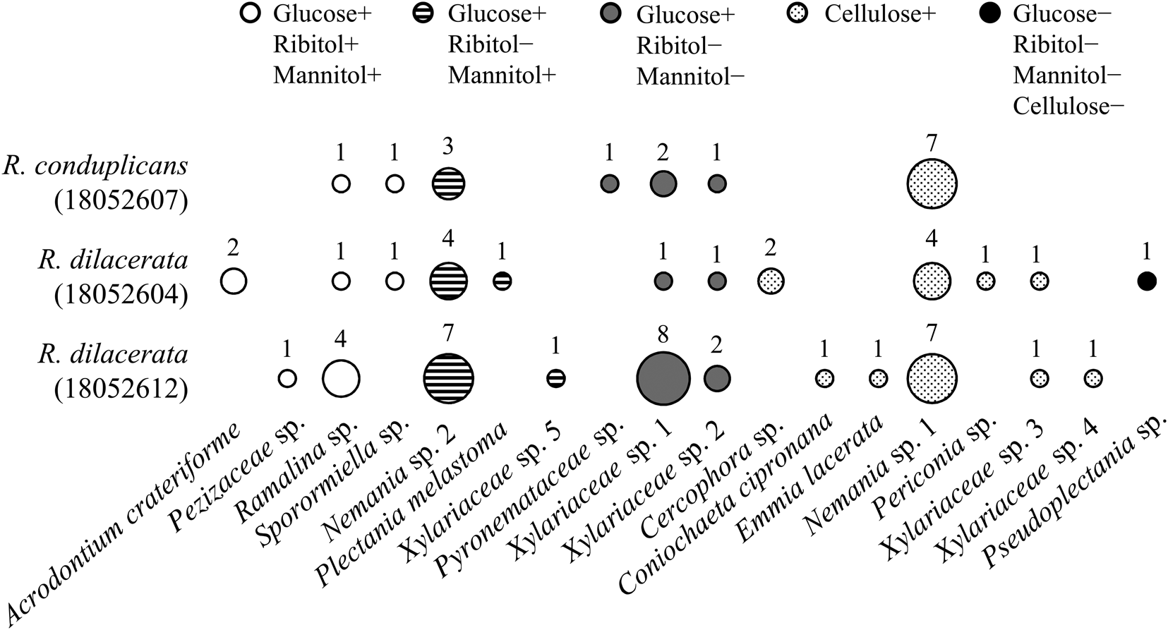
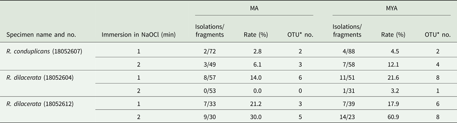
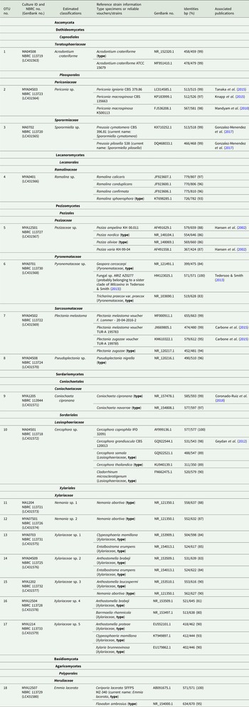
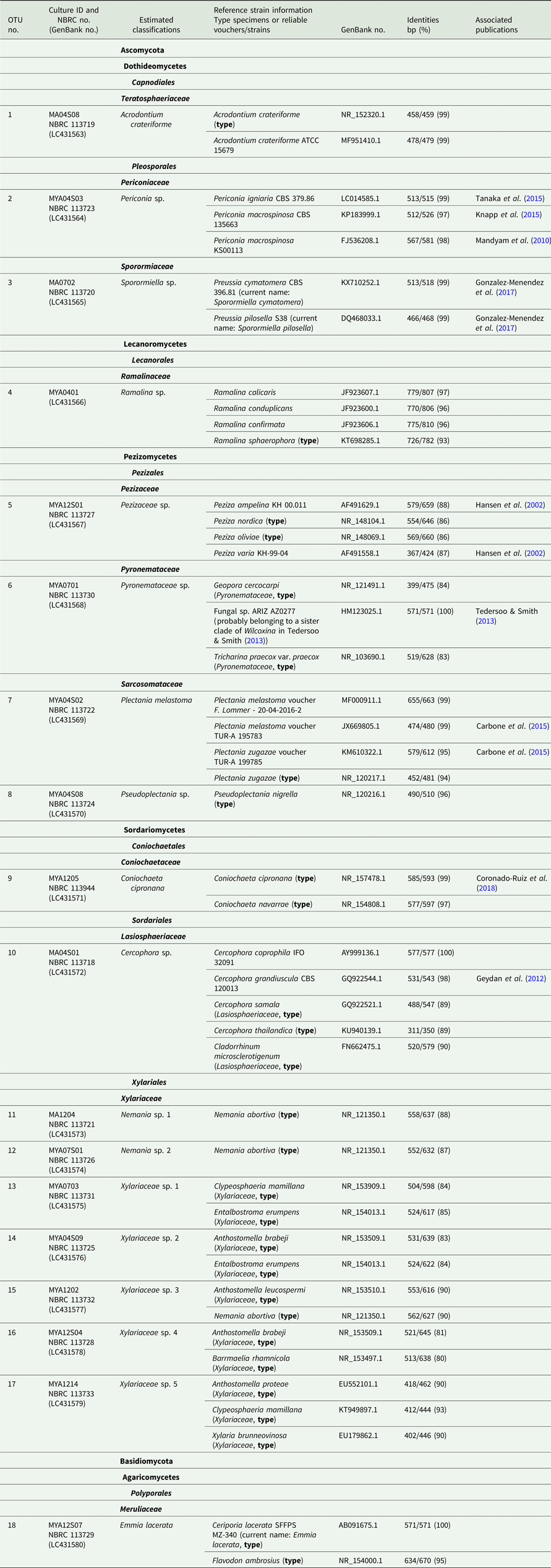
Frequencies of isolation of lichen-inhabiting fungi from fragments of three Ramalina specimens were 0–30.0% on MA and 3.2–60.9% on MYA (Table 2). Most isolates showed hyphal growth within 2 weeks. OTUs were isolated on MA (0–6) and on MYA (1–8). The isolation frequency and OTU numbers obtained were not affected by the sample-soaking time in NaOCl but showed a tendency to be higher on MYA than on the MA medium (Table 2). Three isolates from R. dilacerata (K. Yoshino 18152612), were not used for further experiments as sequencing was unsuccessful and they could not be identified. All isolates were filamentous fungi, including 69 isolates of ascomycetes and 1 isolate of a basidiomycete. The nrITS sequences from 70 isolates yielded 18 OTUs based on 97% similarity (Table 3, Fig. 1). OTU4, which was recovered from all three thalli, belonged to Ramalina sp. The nrITS sequences from these isolates showed high similarity (99–100%), although they were isolated from different species, Ramalina conduplicans and R. dilacerata. Furthermore, OTU4 and the isolation sources (i.e. R. conduplicans and R. dilacerata) showed low similarity (93–97%); therefore, we could not conclude whether OTU4 is a lichenized- or a lichen-inhabiting fungus. The other isolates belonged to three classes of Ascomycota, Dothideomycetes (3 OTUs), Pezizomycetes (4 OTUs) and Sordariomycetes (9 OTUs), and also Agaricomycetes in the Basidiomycota (1 OTU) (Table 3). Xylariaceae, including Nemania spp., was the most dominant family (72.9%, Fig. 1) and showed the highest OTU diversity (Table 3). The strains and sequence data of each OTU used in the assimilation tests are available from the Biological Resource Center, NITE, Chiba, Japan (NBRC, https://www.nite.go.jp/en/nbrc/index.html) and GenBank (https://www.ncbi.nlm.nih.gov/), respectively (Table 3).
Relationship between number of isolates of each OTU from each Ramalina specimen (followed by specimen ID) and the classification based on carbon-assimilation capacity. The values and size of circles indicate the number of isolates of each OTU from each Ramalina specimen. Positive (+) and negative (−) signs indicate presence or absence of assimilation capacity of each carbon source. No isolate was able to assimilate ribitol only. Glucose was used as a positive control.

Isolation frequency of lichen-inhabiting fungi operational taxonomic units (OTUs) from samples of surface sterilized Ramalina spp., with varying immersion times in sodium hypochlorite (NaOCl) and plated on different nutrient media (MA = malt agar, MYA = malt yeast agar). All specimen numbers are preceded by K. Yoshino.
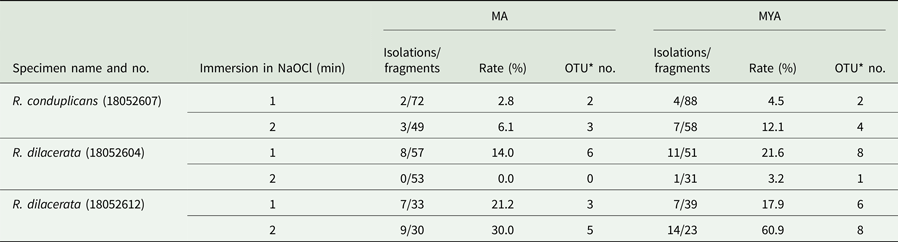
*An OTU was defined as >97% nrITS sequence similarity
Identification of representative isolates of each OTU of lichen-inhabiting fungi derived from surface sterilized samples of Ramalina spp. collected in this study. Identification is based on nrITS sequence similarity with reference strains in GenBank and the same isolates were used in the assimilation ability tests.
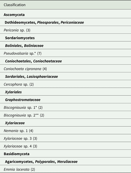
Of the isolates from Flavoparmelia caperata and Peltigera degenii, 12 were used for carbon-assimilation tests: Sordariomycetes sp. (isolated after processing with 70% ethanol), Biscogniauxia sp. 1, Pseudovalsaria sp. (0.5% NaOCl), Hypoxylon perforatum, Nectriaceae sp. and Sarea sp. (20% H2O2) were isolated from F. caperata, and Virgaria sp. 1, Virgaria sp. 2 (20% H2O2), Annulohypoxylon sp., Biscogniauxia sp. 2, Virgaria sp. 3 and Virgaria sp. 4 (0.5% NaOCl) were isolated from P. degenii (Masumoto & Degawa Reference Masumoto and Degawa2019).
Carbon assimilation capacity
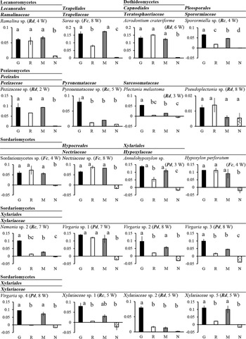
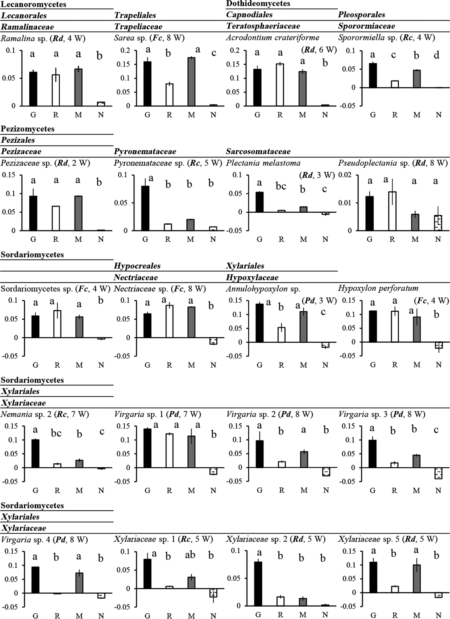
All plates were incubated for a maximum of eight weeks until sufficient mycelia had grown to measure the dry mass. Of the 30 isolates, 18 selected from every OTU group from Ramalina spp. and six each from F. caperata and P. degenii grew well on Lilly and Barnett's medium containing glucose as the sole carbon source, with the exception of Pseudoplectania sp. (Pezizomycetes) which showed limited growth on all agar plates (Fig. 2, dry weights; Supplementary Material Fig. S2, sequential hyphal lengths and plate images after incubation). Ten isolates (one Dothideomycete, eight Sordariomycetes and one Agaricomycete) decomposed the cellulose filter covering mannitol- or ribitol- containing media (Table 4); therefore, the dry mass of these cultures could not be obtained. The other 20 cultures did not decompose the cellulose filter covering the carbon source-containing media. Ramalina sp., which was isolated from R. dilacerata (K. Yoshino 18052604), showed no significant difference in assimilation capacities for the three carbon sources (i.e. glucose, mannitol and ribitol) (Fig. 2). Likewise, ten isolates (Acrodontium crateriforme, Sporormiella sp. (Dothideomycetes), Sarea sp. (Lecanoromycetes), Pezizaceae sp. (Pezizomycetes), Annulohypoxylon sp., Hypoxylon perforatum, Nectriaceae sp., Sordariomycetes sp., Virgaria sp. 1 and Virgaria sp. 3 (Sordariomycetes)) assimilated both mannitol and ribitol. However, the assimilation capacities of these isolates differed between mannitol and ribitol (e.g. Sporormiella sp., Fig. 2). In addition, five isolates (Plectania melastoma (Pezizomycetes), Nemania sp. 2, Virgaria sp. 2, Virgaria sp. 4 and Xylariaceae sp. 5 (Sordariomycetes)) assimilated only mannitol (Fig. 2). By contrast, there were no isolates that assimilated only ribitol. It is noteworthy that the lichen-inhabiting fungi assimilating ribitol (shown as white circles in Fig. 1) were rare in each thallus. Conversely, the fungi decomposing the cellulose filter (shown as stippled circles in Fig. 1) were common in each thallus.
Comparison of lichen-inhabiting fungal isolates based on their growth on media with differing carbon source content. The vertical and horizontal axes indicate mycelial dry mass (g) and carbon sources (G = glucose, R = ribitol, M = mannitol, N = non-carbon source), respectively. Several isolates which decomposed the cellulose filter on carbon source-free media (N) show a negative value of mycelial mass. The isolation source is indicated in brackets (Fc = Flavoparmelia caperata, Rc = Ramalina conduplicans, Rd = Ramalina dilacerata, Pd = Peltigera degenii) followed by incubation weeks (W = weeks). Bars with the same letter are not significantly different based on the Tukey-Kramer test (P < 0.05). Standard errors (n = 3) are also shown in the bar graphs.
Lichen-inhabiting fungal taxa with cellulose-decomposing capacities derived from surface sterilized samples of Ramalina spp., Flavoparmelia caperata and Peltigera degenii. Number of weeks of incubation is indicated in parentheses.
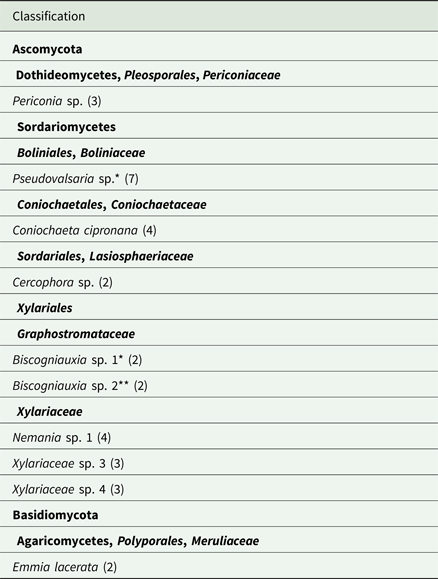
*Isolated from the thallus of Flavoparmelia caperata
**Isolated from the thallus of Peltigera degenii
Discussion
Lichen-inhabiting fungi isolated in this study can be classified into the following five groups based on their ability to assimilate carbon sources: group 1 assimilates both mannitol and ribitol; group 2 assimilates mannitol but does not assimilate ribitol; group 3 assimilates neither mannitol nor ribitol; group 4 decomposes cellulose filters, even on media containing carbon sources; group 5 (i.e. Pseudoplectania sp.) assimilates none of the carbon sources in this study.
Group 1 includes fungi such as Acrodontium crateriforme and Sporormiella sp. (Dothideomycetes) isolated from Ramalina spp., and Sarea sp. (Lecanoromycetes) and Nectriaceae sp. (Sordariomycetes) isolated from Flavoparmelia caperata (Fig. 2). Some of these have previously been isolated from lichens (Li et al. Reference Li, Zhou, Guo and Guo2007; U'Ren et al. Reference U'Ren, Miadlikowska, Zimmerman, Lutzoni, Stajich and Arnold2016; Suryanarayanan et al. Reference Suryanarayanan, Govindarajulu, Rajamani, Tripathi and Joshi2017; Lagarde et al. Reference Lagarde, Jargeat, Roy, Girardot, Imbert, Millot and Mambu2018). Furthermore, most of them are known as saprobes of various substrates. For instance, Sarea can gain nutrition from tree resin (Hawksworth & Sherwood Reference Hawksworth and Sherwood1981) and Sporormiella is a common dung-inhabiting genus (Doveri Reference Doveri2011). In addition, Acrodontium crateriforme can produce cellobiohydrolase (Koukol & Baldrian Reference Koukol and Baldrian2012). Thus, almost all isolates in this group probably utilize diverse organic matter for their growth, including polyols and other recalcitrant organic compounds. Therefore, they might have the ability to change their host from lichens to other nutrient sources such as plant substrates. Few Nectria spp. are known to be lichenicolous fungi (Diederich & Puntillo Reference Diederich and Puntillo1995). It is likely that a small number of species of Nectriaceae, including one isolate obtained from F. caperata (Fig. 2), are obligate parasites or endophytes of lichens and utilize mannitol and ribitol inside the lichen thallus. Since fungi belonging to group 1 were a minority in the lichen thalli (white circles in Fig. 1), even if they ultilise ribitol from the photobionts, their impact on the mycobionts will be limited.
Group 2 (striped circles in Fig. 1) also included possible saprotrophs growing on decaying plants, such as Nemania sp. 2, the second most frequently isolated OTU from Ramalina spp. Lichens have great longevity and it is possible that mature thalli would comprise partially senesced segments. Hence it is likely that the isolates in this group (and possibly also group 1) wait for the mycobiont hyphae to age and utilize mannitol as lichen saprotrophs. Furthermore, group 2 fungi possibly inhabit not only lichen thalli but also plants because mannitol is a polyol produced by lichens, algae and plants (Stoop et al. Reference Stoop, Williamson and Pharr1996; Iwamoto & Shiraiwa Reference Iwamoto and Shiraiwa2005).
Pyronemataceae sp., Xylariaceae sp. 1 and Xylariaceae sp. 2 belonged to group 3 (grey circles in Fig. 1). Their carbon sources are unknown but they probably utilize sources other than mannitol and ribitol in the lichen thallus. Group 4 (stippled circles in Fig. 1, Table 4) also includes Xylariaceae spp., significantly Nemania sp. 1, and was the major group isolated from Ramalina spp. Xylariaceae are regarded as lignin decomposers (Liers et al. Reference Liers, Ullrich, Steffen, Hatakka and Hofrichter2006, Reference Liers, Ullrich, Pecyna, Schlosser and Hofrichter2007; Osono Reference Osono2007). The other lineages in which group 4 isolates occur (Cercophora sp., Coniochaeta cipronana, Emmia lacerata, Periconia sp.) are also known to decompose components of the plant cell wall (i.e. cellulose and lignin). These compounds are much more abundant and more easily accessible than polyols in nature. Our results showed that the members of this group preferred cellulose to polyol. The thalli used in this study were not visibly damaged (Supplementary Material Fig. S1, available online), although this group comprised the highest proportion in each thallus (Fig. 1). Based on these results, we assume these fungi to be plant saprotrophs and opportunistic lichen-inhabitants remaining quiescent within the thallus. Xylariaceae was the most dominant inhabitant in Ramalina spp. However, only one of the 11 Xylariaceae isolates preferred both mannitol and ribitol, as well as glucose, in this study (Fig. 2, Table 4). Furthermore, Xylariaceae is known to comprise endophytes with low host specificity which have been frequently isolated from lichens (Suryanarayanan et al. Reference Suryanarayanan, Govindarajulu, Rajamani, Tripathi and Joshi2017; Lagarde et al. Reference Lagarde, Jargeat, Roy, Girardot, Imbert, Millot and Mambu2018), leaves of different tree species (Osono et al. Reference Osono, Tateno and Masuya2013) and liverworts (Davis et al. Reference Davis, Franklin, Shaw and Vilgalys2003). This suggests that almost all isolates of Xylariaceae in this study might not be specific to lichen thalli due to their preference for carbon sources other than polyols. Furthermore, ascospores of some isolates might have sterilization tolerance. For example, Xylariaceae has brown to dark brown ascospores (Rogers et al. Reference Rogers, Miller and Vasilyeva2008; Wendt et al. Reference Wendt, Sir, Kuhnert, Heitkämper, Lambert, Hladki, Romero, Luangsa-ard, Srikitikulchai and Peršoh2018), Plectania melastoma has ascospores covered with a gelatinous sheath (Carbone et al. Reference Carbone, Loizides and Alvarado2015) and Sporormiella has thick-walled dark ascospores (Doveri Reference Doveri2011). Therefore, some isolates were possibly derived from ascospores only superficially attached to lichens. Similarly, Fernández-Mendoza et al. (Reference Fernández-Mendoza, Fleischhacker, Kopun, Grube and Muggia2017) reported that lichen-inhabiting Sordariomycetes belonging to Xylariaceae and frequently obtained by culture-based experiments, were rarely detected by sequencing metabarcoding methods. It is suggested that such a discrepancy is caused by sterilization tolerant-spores which readily germinate on an agar medium but are too infrequent to be amplified by PCR. To determine how many lichen-inhabiting fungi are present only as spores, further studies are needed based on sequencing, visualization and culture methods including various carbon-assimilation tests.
Some isolates from Ramalina spp. and Flavoparmelia caperata could assimilate ribitol (Fig. 2). Peltigera degenii isolates also assimilated ribitol, even though ribitol should be lacking in the thallus since the photobiont, Nostoc, supplies glucose rather than ribitol to the mycobiont. Therefore, photobiont-influenced differences in lichen-inhabiting fungi caused by their polyol-assimilation capacities were not clear in the present study. However, as mentioned above, group 1 is expected to change their substrate from lichens to other nutrient sources and therefore the lichen-inhabiting fungi of P. degenii might also inhabit other lichens associating with ribitol-releasing algae.
Arnold et al. (Reference Arnold, Miadlikowska, Higgins, Sarvate, Gugger, Way, Hofstetter, Kauff and Lutzoni2009) showed that culturable lichen-inhabiting fungi were rarely isolated from the mycobiont layers in the foliose lichen Umbilicaria mammulata (i.e. medulla and cortices) but rather were preferentially associated within the algal layer. Conversely, Spribille et al. (Reference Spribille, Tuovinen, Resl, Vanderpool, Wolinski, Aime, Schneider, Stabentheiner, Toome-Heller and Thor2016) showed that specific basidiomycetous yeasts were embedded in the cortex of Bryoria spp. Determining the location of lichen-inhabiting fungi and their involvement in the carbon cycle in lichen thalli will elucidate the true ecological role of lichen-inhabiting fungi. Furthermore, polysaccharides such as α-glucans, β-glucans and galactomannans are also present in lichens, and are generally considered to have a fungal origin (Olafsdottir & Ingólfsdottir Reference Olafsdottir and Ingólfsdottir2001). For example, ascomycetous lichen mycobionts have extremely thick cell walls which contain large quantities of glucans (Honegger Reference Honegger and Hock2012). Therefore, there is a possibility that lichen-inhabiting fungi utilize polysaccharides without causing severe damage to the lichen. In addition, lichens contain oxidoreductases and cellulases (Beckett et al. Reference Beckett, Zavarzina and Liers2013). It is therefore necessary to examine the production of these enzymes derived from lichen-inhabiting fungi in order to assess their saprotrophic role within the lichen thallus.
In this study, we isolated lichen-inhabiting fungi belonging to diverse families and classes of Ascomycota, many known to be saprotrophs and plant-endophytes. These lichen-inhabiting fungi were classified into five groups based on different carbon source preferences, which implies they have different life histories and niche segregation of carbon sources within lichen thalli. To elucidate fungal-fungal and fungal-algal interactions, and the function and ecology of lichen-inhabiting fungi, further studies are needed on their carbon sources and the mechanisms of their niche segregation in lichen thalli.
Supplementary Material
To view supplementary material for this article, please visit https://doi.org/10.1017/S0024282919000483
Acknowledgements
We are very grateful to Mr Asato Hosono (Utsunomiya University) for assisting with the field sampling. This study was supported by the Research Fellowships for Young Scientists (No. 17J03039) from the Japan Society for the Promotion of Science (JSPS).