

The hypoxia cascade
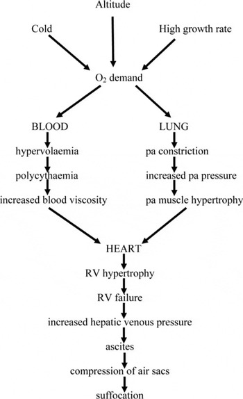
hypoxia and pulmonary vasoconstriction (Figure 1)
Altitude disease is caused by hypoxia. The sequence of events leading from hypoxia to ascites, at the time partially elucidated partially hypothesised, has been explained by Huchzermeyer (Reference HUCHZERMEYER1989). Hypoxia causes contraction of the lung arteries. This can be visualised by lung angiography which entails injecting the lung arteries of sacrificed birds with a contrast medium after which radiographs are taken (Reid, Reference REID1986). The wide pulmonary arteries of normal birds contrast with the contracted arteries of ascitic birds (own unpublished findings).
This pulmonary vasoconstriction is normally designed to shunt blood away from malfunctioning parts of the lung, but in generalised hypoxia it leads to a further exaggeration of the problem, resulting in a vicious cycle. It also increases the pulmonary arterial pressure, which can be measured in normal birds (Guthrie et al., Reference GUTHRIE, CILLIERS, HUCHZERMEYER and KILLEEN1987), however, it is not possible to these measurements in ascitic birds as they tended to die during the procedure.
Chronic pulmonary vasoconstriction causes a thickening (hypertrophy) of the muscular layer of the pulmonary arteries, which can be measured in histological preparations. A study comparing the pulmonary arteries of normal broilers raised at sea level and at high altitude (± 1600 m) with those of ascitic broilers was in progress but remained unfinished.
Hypoxia cascade (pa = pulmonary artery; RV = right ventricle)

right ventricular hypertrophy
Pulmonary arterial vasoconstriction increases the work-load of the right ventricle of the heart resulting in an increase of right ventricular mass. This increase in relation to total heart (ventricular) mass is believed to be in direct proportion to the increase of pulmonary arterial pressure (Cueva et al., Reference CUEVA, SILLAU, VALENZUELA and PLOOG1974). The RRV can be determined post mortem by dissecting and weighing the heart and this is a valuable tool in the evaluation of ascites trials (right ventricular mass divided by total ventricular mass = RRV) (Huchzermeyer and De Ruyck, Reference HUCHZERMEYER and DE RUYCK1986; Julian, Reference JULIAN1987). Normal values range from 0.15 to 0.25 while ascitic birds have RRV values of 0.30 to 0.60. The increase in right ventricular mass is not only caused by a thickening of the wall of the right ventricle; contrary to the situation in mammals the hypertrophic right ventricle of broilers extends further towards the apex of the heart and over a larger proportion of the circumference of the left ventricle (unpublished). A concomitant thinning of the wall of the left ventricle was reported by Julian et al. (Reference JULIAN, FRIARS, FRENCH and QUINTON1987).
hypervolaemia and polycythaemia
As a result of pulmonary arterial vasoconstriction and further aggravated hypoxia, the body tries to compensate by producing more red blood cells (polycythaemia) (Maxwell et al., Reference MAXWELL, ROBERTSON and SPENCE1986) and increasing the blood volume (hypervolaemia). Polycythaemia is determined by calculating the percentage of packed red blood cells in a volume of centrifuged blood (haematocrit, HCT). While normal broilers have HCT values of 18% to 30%, the values of ascitic birds ranged from 35% to 60% (unpublished).
High HCT values increase the viscosity of the blood and add to the overload of the right ventricle of the heart. Using a simple drop method a drastic increase of blood viscosity was found only with near terminal HCT values (unpublished). A much higher viscosity together with HCT values of up to 80% has been reported in hypoxic rats (Ou and Smith, Reference OU and SMITH1984). Blood viscosity has been successfully reduced in ponies by the use of warfarin (Amin et al., Reference AMIN, ALLEN and COLLES1984).
Hypervolaemia is believed to add to the workload of the right heart (Julian, Reference JULIAN1987). Our results showed that blood volume increased in direct relation to the increase of RRV. Plasma volume increased during the early stage of the development of ascites but decreased again in the terminal stage with fluid loss into the peritoneal cavities - ascites (unpublished).
In non-ascitic birds there is a poor relationship between HCT and RRV values because of very large individual variations (own unpublished findings). HCT therefore does not appear to be a useful indicator to identify ascites susceptible birds. At one stage it was thought that there was a correlation between group HCT and RRV values. However, testing the progeny of these groups two years later gave unmatched results (unpublished). Here it must be emphasised that HCT results obtained during summer and/or at sub-maximal growth rates will be unreliable. Our own work was carried out mostly at a moderate altitude of ± 1200m.
ascites
At an advanced stage of right ventricular hypertrophy, excessive blood is dammed back into the portal vein of the liver, probably because of malocclusion of the right atrio-ventricular valve (Julian et al., Reference JULIAN, FRIARS, FRENCH and QUINTON1987). This leads to a severe distension of the portal vein, which could be demonstrated on whole-body crosscuts of frozen ascitic broilers with portal vein diameters in excess of 20 mm (unpublished).
The increased venous pressure within the liver causes the plasma to filter out of the hepatic sinuses into the different peritoneal sacs in the abdominal cavity (Bezuidenhout, Reference BEZUIDENHOUT1988). This accumulation of partially clotted fluid in the peritoneal sacs constitutes ascites. Compression of the abdominal air sacs by the filled peritoneal sacs finally suffocates the bird.
Atrial natriuretic factor (ANF), a hormone that controls blood volume, is deficient in cardiomyopathic hamsters with congestive heart failure (Dlouha and McBroom, Reference DLOUHA and MCBROOM1986). ANF is secreted when the atrial muscle fibres extend as a result of increased blood volume. It then causes an increased production of urine in an attempt to reduce blood volume (Cantin and Genest, Reference CANTIN and GENEST1986). Consequently ANF depletion may lead to the rapid terminal increase in blood volume in ascitic birds due to the cessation of stimulating diuresis.
Ancillary investigations
chemotherapy
In collaboration with Dr J.H. du Preez, an attempt was made to suppress pulmonary vasoconstriction in 3265 broiler chicks at an altitude of 1450 m during winter (July/August, 1986) by continuous administration of nitrofurantoin (200g/t) and a ketanserin analogue (Jansen Pharmaceuticals) (Archer et al., Reference ARCHER, EATON, CHESLER and WEIR1985; Van de Water et al., Reference VAN DE WATER, WOUTERS, XHONNEUX and RENEMAN1985) at 2g/t in pelleted feed. While the nitrofurantoin had no effect, the ketanserin analogue reduced the incidence of ascites from 15.1% in the control birds to 12.73% and the mean RRV from 0.28 to 0.245 without affecting mean live mass and feed conversion ratio at the end of the trial. Wideman et al. (Reference WIDEMAN, ISMAEL, KOCHERA KIRBY, BOTTJE, MOORE and VARDEMAN1995a) achieved similar results with feeding furosemide to broilers at winter temperatures. The incidence of ascites in broilers has also been reduced by the supplementation of l-arginine (Wideman et al., Reference WIDEMAN, KOCHERA KIRBY, ISMAEL, BOTTJE, MOORE and VARDEMAN1995b) and vitamin C (Hassanzadeh and Ladmakhi et al., 1997).
selection for resistance
Evaluation of RRV results of trial flocks led to the discovery that even in highly susceptible groups there were some normal birds and in resistant flocks there were some susceptible birds (Huchzermeyer et al., Reference HUCHZERMEYER, DE RUYCK and VAN ARK1988). In the statistical evaluation of trial results this biphasic distribution of RRV values therefore appears to be at least as important as the actual means and standard deviations.
The discovery of resistant birds in generally susceptible flocks was the basis for the genetic selection in South Africa for ascites resistance. Broiler breeder chicks were reared at altitude (>1600 m) on full ration for maximum growth (‘broilerised’) and the survivors were selected for breeding (unpublished). This resistance did not appear to interfere with other performance traits, especially growth rate and carcass conformation.
hypoxia models
Because of the seasonal incidence of ascites in the field, namely in winter, our work was progressing very slowly. This induced us to look for alternative models.
Hypoxia can be induced by simulating altitude in a chamber ventilated with a mixture of normal air and additional nitrogen (Burton et al., Reference BURTON, SMITH, CARLISLE and SLUKA1969; Besch and Kadono, Reference BESCH and KADONO1978) or from which air is pumped against a controlled leak consisting of a partially opened gas tap. Such a hypobaric chamber was constructed from a 200 l oil drum and a small vacuum pump (25 l/min). The achieved simulated altitude was read from a climbing altimeter suspended behind the plexiglas panel which closed the chamber. Temperature control remained a problem particularly in summer when cold tap water was not available for cooling. Under these conditions growth rates of the birds in the chamber remained sub-maximal. Consequently only a few cases of ascites could be induced.
The predominant role of pulmonary arterial vasoconstriction in association with right ventricular hypertrophy in systemic hypoxia was underscored by our finding that a non-febrile anaemia caused by an infection with Aegyptianella pullorum in broilers (Huchzermeyer et al., Reference HUCHZERMEYER, CILLIERS, DIAZ-LAVIGNE and BARTKOWIAK1987) and with Plasmodium durae in turkeys (Huchzermeyer, Reference HUCHZERMEYER1988) also produced right ventricular hypertrophy and even ascites. The anaemia must be non-febrile, as an undisturbed appetite is necessary to maintain high levels of oxygen consumption. In contrast to human malaria, avian malarias are non-febrile, as no endotoxin is produced by the avian parasites. Endotoxin was not detected in the serum of fowls infected with A. pullorum (unpublished).
We were unable to produce increased RRV values in broiler chicks with coccidial infections (Eimeria tenella and E. necatrix), which cause a severe anaemia but are febrile (unpublished). However, Plasmodium gallinaceum infection could be another suitable hypoxia model in broilers.
Phenylhydrazine is used to induce anaemia in other species (Dornfest et al., Reference DORNFEST, NAUGHTON, JOHNSON and GORDON1983; Long, Reference LONG1926. When we administered phenylhydrazine to broilers, the birds became anorexic and did not develop right ventricular hypertrophy (unpublished).
Primary causes of hypoxia
Further examination of the hypoxia cascade should concentrate on the primary causes of hypoxia. After having achieved a reasonable understanding of the chain of events, our interest was drawn to the following questions:
• Why should broilers die from hypoxia even at sea level?
• Is there an anatomical difference between susceptible and resistant birds?
• Why is ascites limited to broilers while turkeys and ducks achieve comparable growth rates?
growth rate and oxygen consumption
The increase in the incidence of ascites in broilers parallel to increasing growth rates has already been mentioned. A reduction of feed intake by feeding mash instead of pellets, at least during some of the rearing period, is a widely practiced method of controlling ascites-associated mortalities. Similar results can be achieved by feed restriction (Rubio and Lopez Coello, Reference RUBIO and LOPEZ COELLO1986; Suarez and Rubio, Reference SUAREZ and RUBIO1989). A reduced growth rate as consequence of a reduced feed intake is believed to cause a direct reduction in oxygen consumption.
cold
The role of cold as a predisposing factor of ascites mortalities was investigated by measuring oxygen consumption of resting birds at different temperatures. Cold caused a sharp increase in oxygen consumption (Huchzermeyer et al., Reference HUCHZERMEYER, VAN DER COLF and GUINANE1989). An increased incidence of ascites in broilers kept in a cold environment has also been reported by Julian et al. (Reference JULIAN, MCMILLAN and QUINTON1989). Inadequate ventilation of broiler houses during winter (Maxwell et al., Reference MAXWELL, DICK and MITCHELL1989) and emission of carbon dioxide by brooders (Julian and Wilson, Reference JULIAN and WILSON1984) have been cited as alternative causes for the increase of broiler ascites during winter.
respiratory disease
Respiratory disease can interfere with the oxygen transfer efficacy of the lung. As with anaemia, they would have to be non-febrile to cause severe hypoxia. One such condition is pulmonary aspergillosis (Bergmann et al., Reference BERGMANN, MÜLLER-MOLENAR and BIRNBAUM1979; Da Silva, Reference DA SILVA1985). We saw a severe outbreak of ascites with right ventricular hypertrophy in broilers kept on a litter of very dusty hardwood sawdust on a prison farm that used sawdust from the prison workshop as litter (unpublished).
oxygen transfer capacity
All these findings point to the oxygen transfer capacity of the lung as the most likely factor in determining susceptibility or resistance to ascites. In other words, lungs of susceptible birds appear to have an inadequate oxygen transfer capacity. This does not mean that hypoxic birds should breathe more frequently or deeper as postulated by Julian et al. (Reference JULIAN, MCMILLAN and QUINTON1989). In all warm blooded species the respiratory response to chronic hypoxia is weakened. In fact the panting seen in birds kept at elevated temperatures may prevent the induction of hypoxia in spite of the increased oxygen demand seen at high temperature (Huchzermeyer et al., Reference HUCHZERMEYER, VAN DER COLF and GUINANE1989). Consequently neither respiratory rate nor volume can be used as measurement of chronic pulmonary insufficiency, but rather the arterial blood gases paO2 and paCO2, as well as VO2 (Sneddon et al., Reference SNEDDON, VAN DER WALT, SOLEY, HUCHZERMEYER, VERSTER and GUINANE1989).
While fast growing turkeys showed a reduction of relative lung volume when compared with turkeys from slow growing strains (Timmwood et al., Reference TIMMWOOD, HYDE and PLOPPER1987) such a difference did not appear to be present in a preliminary comparison of susceptible and resistant broilers (Sneddon et al., Reference SNEDDON, VAN DER WALT, SOLEY, HUCHZERMEYER, VERSTER and GUINANE1989).
An ultrastructural morphometric comparison of lungs of the domestic fowl (White Leghorn) and its wild ancestor, the red jungle fowl, showed differences of the thickness of the blood-gas barrier as well as in relative exchange surface (Vidyadaran et al., Reference VIDYADARAN, KING and KASSIM1987; Reference VIDYADARAN, KING and KASSIM1988; Reference VIDYADARAN, KING and KASSIM1990). If such differences exist between two different strains of the same species, there is no reason why they could not exist between individuals. These differences could therefore be the anatomical basis for the difference between susceptible and resistant broilers.
A computerised electron microscope for this kind of morphometric study was available at the University of Giessen in Germany, where similar studies had been carried out on lungs of various vertebrate species (Duncker, Reference DUNCKER1972; Duncker and Kriete, Reference DUNCKER and KRIETE1987; Duncker and Güntert, Reference DUNCKER and GÜNTERT1989). During a visit to the University of Giessen in 1989 our research group was allowed access to the facilities , however due to the closure of the Section this offer could not be taken up.
It appears that an anatomical pulmonary insufficiency, the exact nature of which still needs to be determined, does not allow affected high performance broilers to extract sufficient oxygen from the air in order to meet the high metabolic demand, particularly under adverse conditions, e.g. cold. Only when the exact nature of ascites susceptibility has been clarified, will it be possible to search for clinical parameters allowing the selection of live birds for ascites resistance.
The partial success achieved so far and the present availability of resistant stock had induced a certain degree of apathy towards further ascites research in South Africa, which with the closure of the Poultry Section came to a standstill. However, a review of the literature up to that point led to the formulation of a new hypothesis regarding the particular susceptibility of broilers in comparison to turkeys and ducks.
A new hypothesis
Several factors combine to cause hypoxic broiler ascites, in particular an achieved fast growth rate (Julian et al., Reference JULIAN, FRIARS, FRENCH and QUINTON1987), altitude (Sillau et al., Reference SILLAU, CUEVA and MORALES1980; Hernandez, Reference HERNANDEZ1987), cold (Julian et al., Reference JULIAN, MCMILLAN and QUINTON1989) and a genetic predisposition independent of potential growth rate (Huchzermeyer et al., Reference HUCHZERMEYER, DE RUYCK and VAN ARK1988; Smith, Reference SMITH1989). However, two questions remain: Why these birds should not be able to compensate for hypoxia and why this disease should now occur at sea level (Albers and Frankenhuis, Reference ALBERS and FRANKENHUIS1990).
We propose that the explanation lies in the evolutionary history of the domestic fowl, whose ancestor, the red jungle fowl, was adapted to a tropical environment where it was not obliged to perform sustained exercise, neither in flight nor running. The red jungle fowl has, however, a higher anatomical gas diffusion capacity (25%) than the domestic fowl (Vidyadaran et al., Reference VIDYADARAN, KING and KASSIM1990). Taking this into consideration, according to Duncker (Reference DUNCKER1972) only the neopulmo (± 20% of total lung volume) is normally used for resting respiration, there may be a large discrepancy between resting respiratory capacity and body mass in the broiler. Duncker's hypothesis may have been contradicted though by the findings of Holle et al. (Reference HOLLE, HEISLER and SCHEID1978). Another modern bird with a fast growth rate, the domestic turkey, shows a discrepancy between lung and body mass (Timmwood et al., Reference TIMMWOOD, HYDE and PLOPPER1987) without being prone to hypoxic ascites, although a few cases of ascites at high altitude have been reported by Duff et al. (Reference DUFF, VELA and PAREDES1995). Nor does the domestic duck suffer from this condition in spite of a growth performance similar to that of broilers, even though some cases of duck ascites with right ventricular hypertrophy have been reported by Julian (Reference JULIAN1988).
In addition to a possible ventilation mismatch, the domestic fowl shows a week ventilatory response to acute hypoxia (Brackenbury et al., Reference BRACKENBURY, GLEESON and AVERY1982a; Reference BRACKENBURY, GLEESON and AVERY1982b; Reference BRACKENBURY, GLEESON and AVERY1985), while reacting much more strongly to hyperthermia and elevated pCO2 (Brackenbury et al., Reference BRACKENBURY, GLEESON and AVERY1982a; Reference BRACKENBURY, GLEESON and AVERY1983; Estavillo, Reference ESTAVILLO1988).
For a tropical bird, such as the jungle fowl, hyperthermia may be expected to be a major determinant in respiratory control, as hyperthermia would be an early and important consequence of exercise. However, such a mechanism would seriously compromise respiratory control of a non-exercising broiler kept in a cold environment even at low altitude. The increased oxygen demand with cold (Huchzermeyer et al., Reference HUCHZERMEYER, VAN DER COLF and GUINANE1989; Meltzer, Reference MELTZER1987) could further contribute to a hypoxic state, despite the concomitant increase in pCO2, which would tend to stimulate ventilatory activity. Although winter conditions in broiler houses rarely produce extremely low temperatures, additional heating has been found to be effective in reducing the winter incidence of ascites (Hernandez, Reference HERNANDEZ1984).
The ancestor of the domestic turkey evolved in a temperate climate with potentially very cold winter temperatures. Of necessity it must have developed different mechanisms of ventilatory control, thereby enabling it to respond more effectively to hypoxia, particularly if accompanied by cold. This would also apply to the domestic duck, which, as a diving bird of arctic origin with an ancestor capable of sustained flight, can be presumed also to have more efficient respiratory control mechanisms.
In man, chronic exposure to altitude blunts the hypoxic response (Weil et al., Reference WEIL, BYRNE-QUINN, SODAL, FILLEY and GROVER1971). Although not demonstrated, this same adaptation probably also might occur in the domestic fowl, if there are any O2 receptors that could be blunted. Furthermore, obesity is known to hinder ventilatory movements during sleep in man, which thus causes hypoxic pulmonary hypertension (Reeves, pers. comm.). A heavy broiler resting on its sternum could be subject to similar forces.
Conclusions
It is believed that all broilers are compromised by their innate inability to respond to hypoxia. The genetic predisposition of certain lines and families to ascites will be expressed as an aggravated resting pulmonary insufficiency, namely an imbalance between body mass and volume either of the neopulmo or both lung compartments and/or an increased blood/gas barrier. The realisation that the fowl's respiratory control is unable to respond to the increased oxygen demand caused by cold could have far reaching animal welfare implications.
Acknowledgements
My sincere thanks for their contribution to our knowledge are due to my colleagues and co-workers at the time: M.A. Cloryga, A.M.C. de Ruyck, R.S. Verster, J.A. Cilliers, C.D. Diaz-Lavigne, R.A. Bartkowiak, P.R. Guinane and A.J. Guthrie, and in particular A.J. Bezuidenhout and the late J.G. van der Walt, who participated in the formulation of the ‘new’ hypothesis.