Introduction
The lichen genus Diploicia A. Massal. is characterized by an effigurate placodioid thallus which is areolate in the centre, but distinctly lobed along its periphery; the thalli lack a lower cortex and adhere directly to the substratum. The secondary metabolite diploicin (typically associated with atranorin) and biosynthetically related accessory substances (e.g., buellolide, fulgoicin, fulgidin, isofulgidin) are characteristic for this genus. Species of Diploicia have lecideine apothecia with a dark brown hypothecium, Bacidia-type asci with thick-walled ascospores of the Dirinaria-type (with apically thickened spore-walls, ±angular cell lumina separated by ±thick median septum), with spore development following type-B ontogeny (where the septum is inserted after development of apical wall thickening) and bacilliform conidia.
The genus belongs to the Buellia-clade of the Physciaceae (‘calicioid’ Physciaceae ≡ Caliciaceae s. str.; see Wedin et al. Reference Wedin, Baloch and Grube2002; Helms et al. Reference Helms, Friedl and Rambold2003); most representatives of this phylogenetic group have Bacidia-type asci (incl. Biatora-type), a distinctly pigmented hypothecium, and lecideine apothecia (at least during some stage of their apothecial ontogeny).
Given the characteristic secondary chemistry, Diploicia would appear closely related to Endohyalina Marbach (Marbach Reference Marbach2000), which Giralt et al. (Reference Giralt, van den Boom and Elix2010) emended to accommodate species of the Rinodina ericina-group. However, they did not include species with better developed, distinctly areolate, strongly convoluted thalli such as Buellia sequax (Nyl.) Zahlbr. (s. str. non auct., see Giralt et al. Reference Giralt, Bungartz and Elix2011).
Molina et al. (Reference Molina, Crespo, Blanco, Hladun and Hawksworth2002) also demonstrated a close relationship between Diploicia and Diplotomma, and even suggested including Diploicia within Diplotomma. Subsequently, Helms et al. (Reference Helms, Friedl and Rambold2003), Crespo et al. (Reference Crespo, Blanco, Llimona, Ferencová and Hawksworth2004) and Miadlikowska et al. (Reference Miadlikowska, Kauff, Högnabba, Oliver, Molnár, Fraker, Gaya, Hafellner, Hofstetter and Gueidan2014) demonstrated that the two genera were phylogenetically distinct. These genera can also be distinguished by several classical characters: thalli of Diplotomma s. str. (sensu Nordin Reference Nordin2000) do not become distinctly lobate, they are not known to produce diploicin, and, with one possible exception (i.e. B. amabilis B. de Lesd., see Bungartz & Nash Reference Bungartz and Nash2004), they have pluriseptate to submuriform ascospores with a conspicuously thickened perispore, but always lacking apical thickenings. The monotypic Coscinocladium Kunze was included in the Physciaceae by Crespo et al. (Reference Crespo, Blanco, Llimona, Ferencová and Hawksworth2004) and is morphologically similar to Diploicia. Coscinocladium gaditanum (Clemente) A. Crespo et al. is known only from sterile thalli that contain zeorin and an unknown substance, but they lack atranorin (thalli react K−), and, more importantly, do not produce diploicin or related substances. Furthermore, Crespo et al. (Reference Crespo, Blanco, Llimona, Ferencová and Hawksworth2004) showed that C. gaditanum belongs to the Physcia-clade (Physciaceae nom. cons., subfamily Physcioideae ≡ Physciaceae s. str.) rather than the Buellia-clade (Physciaceae nom. cons., subfamily Calicioideae ≡ Caliciaceae s. str.).
In the Physciaceae, distinctly lobate thalli are also observed in Australiaena Matzer et al. (Matzer et al. 1997 Reference Matzer, Mayrhofer and Elixa ) and Dimelaena Norman (Rambold et al. Reference Rambold, Mayrhofer and Matzer1994). The Australasian Australiaena is monotypic, with A. streimannii Matzer et al. characterized by lecanorine to lecideine apothecia that typically have a hyaline, rarely pigmented hypothecium, ‘rinodinoid’ ascospores, a Lecanora-type ascus, and filiform conidia. The thalli do not contain diploicin or atranorin, but instead contain lobaric, gyrophoric and perlatolic acids, and 4,5-dichlorolichexanthone. Dimelaena is characterized by lecideine apothecia that typically do not emerge from the thallus (cryptolecanorine). Like Diploicia, the species have Bacidia-type asci, but ascospores have type A ontogeny and lack conspicuous wall thickenings (Buellia-type); diploicin and related substances have not been reported from this genus.
One species of Dimelaena, D. weberi Sheard, occasionally forms thalli with highly inflated, subglobose areoles. These thalli are superficially similar to Mobergia angelica (Stizenb.) H. Mayrhofer & Sheard, a genus characterized by unusually inflated and often contorted areoles and unique obovate to ellipsoid, often acute conidia (Mayrhofer et al. Reference Mayrhofer, Scheidegger and Sheard1992 a). Diploicin is not known from Mobergia; the species have lecanorine apothecia with Physcia-type ascospores produced in Lecanora-type asci.
Kalb (Reference Kalb2004) recognized Monerolechia Trevis. and included within it a species that lacks diploicin. Monerolechia badia (Fr.) Kalb is initially parasitic, but when well developed, forms distinctly squamulose to lobate, autonomous thalli. Bungartz et al. (Reference Bungartz, Nordin and Grube2007) first noticed that this species was very close to Rinodina californica H. Magn., another typically lichenicolous taxon. Both species have an intermediate ascus type, with an unusually broad, IKI−, central cone (cf. Lecanora-type) which often does not (or barely does) reach the ascus apex. Consequently, Elix (Reference Elix2015) combined Rinodina californica H. Magn. into Monerolechia, also transferring a third species to that genus, M. glomerulans (Müll. Arg.) Elix. All three species lack diploicin.
Diploicia canescens (Dicks.) A. Massal. is the best known species of the genus. It is a common, often abundant, species that occurs worldwide in temperate to Mediterranean, oceanic to suboceanic climates. At present, only three species are generally included in Diploicia (e.g., Matzer et al. 1997 Reference Matzer, Mayrhofer and Ramboldb ; Giralt et al. Reference Giralt, van den Boom and Elix2010); most other species previously described in the genus have been transferred to alternative genera, or remain poorly studied and their taxonomy unresolved (see Appendix 1).
During the inventory of Galapagos lichens (Bungartz et al. 2011 Reference Bungartz, Ziemmeck, Yánez-Ayabaca Ayabaca, Nugra and Aptroota ), we came across several specimens previously identified as Dirinaria or Pyxine that had distinctly lobate margins but lacked a lower cortex and adhered directly to the substratum. Chemical analysis confirmed the presence of diploicin and accessory substances, compounds unknown in either Dirinaria or Pyxine. Although the specimens were characterized by lecanorine apothecia, we suspected that these lichens probably belonged to Diploicia.
In this paper we describe four species of Diploicia from the Galapagos, three of which are new to science and the fourth is transferred from Pyxine. In order to accommodate these species, the generic concept of Diploicia is emended.
Materials and Methods
The study focused primarily on material collected in the Galapagos Islands, but we also reviewed the literature and compiled a table which includes all currently accepted species of Diploicia, as well as species originally described in the genus and subsequently transferred to other genera, and taxa which remain unresolved (see Appendix 1).
Material from the Galapagos was collected mainly during a ten year inventory (2005–2015) in the archipelago, which comprises more than 123 oceanic islands, islets and large rocks that have emerged from the sea as a result of volcanic hot spot activity; fourteen islands are somewhat arbitrarily recognized because of their size as the principal islands (Snell et al. Reference Snell, Stone and Snell1995, Reference Snell, Stone and Snell1996). The Galapagos climate is unusually dry, with a hot and cool season and prevailing winds from the south and south-east (Trueman & d’Ozouville Reference Trueman and d’Ozouville2010). Five principal vegetation zones can be distinguished: coastal, dry, transition, humid, and high altitude dry zone (Tye et al. Reference Tye, Snell, Peck and Adsersen2002; Bungartz et al. Reference Bungartz, Lücking and Aptroot2009; Tye & Francisco-Ortega Reference Tye and Francisco-Ortega2011).
As part of the inventory the following islands were visited, where all vegetation zones were surveyed: Isabela (Volcán Sierra Negra, Volcán Alcedo, Volcán Darwin, Volcán Cerro Azul), Santiago (incl. Rábida, Bartolomé), Santa Cruz (incl. Santa Fé, Plaza Sur, Plaza Norte, Roca Gordon, Pinzón), Pinta, Española, Floreana, and San Cristóbal.
Herbarium collections of the Galapagos lichen inventory are deposited at CDS; historical collections were also examined (COLO, CAS, FH, H, S, B). Only one representative specimen per island is cited below. More comprehensive collection information regarding all specimens examined can be downloaded from the CDF Collections Database online at http://www.darwinfoundation.org/datazone/collections/. For comparison, type material from FH, KASSEL and G was also examined.
All specimens were examined with a Zeiss Stemi DV4 dissecting microscope and a Zeiss Imager A1 compound microscope equipped with differential interference contrast. Macrophotographs were taken with a Nikon D300 and/or D7000, 62 mm Nikkor Micro Lens and R1C1 macro flash directly in the field, or using a Novoflex macro-table to take images of herbarium specimens; for photographic magnifications higher than 1:1 an extension tube or Novoflex bellows was used. For microphotographs the compound microscope was equipped with a Nikon DSLR phototube. Photographs in the laboratory were taken using ControlMyNikon 5.2 (http://www.controlmynikon.com); all photographs were databased with the program IDimager 5 using the Darwin Core XML schema to embed collection and identification information as XMP metadata (http://owl.phy.queensu.ca/~phil/exiftool/TagNames/DarwinCore.html). Photographs were processed with Photoshop CS6.
Secondary metabolites from a selection of specimens were examined using standardized high-performance thin-layer chromatography (HPTLC) following Arup et al. (Reference Arup, Ekman, Lindblom and Mattsson1993). We routinely used solvent C, but also solvents A or B’ for selected specimens (Orange et al. Reference Orange, James and White2001, Reference Orange, James and White2010). Selected specimens were also analyzed by high performance liquid chromatography (HPLC) following Elix et al. (2003).
Standard spot tests with reagents P, K, and C were routinely carried out using methods described in Bungartz (Reference Bungartz2002), and we studied the UV-fluorescence of thalli under long wave UV light (λ365 nm). Lugol’s iodine was used to study asci following a routine protocol outlined in Bungartz (Reference Bungartz2002).
Results
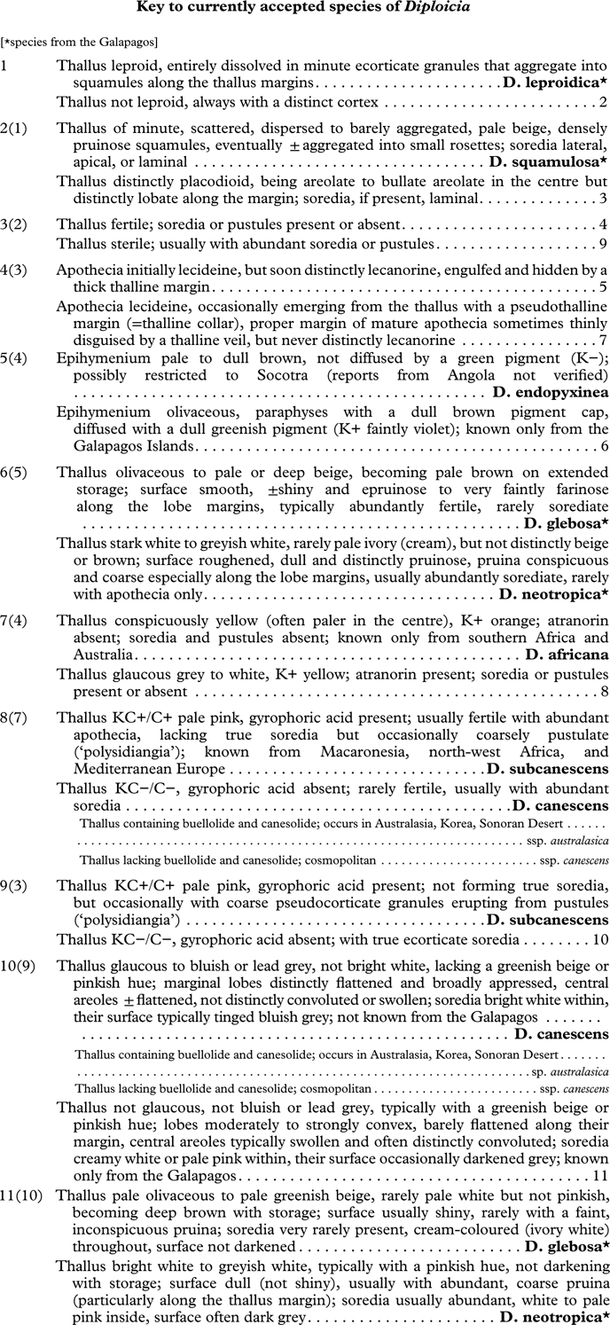
Detailed descriptions of the four species of Diploicia occurring in the Galapagos and a key to all species currently accepted in this genus are provided below. The generic circumscription of Diploicia is emended to include species with lecanorine apothecia as well as those with squamulose-placodioid or leproid-placodioid thalli. Previous reports of D. canescens from the archipelago are rejected. A brief overview of species previously described in the genus is also presented, including brief descriptions of Buellia coquimbensis and B. taltalensis, two species described in section Diploicia by Dodge (Reference Dodge1966) from Chile.
Diploicia A. Massal. [emend. Bungartz, Elix, Kalb & Giralt]
Type: Diploicia canescens (Dicks.) A. Massal.
Thallus crustose to subfoliose, placodioid, forming distinct rosettes with elongate marginal lobes, or rosettes composed of ecorticate granules (leproid-placodioid) or squamules that subsequently aggregate (squamulose placodioid), or rosettes that are areolate-lobate (central areoles extending into distinct marginal lobes); surface ecorticate, pseudocorticate or phenocorticate, many species forming distinct soralia, rarely entirely dissolved into sorediate granules.
Apothecia often present, emergent, soon sessile; disc black, pruinose or not; margin usually lecideine or more rarely surrounded by a distinct thalline collar or indistinct thalline veil when emerging from the thallus, sometimes enclosed by a thalline margin and becoming lecanorine; proper exciple distinct, ±carbonized by fuscous brown pigments, sometimes excluded in convex apothecia, occasionally with adhering thallus fragments, or becoming reduced and surrounded by a thalline exciple with cortex, photobiont layer and medulla; epihymenium of paraphyses with fuscous brown pigment caps (K−), sometimes inspersed with granules and/or a dull green pigment (K+ faintly violet); hymenium hyaline, ±with a subhymenium which is not clearly differentiated from the darkened hypothecium, laterally extending into the exciple and at the base extending to form a stipe; these layers (hymenium, subhymenium and hypothecium) are usually not inspersed or rarely so with sparse to moderately abundant oil droplets; asci clavate, similar to the Bacidia-type (central, unstained part cylindrical, but stained flanks merging at the tip); ascospores with moderate median and ±distinct apical thickenings, cell lumina initially angular to ±square, but with age becoming thin-walled (±Dirinaria-type). Spore development following ontogeny of type-B.
Pycnidia immersed, flask-shaped; conidia simple, bacilliform (often slightly tapered at one end, ±lacrimiform).
Secondary Chemistry. Thallus with diploicin or biosynthetically closely related orcinol depsidones and diphenyl ethers, often with additional atranorin and secalonic acid derivatives.
Notes. The emended generic concept differs from the original by including species containing diploicin and related substances, but forming placodioid rosettes derived from squamules or leproid granules, as well as species which develop lecanorine apothecia, either as a thalline collar or veil, or where the lecideine margin is enveloped by a distinct thalline margin early during the ontogeny and the proper exciple is almost completely reduced.
Description of Diploicia species from the Galapagos
Diploicia glebosa (Tuck.) Bungartz, Elix & Kalb comb. nov.
MycoBank No.: MB 816803
Basionym: Pyxine glebosa Tuck. Syn. N. Amer. Lich. (Boston) 1: 79 (1882), published in footnote no. 1, not the main text; type: Ecuador, Galapagos [specific locality and habitat not recorded], Hassler Expedition, 1872, Hill s. n. (FH-TUCK 197448—lectotype selected here!).
Protologue (text of footnote 1). “…And a still more marked reduction of this type is presented by a lichen from volcanic rocks of the Galapagos Islands (Dr. Hill in Hassler Exp.) in which while the apothecia offer no differences unless possibly rather smaller spores, the thallus has passed wholly into convex, glebous areoles, somewhat lobed only at the circumference (P. glebosa, Mihi, herb.)…”
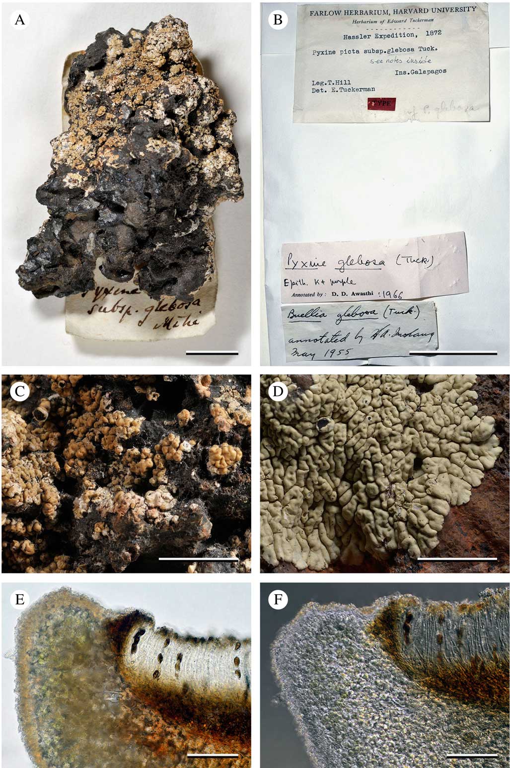
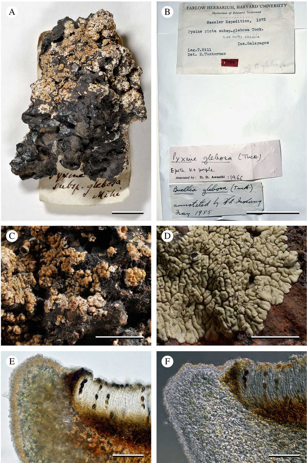
Diploicia glebosa, lectotype specimen (Hill, T. s. n., FH 197448). A, overview of the type material on a lava rock; B, label with annotations by Imshaug (Reference Imshaug1955, as Buellia glebosa), and Awasthi (1966, as Pyxine glebosa); C, close-up of damaged thallus with gnarled lobes and one apothecium; D, recent collection of a largely intact, placodioid thallus, completely adhered to the substratum, medulla white (Bungartz, F. 4513, CDS 28599); E & F, section of apothecium, prominent, dark brown proper exciple enclosed by a thick thalline exciple (Bungartz, F. 7020, CDS 36527); E, phenocortex and photobiont layer abundantly filled with minute crystals (regular light, in H2O); F, crystals almost completely dissolved (differential interference contrast, in K). Scales: A & B=5 mm; C & D=3 mm; E & F=50 µm.
Thallus saxicolous, crustose to subfoliose, placodioid, the centre of elongate, typically distinctly convex, convoluted, gnarled, contiguous to ±separate areoles, irregularly radiating towards the margin and extending into contiguous to ±separate, apically broadened, convex to ±flattened marginal lobes; surface faintly olivaceous grey to pale or deep beige, becoming more intensely brown on extended storage, rarely darkened along the lobe margins, ±shiny, with a smooth texture, mainly epruinose, but usually with a very faint, farinose pruina confined to the lobe margin and lining the edges of central areoles; central areoles usually abundantly fertile, very rarely with sparse soralia; soralia very rarely present, convex, erumpent, circular to irregular, occasionally ±confluent, developing from the thallus surface; soredia ivory white, typically paler than the thallus surface, granular compact, ecorticate with sparse, short protruding hyphae, (45–)61–91(–122) µm diam.; cortex phenocorticate, with a 7·5–10·0 µm thick epinecral layer, interspersed with fine crystals and a pale yellowish brown pigment (both dissolving in K), phenocortical layer prosoparenchymatous (c. 10 µm wide), interspersed with minute crystals (dissolving in K) and a few dead algal cells; medulla of ±densely interwoven, white hyphae, typically with a dirty orange-brown pigment near the substratum (unidentified secalonic acid derivatives), IKI−, filled with minute crystals that dissolve in K and sparse, large crystals insoluble in K, no needles formed in H2SO4 (calcium oxalate absent).
Apothecia emergent, soon sessile; disc black, typically epruinose to very faintly pruinose, rarely with a bluish tinge caused by a strong white pruina; initially lecideine with a thin proper margin, soon becoming lecanorine, engulfed in and obscured by a thick, often ±discontinuous thalline margin which becomes irregularly crenate with age; thalline exciple with cortex, photobiont layer and medulla, filled with abundant minute crystals (same reactions as the thallus); proper exciple initially conspicuous, dark brown to strongly carbonized, with age becoming reduced, fading to brown or becoming hyaline, extending from a deep, fuscous to reddish brown hypothecium that extends from the base into a short, fuscous brown to reddish brown stipe; subhymenium not distinctly differentiated from the hypothecium, not inspersed; hymenium hyaline, not inspersed; epihymenium olivaceous, interspersed with hyaline to brownish granules and a diffuse, dull greenish pigment (K+ faintly violet); paraphyses with brownish pigment cap; asci clavate, 8-spored, similar to the Bacidia-type (central, unstained part cylindrical, but stained flanks merging at the tip); ascospores 1-septate, olive-grey then deep brown, smooth, ellipsoid to narrowly oblong, with age ±constricted at septum and often with ±pointed ends, with moderate median and indistinct to soon distinct apical thickenings; cell lumina initially ±angular, but with age becoming thin-walled (±Dirinaria-type), (12·0–)13·5–19·5(–28·5) × (5–)6–8(–10) μm (n=45); spore wall smooth, not ornamented.
Pycnidia immersed, flask-shaped with colourless wall, and deep fuscous to olivaceous ostiole; conidia simple, bacilliform (slightly tapered at one end) (3·0–)4·0–6·0(–6·5) × c. 1·0–1·5 μm (n=15).
Secondary Chemistry. Thallus P+ yellow, K+ yellow, C−, KC−, UV− (dull yellowish); medulla P−, K−, KC−, C−, UV− (dull yellowish); atranorin (major), chloroatranorin (major or minor), diploicin (major), dechlorodiploicin (trace), buellolide (major), norbuellolide (major), fulgidin (minor), isofulgidin (minor or submajor), norcanesolide (minor), unknown secalonic acid derivatives (major, minor or trace).
Etymology. From the Latin ‘glebous’, meaning lumpy, cloddy, gnarled.
Ecology and distribution. Occurs in the Galapagos from the coastal to the transition zone, in sunny to semi-shaded, rarely ±shaded, mostly wind- and rain-exposed habitats such as the top of boulders or the front of cliffs.
Notes. Specimens of D. glebosa are fairly uniform in their overall appearance. Recently collected specimens are pale olivaceous grey to pale greenish beige, and their surface is shiny though sometimes barely dulled by a very fine, inconspicuous pruina. With storage in the herbarium, the colour of the thalli increasingly darkens and the lectotype specimen in FH has become pale brown.
The species is aptly named because the convoluted central areoles typically appear knotty, gnarled, and lumpy. The systematic position of this species has caused considerable confusion. Tuckerman (Reference Tuckerman1882) described it within Pyxine, which is not surprising as superficially it closely resembles that genus. Thalli of D. glebosa form distinct marginal lobes which appear similar to those of P. endoleuca (Müll. Arg.) Vain. or P. petricola Nyl., but these lobes as well as the central areoles of D. glebosa always adhere directly to the substratum. The thalli do not develop a lower cortex and lack rhizines characteristic of Pyxine and hapters characteristic of Dirinaria.
Imshaug examined and annotated the type material in FH (Fig. 1A). He must have observed this difference when he annotated the species as Buellia glebosa, comparing it with B. straminea Tuck., a species with much finer lobed, placodioid thalli that are distinctly yellow due to the presence of xanthones.
According to his annotation, Weber studied the type specimen at FH in 1964, and added a lengthy note, writing: “Contrary to Imshaug’s annotation and his notes in Bryologist 58: 283. 1955 [Imshaug Reference Imshaug1955], the type specimen is a Pyxine and not a Buellia...”. Awasthi subsequently examined the FH material and apparently followed Weber, annotating the specimen as “Pyxine glebosa (Tuck.) Epithet. K+ purple”.
Weber (Reference Weber1986) later published his observations, but his statements in that publication are contradictory. For P. glebosa, Weber (Reference Weber1986, p. 462) states: “Nothing like it has been collected recently”. This was incorrect as he collected specimens of Diploicia glebosa (e.g., COLO 294551; L-62971, COLO 294659; L-62862, cited below), but identified them as Dirinaria caesiopicta (Nyl.) D. D. Awasthi. Among the COLO collections identified as D. caesiopicta, several do not belong to Dirinaria; they lack a lower cortex and hapters. Most of these specimens with a lower medulla directly adhering to their substratum are identified here as the newly described Diploicia neotropica, but a few belong to D. glebosa.
It is no surprise that these two Diploicia species have been confused with Pyxine endoleuca and Dirinaria caesiopicta. Whereas the typical, shiny, smooth, beige forms of D. glebosa more closely resemble a Pyxine, the strongly pruinose forms of D. neotropica are superficially similar to those of Dirinaria caesiopicta.
Additional specimens examined. Ecuador: Galapagos: Bartolomé, N-side of island, near Pinnacle Rock, 0°17'2''S, 90°33'15''W, 10 m alt., coastal zone, 2006, Bungartz 5323 A (CDS 29539); Floreana, lava flow near the shore just N of Black Beach, dry zone, 1976, Weber s. n. (COLO 294551; L-62971); Pinzón, along the trail going up from Playa Escondida, N- to W-facing cliff above a crater, 0°36'29''S, 90°40'14''W, 318 m alt., transition zone, 2006, Aptroot 64028 A (CDS 30589); Rábida, E-side of the island, dry zone, 1976, Weber s. n. (COLO 294659; L-62862); San Cristóbal, hills S of Punta Pit at the NE-coast of the island, 0°43'8''S, 89°14'36''W, 44 m alt., dry zone, 2007, Bungartz 6142 (CDS 34354); Santa Cruz, at the East coast of the island, c. 400 m N of Cerro Colorado, on cliff, 0°34'47''S, 90°10'21''W, 21 m alt., dry zone, 2006, Aptroot 64367 (CDS 30932); Santiago, at the W-coast of the island in Bahía Ladilla, 0°18'8''S, 90°49'56''W, 10 m alt., coastal zone, on cliff, 2006, Bungartz 5367, 5387, 5374 A (CDS 29583, 29603, 29590); Isabela, Volcán Alcedo, cinder cone c. 100 m from the shore and a little N of Islote Cowley, 0°22'54''S, 91°0'4''W, 17 m alt., coastal zone, 2006, Bungartz 4513, 4501 A (CDS 28599, 55384); Volcán Sierra Negra, Las Tintoreras, small island in front of Puerto Villamil at the S-coast of Isabela, 0°58'13·6''S, 90°57'39''W, 13 m alt., coastal zone, 2007, Bungartz 7020 (CDS 36527).
Diploicia leproidica Bungartz & Elix sp. nov.
MycoBank No.: MB 816799
Thallus forming minute rosettes with ecorticate sorediate granules in the centre and pseudocorticate lobes along the margin; containing atranorin, chloroatranorin, diploicin and unidentified secalonic acid derivatives.
Type: Ecuador, Galapagos, Floreana, c. 500 m S of La Lobería, c. 200 m inland from coast, 1°17'12·7''S, 90°29'36''W, 20 m alt., dry zone, open Bursera forest with Waltheria ovata, few shrubs of Scalesia affinis and some grasses, on rock below overhang, semi-shaded, wind- and rain-sheltered, 16 January 2011, Bungartz 9761 (CDS 47078—holotype).
Thallus saxicolous, leproid-placodioid, forming minute rosettes with sorediate granules in the centre and minute, squamulose lobes along the margin, adhering to the substratum by sparse, protruding hyphae forming an indistinct prothallus; upper surface white to greyish white; granules in the thallus centre compact, ecorticate with sparse, short protruding hyphae, (61–)76–91(–121) µm diam., but aggregating into coarse, compact, pseudocorticate lobes, (136–)152–212(–273) µm width, towards the margin, photobiont layer and medulla not distinctly differentiated, unpigmented, IKI−, filled with minute crystals that dissolve in K and sparse, large crystals that are insoluble in K, no needles forming in H2SO4 (calcium oxalate absent). Photobiont green, trebouxioid, 7–10 μm diam.
Apothecia not known.
Pycnidia not known.
Secondary Chemistry. Thallus P+ yellow, K+ yellow, C−, KC−, UV− (dull yellowish); medulla P−, K−, KC−, C−, UV− (dull yellowish); atranorin (major), chloroatranorin (major or minor), diploicin (major) and unknown secalonic acid derivatives (minor).
Etymology. Named for its rosettes composed of leproid granules.
Ecology and distribution. Known from a single locality along ±sheltered rock along the coast of Floreana Island.
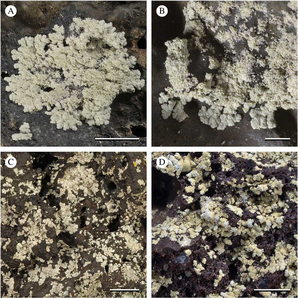
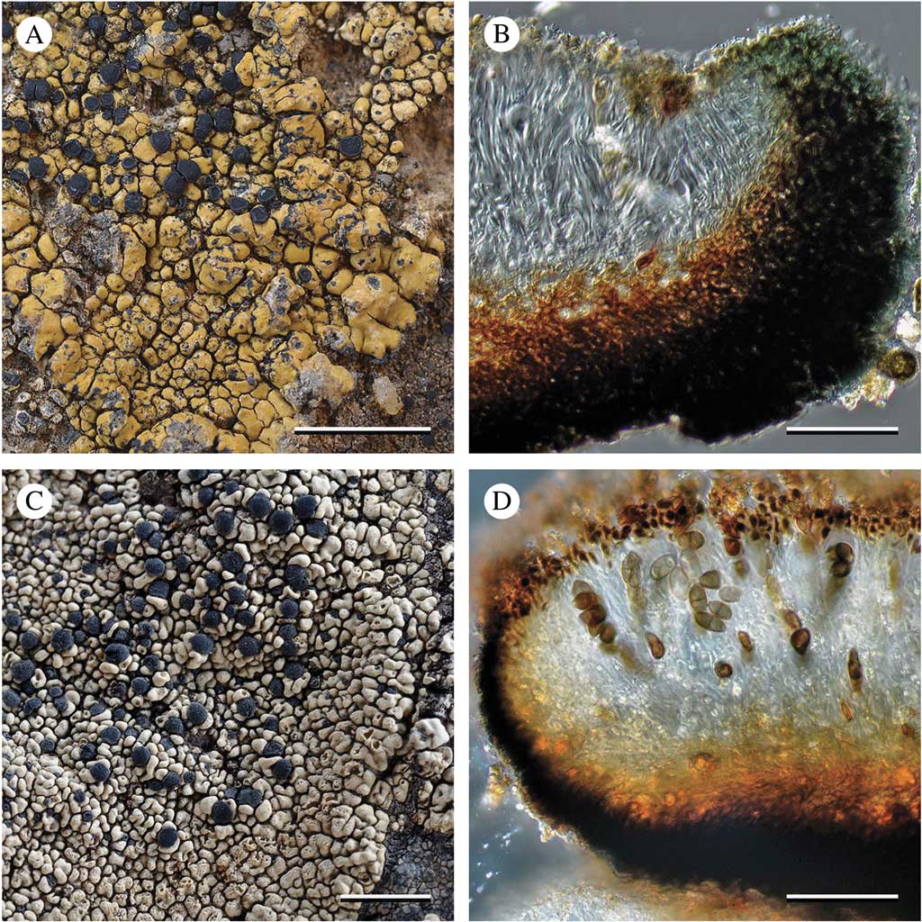
Notes. The small rosettes of D. leproidica may be confused with those of D. squamulosa, described below. The thalline rosettes and individual lobes of the two species are similar in size, but the pale white to greyish surface of the marginal lobes of D. leproidica is rough and lacks a distinct cortex (pseudocorticate), and towards the centre the lobes irregularly disintegrate into coarse, ecorticate granules. The granules and lobes are not clearly differentiated internally. This contrasts with the ivory to pale beige, compact, dull upper surface of D. squamulosa. This species forms distinctly delimited, erumpent soralia, which may become confluent with age. Although it is sometimes difficult to discern, the squamules of D. squamulosa are anatomically clearly differentiated into phenocortex, photobiont layer, and medulla.
The two species are ecologically similar, growing in sheltered habitats typically protected below overhangs, unlike the much larger thalli of D. glebosa and D. neotropica, which are commonly found in exposed habitats. Nevertheless, habitat preferences of the two species are distinct. Diploicia leproidica grows in close proximity of the coast, whereas D. squamulosa occurs in the upper transition zone and the high altitude transition zone.
Diploicia neotropica Kalb, Elix & Bungartz sp. nov.
MycoBank No.: MB 816800
Similar to Diploicia glebosa but differs in having a much rougher, white to grey upper surface that is usually strongly and coarsely pruinose and more flattened, with fewer convoluted lobes.
Type: Ecuador, Galapagos, Santa Cruz, along shore E of Puerto Ayora near Charles Darwin Research Station, 0°44'45''S, 90°17'39''W, 20 m alt., coastal zone, barren coastal cliffs, growing on basalt, 29 May 2005, Aptroot 63280 (CDS 30020—holotype).
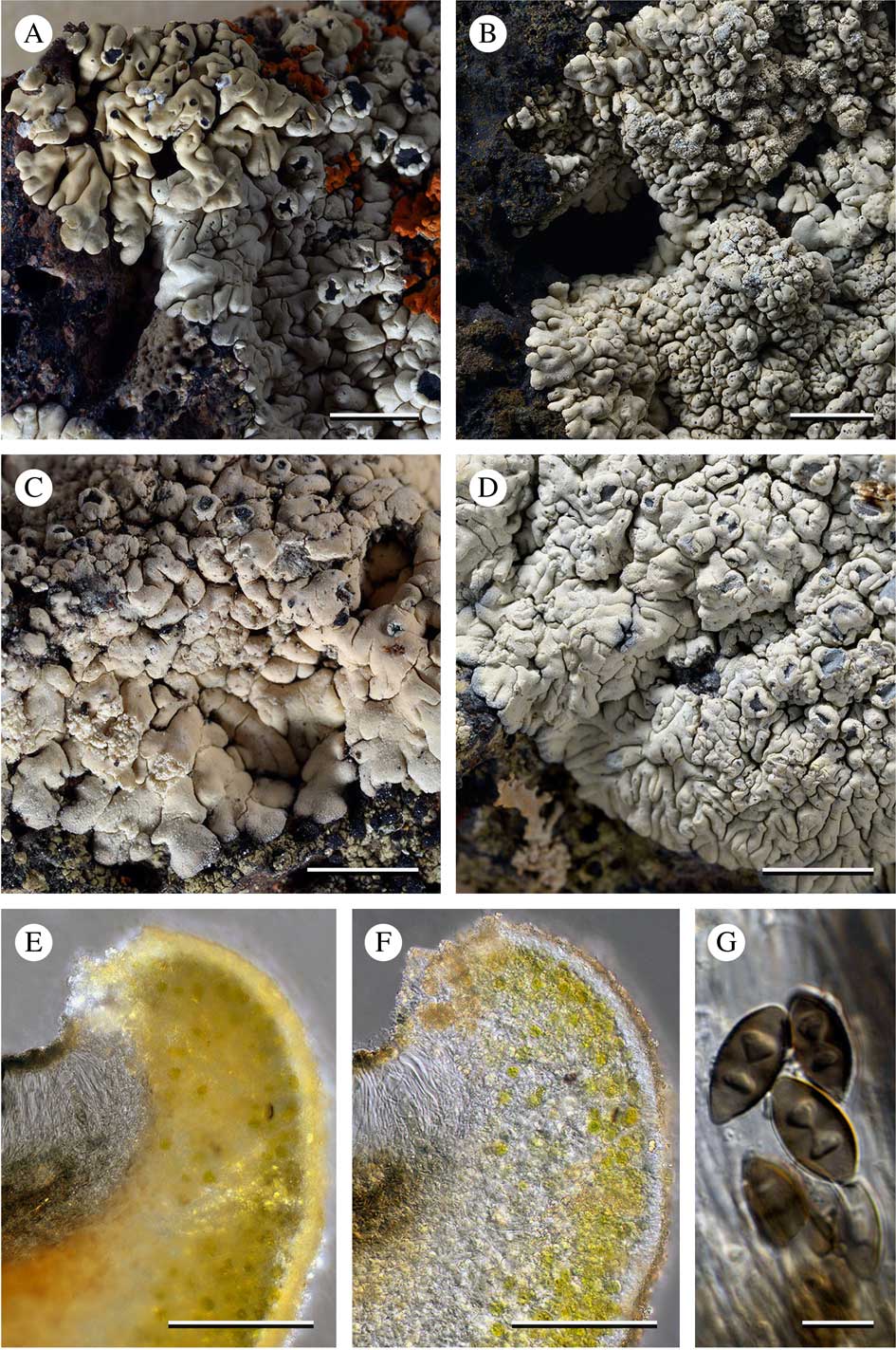
Variation in Diploicia neotropica; A, pale beige, epruinose, placodioid thallus of D. glebosa (upper left; Bungartz, F. 4499 B, CDS) overgrowing pale white, finely pruinose, placodioid thallus of D. neotropica (lower right; Bungartz, F. 4499 A, CDS 28585), apothecial disc of both specimens epruinose; B, ±ivory white, finely pruinose, placodioid thallus with erumpent soralia with greyish tinge (Bungartz, F. 6037, CDS 33716); C, ±ivory white, finely pruinose, placodioid thallus, lobes ±flattened, increasingly darkened and more abundantly pruinose towards their margin, thallus centre with laminal, erumpent soralia and lecanorine apothecia, disc barely pruinose (Aptroot, A. 63280, CDS 30020, holotype); D, pale white specimen with abundant, coarse pruina, placodioid thallus with ±flattened lobes, increasingly darkened and very strongly pruinose towards their margin, apothecial discs with bluish tinge caused by conspicuous pruina (Bungartz, F. 7029, CDS 36536); E & F, section of the apothecium, proper exciple strongly reduced to a few hyaline hyphae, enclosed by a thick thalline exciple (Bungartz, F. 7029, CDS 36536); E, phenocortex and photobiont layer abundantly filled with minute crystals (differential interference contrast, in H2O); F, crystals almost completely dissolved (differential interference contrast, in K, the diffuse greenish, weak K+ violet epihymenium pigment washed out, dull brown pigment persistent); G, ascospores (Bungartz, F. 7029, CDS 36536). Scales: A–D=3 mm; E & F=50 µm; G=10 µm.
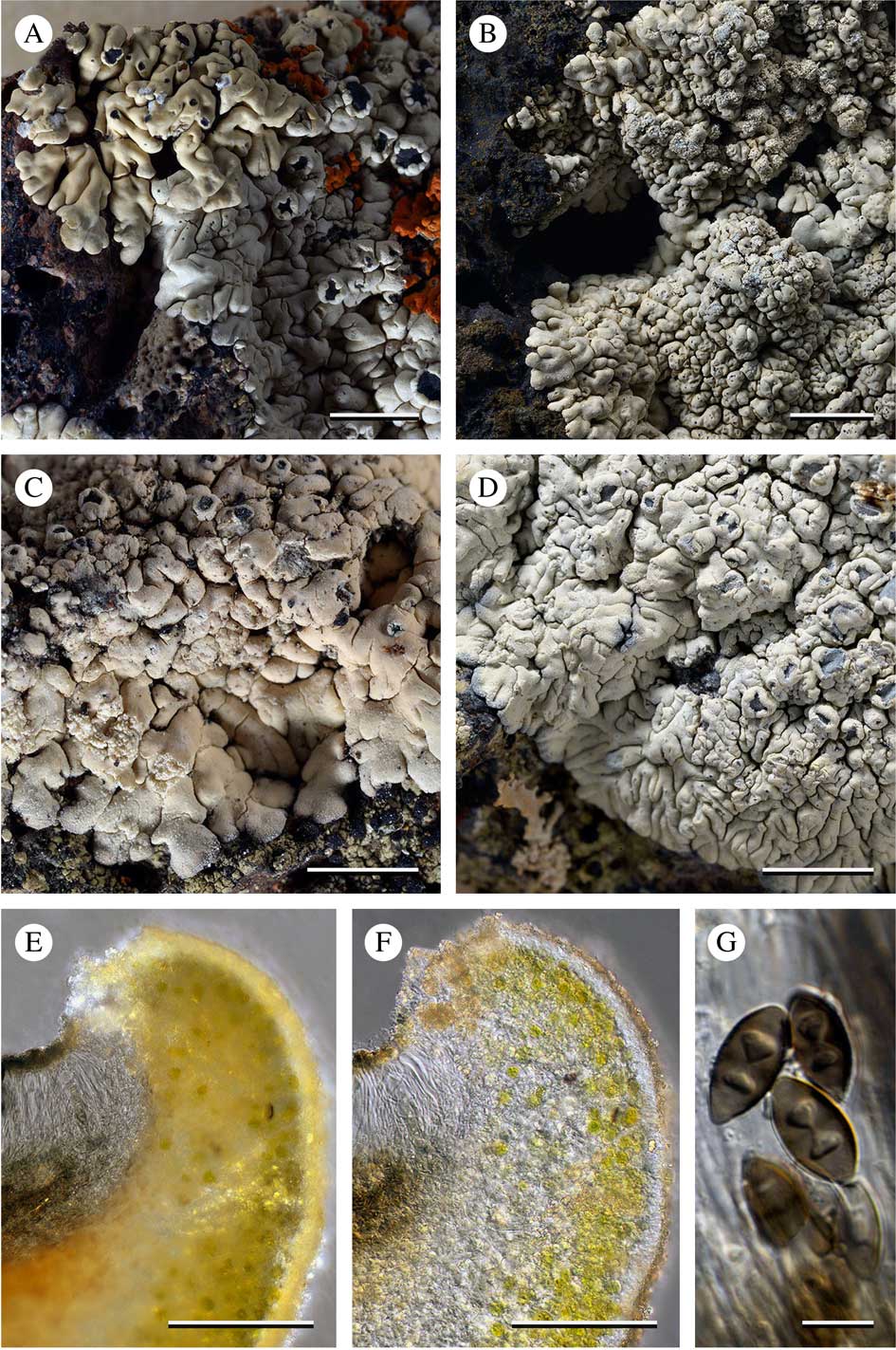
Thallus saxicolous, crustose to subfoliose, placodioid, with elongate, sometimes slightly convex, rarely convoluted, contiguous areoles in the centre, but radiating, contiguous, fan-shaped, apically broadened, flattened marginal lobes; surface white to greyish white, rarely ±cream-coloured or with a faint pinkish tinge, but not beige or brownish, often conspicuously darkened along the lobe margins (tan to dark grey or blackened), usually dull, with a conspicuously roughened texture, rarely ±smooth, completely and evenly covered by moderate to very strong, coarse to very coarse pruina, which is especially pronounced along the lobe margins; central areoles often sorediate or only with apothecia, rarely with both; soralia, when present, convex, erumpent, circular to irregular, occasionally ±confluent, developing from the thallus surface; soredia white, but often with a pinkish to dark grey tinge, granular, compact, ecorticate with sparse, short protruding hyphae, (45–)61–85(–106) µm diam.; cortex phenocorticate, with a 7·5–10·0 µm thick epinecral layer, interspersed with fine crystals (most, but not all, dissolving in K) and a pale pinkish brown pigment (±persistent in K), phenocortical layer prosoparenchymatous (c. 10 µm wide) filled with minute crystals (soluble in K) and a few dead algal cells, with the photobiont layer below; medulla of densely interwoven hyphae, white throughout, rarely dirty orange-brown near the substratum (unidentified secalonic acid derivatives), IKI−, filled with minute crystals that dissolve in K and sparse, large crystals that are insoluble in K, usually not forming needles in H2SO4 (only one specimen collected on limestone rather than lava contained calcium oxalate and formed characteristic colourless needles in H2SO4).
Apothecia emergent, soon sessile; disc black, typically epruinose to very faintly pruinose, rarely with a bluish tinge caused by a dense white pruina; initially lecideine with a thin proper margin, soon lecanorine, engulfed in and obscured by a thick, often ±discontinuous thalline margin which becomes irregularly crenate with age; thalline exciple with cortex, indistinct photobiont layer and medulla, filled with abundant minute crystals (same reactions as the thallus); proper exciple initially conspicuous, dark brown to strongly carbonized, progressively reduced with age, fading to brown or even hyaline, extending from a deep, fuscous brown, or very rarely reddish brown hypothecium that extends into a short, fuscous brown to rarely reddish brown stipe at the base; subhymenium not distinctly differentiated from the hypothecium, inspersed with a few, inconspicuous oil droplets; hymenium hyaline, not (or very sparsely) inspersed with a few oil droplets; epihymenium olivaceous, interspersed with hyaline to brownish granules and a diffuse, dull greenish pigment (K+ violet); paraphyses with brownish pigmented caps; asci clavate, 8-spored, similar to the Bacidia-type (central, unstained part ±cylindrical, but stained flanks merging at the tip); ascospores 1-septate, soon deep olive-brown, smooth, ellipsoid to narrowly ellipsoid, often with ±pointed ends, with moderate median and initially indistinct but soon distinct apical thickenings, cell lumina initially ±angular, but becoming thin-walled with age (±Dirinaria-type), (11·0–)14·0–18·0(–22·5) × (5·0–)6·0–7·5(–9·0) μm (n=73); spore wall smooth, not ornamented.
Pycnidia immersed, flask-shaped with colourless wall, and deep fuscous to olivaceous ostiole; conidia simple, bacilliform (slightly tapered at one end) (4·0–)4·5–5·5(–6·0) × c. 1·0–1·5 μm (n=10).
Chemistry. Thallus P+ yellow, K+ yellow, C−, KC−, UV− (dull yellowish); medulla P−, K−, KC−, C−, UV− (dull yellowish); atranorin (minor), chloroatranorin (minor or trace), diploicin (major or minor), dechlorodiploicin (major or minor), dechloro-O-methyldiploicin (trace), O-methyldiploicin (trace), buellolide (major), norbuellolide (major), fulgidin (minor), isofulgidin (minor or submajor), norcanesolide (minor), rarely also with unknown secalonic acid derivatives (minor).
Etymology. A neotropical species (presently known only from the Galapagos, but possibly more widely distributed in the Neotropics, see below).
Ecology and distribution. This species occurs from the coastal to the lower transition zone, in sunny to semi-shaded, rarely ±shaded, mostly wind- and rain-exposed habitats such as the top of boulders or the front of cliffs; one specimen was collected from limestone from an ancient marine deposit, uplifted above the sea, a substratum rarely found in Galapagos (Aptroot, A. 64356, CDS 30921).
Notes. Diploicia neotropica is a morphologically variable species and three different forms can be distinguished informally: the first is strongly pruinose with bright white, somewhat ‘frosted’ thalli with densely pruinose apothecial discs with a bluish tinge (Fig. 2D). This morphotype is typically fertile, although a few specimens are also sorediate. The second, more common morphotype has weakly pruinose thalli with a faint pinkish surface and typically epruinose or faintly pruinose discs (Fig. 2C). Usually these thalli have both soredia and apothecia, although apothecia may be sparse or even absent in some specimens (Fig. 2B). Both forms have ±flattened marginal lobes with increasingly coarse pruina towards their tips. The lobes usually have darkened (fuscous) margins. These moderate to densely pruinose morphotypes of D. neotropica can readily be distinguished from the shiny, epruinose thalli of D. glebosa. Unfortunately, pruina are not completely absent from all forms of D. glebosa. Although specimens of D. glebosa are generally uniform in their appearance, a few have fine, faint, white pruina. Where such pruina cover the surface, the otherwise shiny cortex appears dulled. These specimens could then be confused with a third morphotype of D. neotropica with fine, less abundant pruina. Nevertheless, these superficially similar thalli can be distinguished even when they grow in close proximity (Fig. 2A).
Despite their variability, all morphotypes of D. neotropica are characterized by a surface which remains bright white or rarely has a faint pinkish hue, and these specimens do not darken even with prolonged herbarium storage. All have a roughened, distinctly pruinose surface. Even if the pruina of D. neotropica varies from fine to coarse, faint to abundant, it covers a distinctly roughened surface and, unlike D. glebosa, the upper surface of D. neotropica is never shiny.
These differences in pruinosity, texture and colour are conspicuous, but a detailed examination of the cortical anatomy of the two species failed to reveal any correlating differences. The internal cortical structure, and the presence, size and distribution of crystals are similar in both species. In section, the outer cortex of both species is typically pale brown, even in specimens of D. neotropica with a bright white, strongly pruinose thallus. We suspect that the cortical pigments are different, because only specimens of D. glebosa darken conspicuously with prolonged storage. Nevertheless, pigment concentrations are generally so low that the alternative reactions in K and HNO3 (following Meyer & Printzen Reference Meyer and Printzen2000) were not observed.
Thalli of D. glebosa are typically not sorediate and usually abundantly fertile, whereas those of D. neotropica display greater variation. Most have abundant soredia with many, few or no apothecia, but a few are abundantly fertile with sparse or no soredia.
The considerable morphotypical variation of D. neotropica is reflected by an equally diverse chemistry. Unfortunately the chemical variations did not correlate with the three morphotypes described above.
Specimens examined. Ecuador: Galapagos: Bartolomé, N-side of island, near Pinnacle Rock, 0°17'2''S, 90°33'15''W, 10 m alt., coastal zone, 2006, Bungartz 5323 B (CDS 55385); Española, along S-coast of the island, near Punta Suárez, 1°22'32·10''S, 89°44'16·89''W, 26 m alt., coastal zone, 2010, Bungartz 8856, 8859 (CDS 45674, 45677), Yánez-Ayabaca 1568 (CDS 45450); Gardner (near Española), SW-part of the island, 1°20'46·39''S, 89°38'54''W, 7 m alt., coastal zone, 2010, Bungartz 9170 (CDS 45988); Floreana, lava flow c. 200 m N of Puerto Velasco Ibarra, at the W-coast of the island, c. 100 m inland, 1°16'10·1''S, 90°29'17·80''W, 11 m alt., coastal zone, 2011, Bungartz 9864 (CDS 47202); Genovesa, edge of new lava flow N of Darwin Bay, dry zone, 1964, Smith s. n. (COLO 192017; L-41138); Pinta, along the S-coast, W of Cabo Ibbetson, 0°32'42" N, 90°44'12''W, 2 m alt., coastal zone, 2007, Bungartz 6037 (CDS 33716); San Cristóbal, Cerro Colorado, enclosure for C. galapagosa near the viewpoint on the top, 0°54'58''S, 89°26'5''W, 130 m alt., transition zone, 2007, Bungartz 6729 (CDS 34973); Santa Cruz, at the E-coast of the island, c. 200 m N of Cerro Colorado, 0°34'49''S, 90°10'21''W, 1 m alt., coastal zone, 2006, Aptroot 64356 (CDS 30921); Caamaño (near Santa Cruz), dry zone, 1964, Weber 133 (COLO 190108; L-40992); Plaza Norte (near Santa Cruz), 0°34'47''S, 90°9'47''W, 1 m alt., coastal zone, 2006, Bungartz 3813 (CDS 27695); Plaza Sur (near Santa Cruz), 0°34'59''S, 90°9'54''W, 1 m alt., coastal zone, 2006, Bungartz 3757, 3758, 3762 (CDS 27639, 27640, 27644); Santa Fé, top of steep lava cliff above the sea, 0°47'59·5''S, 90°2'39·20''W, 16 m alt., coastal zone, 2007, Bungartz 7283 (CDS 37767); Santiago, at the W-coast of the island in Bahía Ladilla, 0°18'8''S, 90°49'56''W, 10 m alt., coastal zone, 2006, Bungartz 5396, 5374 B (CDS 29612, 55386); Isabela, Volcán Alcedo, cinder cone c. 100 m from the shore and a little N of Islote Cowley, 0°22'54''S, 91°0'4''W, 17 m alt., coastal zone, 2006, Bungartz 4499, 4501 B (CDS 28585, 28587); Volcán Sierra Negra, Muro de las Lagrimas, c. 5 km W of Puerto Villamil, 0°57'52·70''S, 91°0'46·79''W, 78 m alt., dry zone, 2008, Bungartz 8431 (CDS 41077).
Diploicia squamulosa Bungartz & Elix sp. nov.
MycoBank No.: MB 816801
Thallus of minute, finely pruinose, ivory to beige, corticate squamules with marginal or laminal soredia, occasionally aggregating into minute placodioid rosettes; containing atranorin, chloroatranorin, alectoronic acid, diploicin and unidentified secalonic acid derivatives.
Type: Ecuador, Galapagos, Isabela, Volcán Darwin, c. 1·5 km from the south-western crater rim, 0°12'20·5''S, 91°18'52·8''W, 1280 m alt., high altitude dry zone, basalt outcrop for the most part bare of vegetation, on rock below W-exposed overhang of basalt outcrop, shaded, wind- and rain-sheltered, 14 November 2007, Bungartz 7597 (CDS 38093).
Diploicia leproidica and D. squamulosa. A & B, Diploicia leproidica (Bungartz, F. 9761, CDS 47078, holotype); A, small thallus rosette composed of leproid granules; B, close-up of thallus with intact, granular lobes along the margin, almost completely dissolved in the granular-leproid centre. C & D, Diploicia squamulosa; C, growth aspect of dispersed thallus squamules, occasionally growing into small rosettes (Bungartz, F. 7749, CDS 38255); D, close-up of phenocorticate squamules with erumpent soredia at lobe apices, along the margins, or laminal (Bungartz, F. 7597, CDS 38093, holotype). Scales: A=2 mm; B=1 mm; C & D=3 mm.
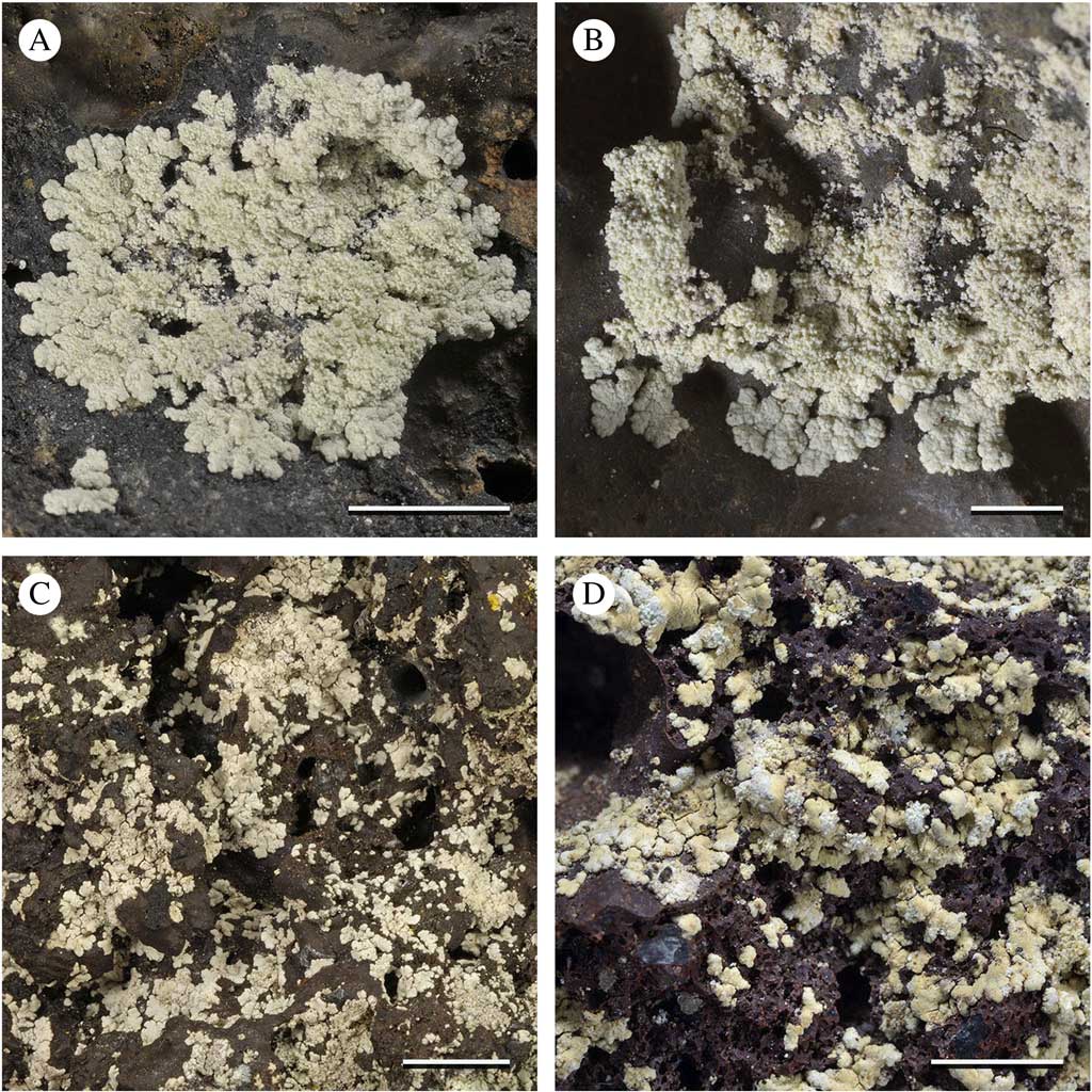
Thallus saxicolous, squamulose to minutely placodioid, of scattered sorediate squamules, (273–)303–394(–455) µm wide, that when well developed, aggregate into minute rosettes (2–5 mm diam.); prothallus indistinct; surface pale cream to pale beige, dull with a ± roughened texture, moderately covered by farinose pruina which is more pronounced and slightly coarser along the edges of the squamules; cortex phenocorticate, prosoplectenchymatous, indistinctly delimited, interspersed with dead photobiont cells, the upper part disintegrating into an indistinct epinecral layer, densely interspersed throughout by minute crystals, most of which dissolve in K, but some are persistent and remain irregularly dispersed; medulla of densely interwoven, white hyphae, typically dirty orange-brown near the substratum (unidentified secalonic acid derivatives), IKI−, filled with minute crystals that dissolve in K and sparse, large crystals insoluble in K, no needles forming in H2SO4 (calcium oxalate absent); soralia convex, erumpent, circular to irregular, occasionally ±confluent, forming at the lobe apices, along the margin or on the thallus surface; soredia white (distinctly paler than the thallus surface), granular compact, ecorticate with sparse, short protruding hyphae, (61–)91–110(–150) µm diam. Photobiont green, trebouxioid, 7–10 μm diam.
Apothecia not known.
Pycnidia not known.
Secondary Chemistry. Thallus P+ yellow, K+ yellow, C−, KC−, UV− (dull yellowish); medulla P−, K−, KC−, C−, UV− (dull yellowish); atranorin (major), chloroatranorin (major or minor), alectoronic acid (major), diploicin (major or minor) and unknown secalonic acid derivatives (major or minor).
Etymology. Named for its thalline squamules.
Ecology and distribution: Currently known only from shaded, sheltered rocks in the upper transition zone and high altitude transition zone on the western slope of Volcán Darwin on Isabela Island.
Notes. Diploicia squamulosa is most similar to D. leproidica (see comments above). This is the only species of Diploicia in the Galapagos known to occur at high altitudes, in dry, sheltered habitats.
Additional specimen examined. Ecuador: Galapagos: Isabela, Volcán Darwin, south-western slope, above Tagus Cove, 0°13'28·2''S, 91°19'17·9''W, 872 m alt., transition zone, 2007, Bungartz 7749 (CDS 55297).
Species of Diploicia excluded from the Galapagos checklist
Diploicia canescens (Dicks.) A. Massal.
Ric. Auton. Lich. Crost. (Verona): 86 (1852).
Buellia canescens (Dicks.) De Not., G. Bot. Ital. 2(1.1): 197 (1846).
Diploicia canescens was first reported from the Galapagos by Weber (Reference Weber1986) and included in all subsequent updates of his species list (e.g. Elix & McCarthy Reference Elix and McCarthy1998). The single specimen on which this record was based (Weber & Lanier s. n., L-63018, COLO 291533) consisted of a few, inconspicuous squamules growing among abundant Chrysothrix xanthina (Vain.) Kalb; the material certainly does not belong to D. canescens, but is most likely a poorly developed specimen of D. neotropica.
Diploicia endopyxinea (Müll. Arg.) Kalb, Elix & Bungartz comb. nov.
MycoBank No.: MB 817125
Basionym: Physcia endopyxinea Müll. Arg., Proc. R. Soc. Edinb. 11: 459 (1882); Dirinaria endopyxinea (Müll. Arg.) C. W. Dodge, Beih. Nova Hedwigia 38: 181 (1971); type: Africa, Yemen, Socotra Islands, ‘ramulicola partim cum Pyxine convexa Müll. Arg. crescens’, 1881, Balfour 1359 (‘BCS’=Balfour, Cockburn & Scott) (G–66116 A, collector’s number 1359 red-underlined, selected here as lectotype!), 1400 (‘BCS’) (G–66116 B syntype, number cited in protologue), 1563 (‘BCS’) (G–66116 C syntype, number cited in protologue).
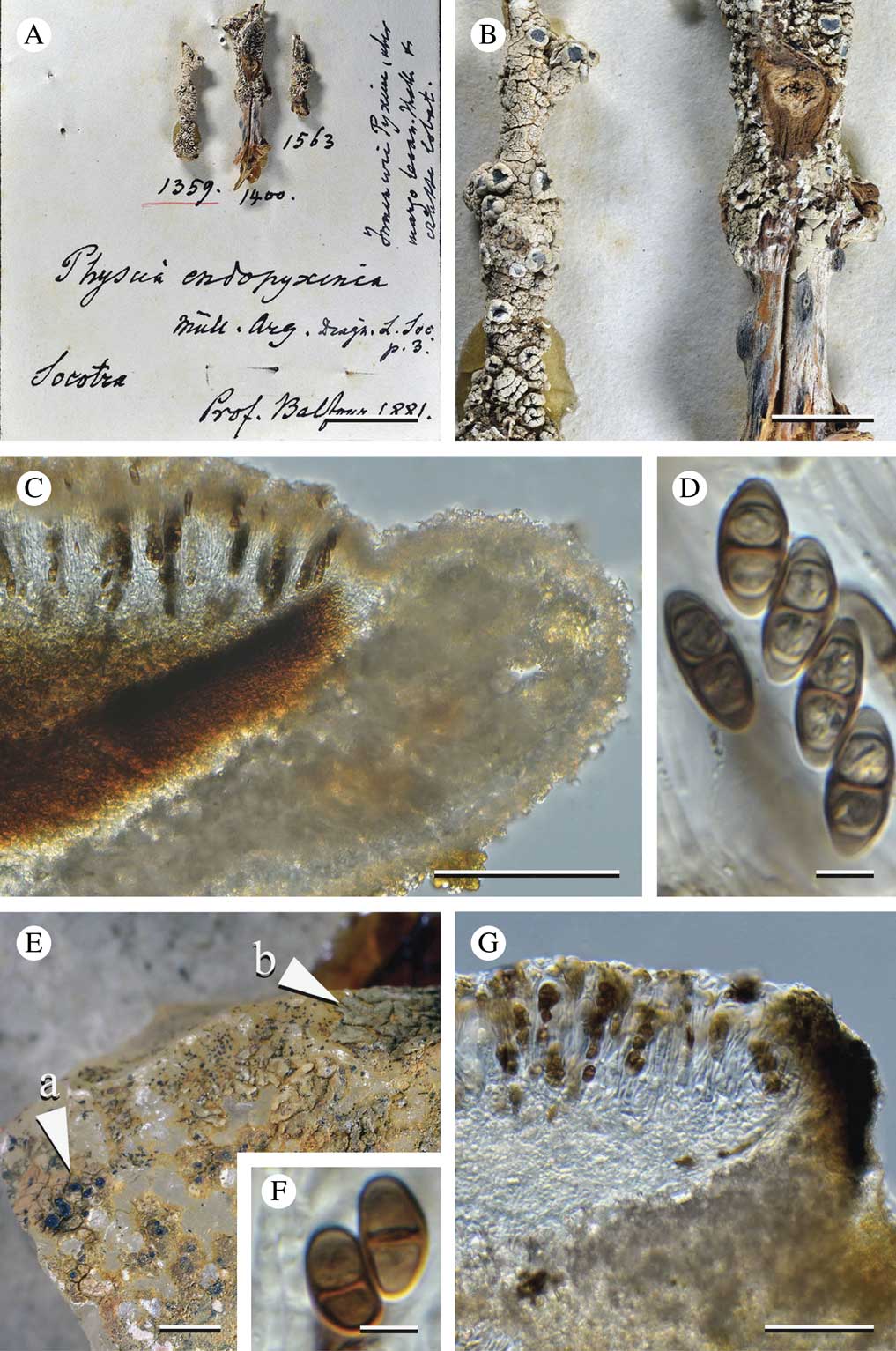
Diploicia endopyxinea and Buellia stanleyi. A−D, Diploicia endopyxinea; A, overview of the type material [collected in Socotra by ‘BCS’ Balfour, Cockburn & Scott; G 66116]; on the left specimen Balfour, I. B. 1359 (number red-underlined, selected here as the lectotype), middle and right: syntype specimens Balfour, I. B. 1400 & 1563 (syntypes); B, close-up of the lectotype (on the left) and one of the syntype specimens; C, section of the apothecium with a pale brown epihymenium, phenocortex and photobiont layer abundantly filled with minute crystals (differential interference contrast, in H2O); D, ascospores. E−G, Buellia stanleyi (Ledien, F. 346, G 47580); E, syntype, specimen on the left an areolate thallus with minute lecideine apothecia (arrow ‘a’), specimen on the right a placodioid sterile thallus (arrow ‘b’); F, thin-walled, broadly ellipsoid ascospores (specimen ‘a’); G, section of the apothecium with aethalea-type exciple and hyaline hypothecium (specimen ‘a’). Scales: A=15 mm; B=5 mm; C=100 µm; D & F=5 µm; E=1 mm; G=50 µm.
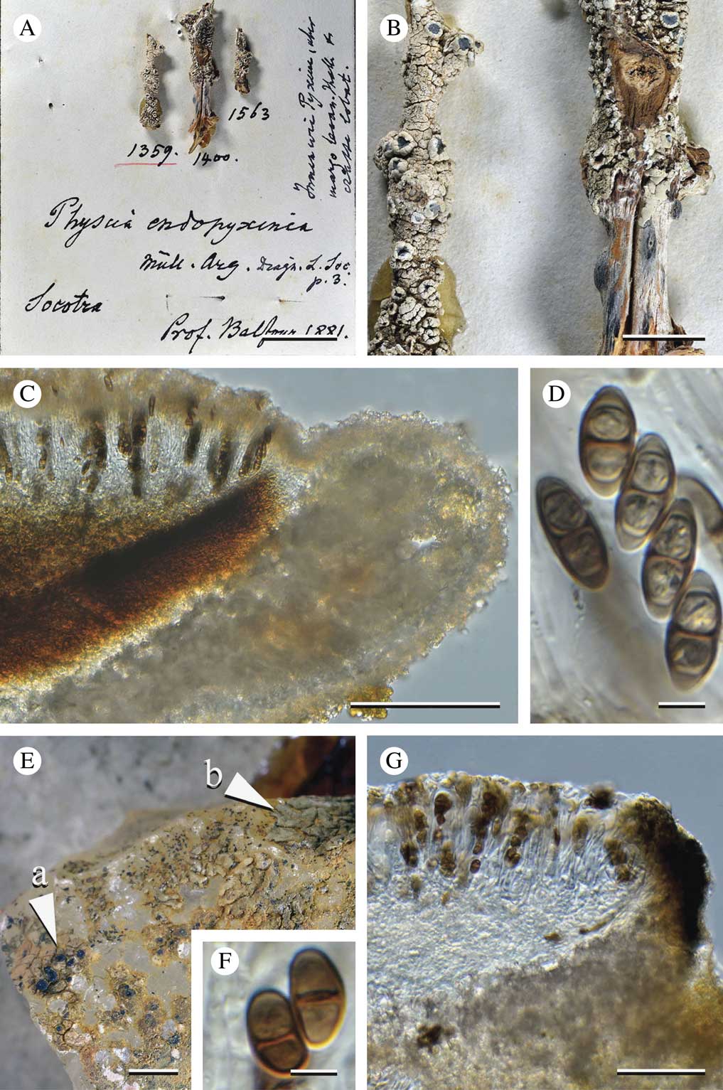
With its weakly pruinose thallus, a pale white surface and weakly pruinose discs, this African species appears rather similar to D. neotropica (especially the second, most commonly found morphotype). Superficially the two taxa are difficult to distinguish, although thalli of D. neotropica typically have a pinkish hue and those of D. endopyxinea are ivory white to pale beige. Furthermore, D. neotropica is a saxicolous species whereas D. endopyxinea is epiphytic.
The type of D. endopyxinea differs from D. neotropica in having a subhymenium that consistently lacks oil droplets and an epihymenium that is brown, rather than olivaceous. The epihymenium of D. endopyxinea lacks the diffuse greenish pigment present in D. neotropica which reacts K+ violet. Both species have ascospores with ±pointed ends similar to the Dirinaria-type, initially with angular cell lumina, but their spore walls soon become thin. Although the ascospore dimensions of the two species fall into the same range, the spores of D. endopyxinea are generally more narrowly elongate. In the Galapagos, all specimens of D. neotropica occur exclusively on rock. The Socotran species is known only as an epiphyte (the substratum of the type material is decorticated branches).
Chemistry. Thallus P+ yellow, K+ yellow, C−, KC−, UV− (dull yellowish); medulla P−, K−, KC−, C−, UV− (dull yellowish); atranorin (minor), chloroatranorin (minor or trace), diploicin (major); Swinscow & Krog (Reference Swinscow and Krog1978) examined the type and confirmed that the specimen contains the same secondary metabolites as Diploicia canescens, rather than divaricatic acid as originally suggested by Awasthi (Reference Awasthi1975). We did not re-analyze this material.
Ecology and distribution. The checklist of lichens included in Brown & Mies (Reference Brown and Mies2012) suggests that ‘Physcia endopyxinea Müll. Arg.’ is endemic to Socotra, even though Awasthi (Reference Awasthi1975) cites one collection from Angola. The identity of this Angolan specimen appears doubtful considering Awasthi’s confusion regarding the chemistry of the specimens.
Taxa originally described in Diploicia
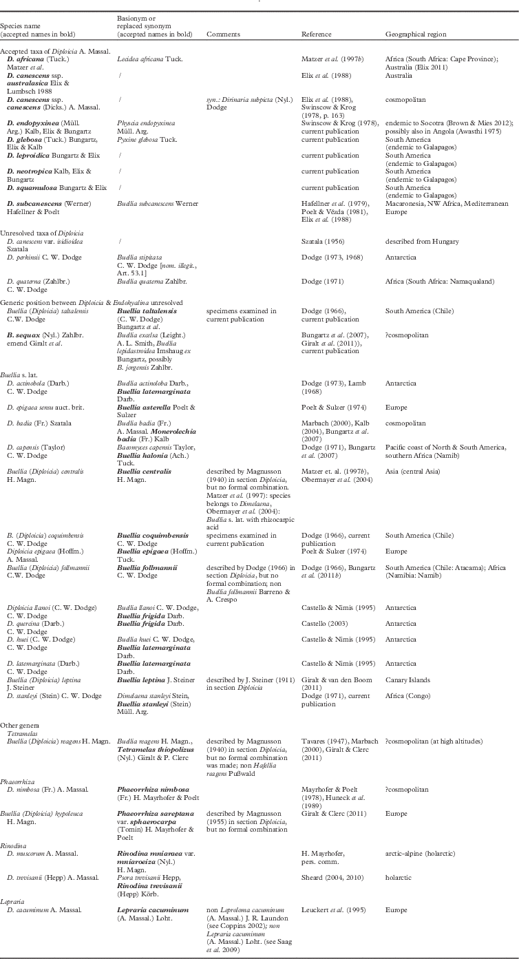
The list of taxa described in the genus or as part of section Diploicia still includes several unresolved names (see Appendix 1) although none of these names refer to species reported from the Galapagos. One is an unresolved isidioid variety of D. canescens described from Hungary. In addition, a taxon reported by Dodge (Reference Dodge1973) from Antarctica, Diploicia perkinsii C. W. Dodge, remains unresolved. The name was introduced as a replacement for the illegitimate Buellia stipitata C. W. Dodge (non Riddle). The type material has not been re-examined and the placement of this species in Diploicia cannot be confirmed, but the taxon certainly does not occur in the Galapagos.
Another species was described by Steiner (Reference Steiner1911) in section Diploicia as Buellia leptina J. Steiner. This taxon was considered by Şenkardeşler (Reference Şenkardeşler2010) to be conspecific with Diploicia subcanescens, resulting in a proposal to conserve B. subcanescens Werner against B. leptina (Şenkardeşler et al. Reference Şenkardeşler, Lökös and Fuat Calba2011). Having examined the chemistry of the type, Giralt & van den Boom (Reference Giralt and van den Boom2011) recently reported that the material contains hypostictic acid rather than diploicin. Accordingly this species is not related to Diploicia. Instead, it shares many characteristics with Buellia caloplacivora Llimona & Egea, a species with affinities to the B. subalbula-group (Bungartz & Grube Reference Bungartz and Grube2005; Bungartz et al. 2011 Reference Bungartz, Grube, Elix, Heininger and Mayrhoferb ).
Dodge (Reference Dodge1971) transferred Dimelaena stanleyi Stein from the Congo region into Diploicia. Although it appeared unlikely that a species from continental central Africa would occur in the Galapagos, we have examined the type (Ledien, F. 346, G-47580). The specimen is extremely poor, consisting of a few irregular areoles with minute apothecia (Fig. 4E, specimen ‘a’) growing alongside a few, isolated lobes of an unidentified sterile, isidiate, placodioid crust (Fig. 4E, specimen ‘b’). The growth form of the latter would explain why Stein described this species as belonging in Dimelaena and why Dodge (Reference Dodge1971) transferred it into Diploicia. The identity of the material, however, remains unresolved. The thalli included as part of the type clearly do not belong to the same species; only the part that is fertile, lacks isidia and does not grow placodioid (Fig. 4E, specimen ‘a’) has lecideine apothecia with a simple, aethalea-type exciple, ascospores that lack wall thickenings, and a hyaline hypothecium. Whether the other specimen even belongs in Physciaceae (Fig. 4E, specimen ‘b’) cannot be determined with certainty. Given the paucity of this material, we did not analyze the chemistry of either one of the specimens.
The identity of Buellia quaterna Zahlbr. at present also remains unresolved. Dodge (Reference Dodge1971) transferred this taxon into Diploicia. It has been described from coastal areas of South Africa and Namibia (Namaqualand) and is characterized by 4-spored asci (Dodge Reference Dodge1971). The two fertile Galapagos species are quite different as they have eight spores per ascus.
In addition, a further 16 species have at some point in time been included in Diploicia; however, they do not belong in this genus and are currently included in Buellia s. lat., Tetramelas, Phaeorrhiza, Rinodina, and even Lepraria (see Appendix 1).
This leaves two species described by Dodge (Reference Dodge1966) in Buellia section Diploicia from southern South America (Chile) to be considered, Buellia (Diploicia) coquimbensis and B. (Diploicia) taltalensis. Previously, Weber et al. (Reference Weber, Gradstein, Lanier and Sipman1977) had suggested that saxicolous species along the Galapagos coast and in the dry lowlands showed strong affinities with species from Chile. Indeed, recent studies of Roccella by Tehler et al. (Reference Tehler, Irestedt, Bungartz and Wedin2009) and of Redonographa by Lücking et al. (Reference Lücking, Tehler, Bungartz, Rivaz Plata and Lumbsch2013) supported this hypothesis. Since it appeared possible that several Galapagos endemics might have their closest relatives growing along the Chilean coast, we examined material of the two Chilean species, B. taltalensis and B. coquimbensis, and provide brief descriptions here. The first taxon is closely related to Buellia sequax, whereas the second appears related to B. halonia. Although thalli of B. sequax and especially B. taltalensis can become quite exuberant, they do not become placodioid.
Buellia coquimbensis C. W. Dodge
Nova Hedwigia 12: 349 (1967) [1966]; type: Chile, Elqui Prov. (“Coquimbo”), La Serena, on coastal rocks, 1965, Follmann 14765 (holotype not seen; herbarium not mentioned in the protologue).
Diploicia coquimbensis and Buellia taltalensis. Diploicia coquimbensis (Follmann, G. 16228, KASSEL, topotype); A, effigurate thallus of flattened to barely convex areoles, marginal areoles subsquamulose; B, leptocline-type exciple of mesodermatous intricately interwoven hyphae strongly carbonized by aeruginose pigment. C & D, Buellia taltalensis (Follmann, G. 14686, KASSEL, holotype); C, effigurate thallus with convex, convoluted areas delimited by a blackish prothallus line; D, dispersa-type exciple, pale inside, hymenium inspersed with few oil droplets. Scales: A & C=3 mm; B & D=50 µm.
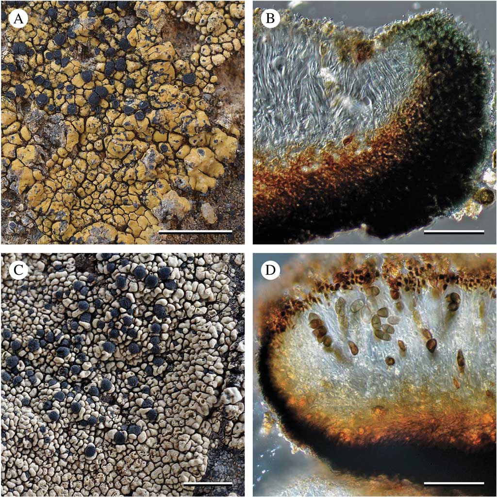
Thallus greenish beige (mustard-coloured), crustose-areolate, central areoles flattened to ±convex, marginal areoles subsquamulose to squamulose, ±effigurate, but not distinctly lobate or placodioid, surrounded by a compact black prothallus.
Apothecia lecideine; exciple strongly carbonized, leptocline-type, aeruginose throughout (K−, N+ purple); epihymenium aeruginose (Cinereorufa-green, K−, N+ purple); hymenium hyaline, not inspersed; hypothecium reddish brown (K± dull brown); asci clavate, 8-spored, Bacidia-type; ascospores ellipsoid, 1-septate, with inconspicuous medial thickenings (±Physconia-type), (12–)13–15(–16) × (5·0–)6·5–8·0(–9·0) μm (n=20); spore wall smooth to faintly ornamented (only visible with DIC).
Conidia bacilliform (not tapered), (5·0–)5·5–6·5(–7·0) × c. 1·5–2·0 μm (n=15).
Secondary Chemistry. Thallus P−, K−, KC−, C−, UV− (pale); arthothelin (minor), 6-O-methylarthothelin (major).
Etymology. The specific epithet refers to the type locality.
Notes. Buellia coquimbensis is definitely not a Diploicia. Thalli of the species are barely effigurate and certainly not placodioid, with marginal areoles becoming subsquamulose to barely squamulose, not elongating into distinctly radiating lobes. The species appears most closely related to B. halonia (Ach.) Tuck. Both species are similar in colour and overall appearance and share anatomical characters: a proper exciple of the leptocline-type strongly carbonized by an aeruginose pigment, and faintly ornamented ascospores of the Physconia-type (see Bungartz et al. Reference Bungartz, Elix and Nash2004, Reference Bungartz, Nordin and Grube2007).
Specimen examined from the type locality. Chile: Elqui Prov. (labelled “Coquimbo”): La Serena, on coastal rocks, 1965, Follmann 16228 [KASSEL; erroneously labelled “type”].
Buellia taltalensis C. W. Dodge
Nova Hedwigia 12: 351 (1967) [1966]; type: Chile, Antofagasta Prov. (“Atacama”), Taltal, on coastal rocks, 1965, Follmann 14686 & Follmann-Schrag (holotype—KASSEL!).
Thallus pale creamish white, crustose-areolate, central areoles ±convoluted, barely subsquamulose at the margins, never lobate or placodioid, surrounded by a compact black prothallus.
Apothecia lecideine, with pruinose discs; exciple barely carbonized, dispersa-type, fuscous along the outer part (K−, HNO3−), with a hyaline inner part; epihymenium fuscous (K−, HNO3−); hymenium hyaline, strongly inspersed with minute oil droplets; hypothecium dull brown (K−, HNO3−); asci clavate, 8-spored, Bacidia-type; ascospores ±citriform, 1-septate, juvenile with weak apical wall thickenings and pronounced median wall thickenings, the cell lumen ±angular (Dirinaria-type) or with median wall thickenings and no apical thickenings (Physconia-type), the lateral wall sometimes weakly dilated but not forming distinct Callispora-type spores; all wall thickenings almost inapparent in mature ascospores (±Buellia-type), (10–)11–14(–15)×(5·0–)6·5–8·0(–9·0) μm (n=45); spore wall distinctly ornamented (microrugulate).
Conidia bacilliform (slightly tapered at one end, ±lacrimiform), 4–5(–6) × c. 1·0–1·5 μm (n=15).
Secondary Chemistry. Thallus P+ yellow, K+ yellow, KC−, C−, UV− (pale); atranorin (major), diploicin (major), isofulgidin (minor).
Etymology. The specific epithet refers to the type locality.
Notes. Buellia taltalensis is closely related to B. sequax as emended by Giralt et al. (Reference Giralt, Bungartz and Elix2011). Specimens of B. taltalensis are clearly distinguished from that species by much thicker, more exuberant, almost ‘cerebriform’ thalli with pruinose apothecia and distinctly microrugulate to rugulate ascospores (their ornamentation is clearly visible at ×1000 in conventional transmitted light microscopy, and is particularly conspicuous in differential interference contrast). Buellia jorgensis Zahlbr., also described from coastal Chile, more closely resembles B. sequax (Giralt et al. Reference Giralt, Bungartz and Elix2011, p. 117: an assessment based on a single specimen in M (M-0061335); the type unfortunately could not be located).
Specimens examined from the type locality. Chile: Antofagasta Prov. (“Atacama”): Taltal, on coastal rocks, 1965, Follmann 16182, 16183 (KASSEL; erroneously labelled “type”); 16185 (COLO 216064, L-45771; erroneously labelled “isotype”).
Discussion
The four Galapagos species discussed above cannot be accommodated within the traditional concept of Diploicia. Consequently, we have emended the generic concept to include species with diploicin and related substances, which develop lecanorine apothecia, and species forming placodioid rosettes derived from squamules or leproid granules.
Emending the generic concept is not problematic when the ontogeny of the thalline margin is compared with that of the lecideine margin present in species traditionally included in Diploicia. Poelt & Vězda (Reference Poelt and Vězda1981, p. 146) previously described the apothecia of Diploicia as “...zuerst vom Lager bedeckt, dann frei...”, which described an ontogeny where the lecideine margin remains at least temporarily obscured by thalline material when the apothecia emerge. Bungartz et al. (Reference Bungartz, Nordin and Grube2007) introduced the term ‘thalline collar’ to describe such species. They also used the term ‘thalline veil’ to describe a phenomenon where a lecideine margin remains thinly disguised by a necrotic thalline layer. A thalline collar can sometimes be observed in immature, emerging apothecia of Diploicia species that otherwise form distinctly lecideine apothecia (e.g. D. canescens).
A further step in this scenario can be easily imagined, whereby the thalline material remains well developed to form a distinct thalline margin, while the proper exciple successively diminishes. Pyxine, a closely related genus in the Caliciaceae s. str. (Kalb Reference Kalb1987, Reference Kalb2002), has three different types of exciple ontogeny. Some species form distinctly lecanorine apothecia (physciaeformis-type); the development is clearly analogous to that of Diploicia glebosa and D. neotropica. Other Pyxine species have apothecia that remain distinctly lecideine throughout their ontogeny (obscurascens-type), corresponding to most other Diploicia species. Finally, a third group is characterized by an ontogeny changing from lecanorine to lecideine (cocoës-type), the thalline margin becoming excluded with age. An ontogeny similar to the cocoës-type is also known from Buellia s. lat. (e.g. the mamillana-type, Bungartz et al. Reference Bungartz, Nordin and Grube2007), but has not yet been observed in Diploicia.
Including species with modified thalline morphology is more problematic. Giralt et al. (Reference Giralt, van den Boom and Elix2010) noted that Endohyalina was closely related to Diploicia, with most species containing diploicin but distinguished by their inconspicuous crustose or lichenicolous growth.
Thalli of Diploicia are distinctly subfoliose, but because of their placodioid growth form they appear foliose, despite having an ecorticate lower surface which adheres to the substratum. Thus, thalli of Diploicia are crustose, although they superficially look foliose.
This is in stark contrast to Endohyalina, which generally forms very inconspicuous thalli. Some species are epi- or even endokapylic lichenicolous fungi, their thalli almost entirely reduced. Most species are rimose to granular-verrucose, few form autonomous, subsquamulose, but not placodioid thalli: Endohyalina interjecta (Müll. Arg.) Giralt (Mayrhofer et al. Reference Mayrhofer, Scheidegger and Sheard1992 b; Giralt et al. Reference Giralt, van den Boom and Elix2010) and E. arachniformis Elix & Kantvilas (Elix & Kantvilas Reference Elix and Kantvilas2015).
Two other diploicin-containing species of interest are Buellia sequax (Nyl.) Zahlbr. emend Giralt et al. and B. taltalensis, as described above. Given its thick central areoles, Giralt et al. (Reference Giralt, Bungartz and Elix2011) hesitated to include Buellia sequax in Endohyalina, arguing that the species could be considered an intermediate between the distinctly placodioid thalli of Diploicia and the poorly developed or absent ones encountered in Endohyalina. In addition, B. sequax has a much better developed, dispersa-type exciple. By contrast, Endohyalina is characterized by a rather poorly developed, aethalea-type exciple (Giralt et al. Reference Giralt, Bungartz and Elix2011). Buellia taltalensis is closely related to B. sequax. Both species have the same exciple type and distinctly crustose thalli that lack the subfoliose appearance of Diploicia. Although their thalli are effigurate, with a margin composed of closely adjoining, subsquamulose areoles, these areoles do not become elongated into lobes and the thalli therefore do not become placodioid. They are instead usually delimited by a black prothallus (Fig. 5C) which further distinguishes these species from Diploicia as currently circumscribed.
Because of their apically thickened ascospores (during part of the ontogeny resembling those of Diploicia), and the presence of diploicin in the thalli, Nordin (Reference Nordin2000) argued that B. pallidomarginata A. Nordin and B. proximata H. Magn., two species with 3-septate spores, could be placed in Diploicia. In addition, this group with 3-septate spores and diploicin also includes Buellia lauricassiaeoides Aptroot (J. A. Elix: atranorin and diploicin confirmed, unpublished data). Buellia pallidomarginata forms grey to brownish, effuse, rather inconspicuous thalli, so it may perhaps better be accommodated in Endohyalina. Thalli of B. proximata and B. lauricassiaeoides are thicker, more exuberant, typically with a black prothallus, although perhaps not quite as well developed as those of B. sequax or B. taltalensis. Diploicin is often also present in species of Buellia s. str. (i.e. “Hafellia”), some with 1-septate spores (e.g. Buellia demutans (Stirt.) Zahlbr., B. parastata (Nyl.) Zahlbr. or B. procellarum A. Massal., cf. Elix Reference Elix2011), others with pluriseptate spores (e.g. B. oidalea (Tuck.) Tuck. and B. oidaliella A. Nordin; cf. Nordin Reference Nordin2000; Bungartz et al. Reference Bungartz, Nordin and Grube2007). In contrast to Diploicia, which is characterized by ±Dirinaria-type ascospores, these species of Buellia s. str. all have Callispora-type ascospores (i.e., forming subapical and not apical wall thickenings during their spore ontogeny).
When well developed, both D. squamulosa and D. leproidica form distinctly placodioid rosettes. We include them in Diploicia even if some areolate or granular specimens are not so different from the strictly crustose, non-placodioid species discussed above. Clearly the distinction between Diploicia, Endohyalina and Buellia sequax is not entirely unequivocal.
Resolving the taxonomy of the Physciaceae/Caliciaceae remains a challenge. Boundaries based on classical generic characters in this family are increasingly challenged as these traditional characters offer insufficient resolution. For a sound phylogenetic re-assessment, it will be necessary to use molecular tools and examine a broad, truly representative range of the diverse species in this group.
We dedicate this publication to Irwin M. Brodo, our mentor, colleague and friend on occasion of his 80th birthday. Ernie continues to be an inspiration for lichenology worldwide. We thank Frauke Ziemmeck for managing cryptogam collections at CDS. Diana Flores assisted with HTLC. We acknowledge support by successive CDF science directors (Alan Tye, Mark Gardener, Rodolfo Martinez, Ulf Härdter, and Noëmi d’Ozouville) and are indebted to the Directorate of the Galapagos National Park (particularly Galo Quezada and Victor Carrión, granting specimen export permits). The Census of Galapagos Biodiversity and the CDF Checklist of Galapagos Species received several grants (donors and contributing scientists cited at http://www.darwinfoundation.org/datazone/checklists/). The lichen inventory received funds from the Paul and Bay Foundations, and the Erwin Warth Stiftung. An international workshop was supported in 2010 by the US National Science Foundation (DEB 0715660, PI Robert Lücking; DEB 0841405, PI James Lawrey, local coordinator F. Bungartz). Taxonomic research with the objective to publish an IUCN red list of endemic Galapagos lichens is funded by the Mohamed bin Zayed Species Conservation Fund (project 152510692). Research by the last author (MG) was financially supported by the Catalan Government (project 2014SGR 414 GRC). The Galapagos lichen herbarium is indebted to the curators of: B, COLO, CAS, FH, H, and S. Michaela Schmull (FH), Peter Mansfeld (KASSEL) and Philippe Clerc (G) kindly provided type material on short notice. A visit to FH by the first author (FB) in 2009 was generously supported by a Friends of the Farlow Fellowship. Finally, we appreciate the support of Bodo Möseler, Eberhard Fischer and Rolf Wißkirchen from the Naturhistorische Verein der Rheinlande in facilitating specimen loans to NHV. We are very grateful to anonymous reviewers and the editors of The Lichenologist for valuable comments. This is contribution no. 2134 of the Charles Darwin Foundation for the Galapagos Islands.
Appendix 1
Overview of all taxa originally described in the genus Diploicia and species transferred into the genus Diploicia by different authors; the currently accepted names are in bold