Patent ductus arteriosus is one of the most common congenital diseases representing 5%–10% of all congenital heart lesions. Reference Schneider and Moore1 The trans-catheter closure of patent ductus arteriosus is the treatment of choice for suitable patients, being less invasive than the alternative surgical procedure. Reference Porstmann, Wierny, Warnke, Gerstberger and Romaniuk2
The standard transcatheter technique to profile the duct and assess device deployment requires arterial access through the femoral artery and is associated with arterial complications particularly in small children. Systemic heparinisation decreases femoral artery thrombosis; however, it remains a significant complication especially in patients less than 10 kg. Reference Kulkarni and Naidu3,Reference Vitiello, McCrindle, Nykanen, Freedom and Benson4
Apart from vascular complications, the use of arterial access is associated with longer fluoroscopic time, contrast volume, increase the risk of bleeding, and longer hospital stay. Reference Uppal, Rohit and Barwad5 Although it is not common, the use of iodinated contrast media may result in contrast-induced acute kidney injury. Reference Laville and Juillard6,Reference Pasternak and Williamson7
This study presents a comprehensive review of transcatheter patent ductus arteriosus device closure in 320 patients over a period of 2 years, aiming to compare exclusive transvenous access with the standard procedures for patent ductus arteriosus closure regarding feasibility, safety, and advantages and evaluate whether exclusive venous approach is a safe and effective alternative compared to the standard technique.
Methods
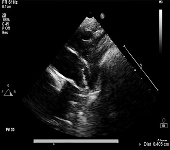
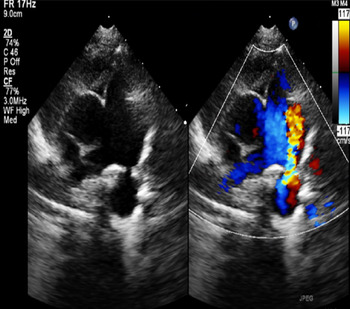
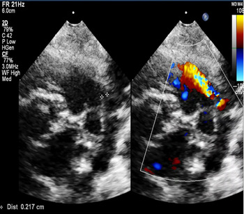
The records of all patients who underwent transcatheter closure of patent ductus arteriosus from July 2018 to July 2020 at Aswan Heart Centre, Magdi Yacoub Heart Foundation, Egypt, were reviewed. All cases were done at the cardiac catheterisation laboratory under echocardiographic and fluoroscopic guidance without angiography. A total of 320 patients were included. A detailed echocardiographic evaluation of the duct morphology based on two-dimensional and color Doppler parameters was performed for all patients. The duct diameter was measured in the standard parasternal short axis view, the high parasternal short axis view (known also as ductal view), and in the supra-sternal long axis view (Figs 1, 2 and 3). The point where the duct opened into the pulmonary artery (the pulmonary end) was measured, and the duct morphology and the shape of the ductal ampulla were also evaluated and measured.
Tubular PDA from suprasternal long axis view.

How to measure the pulmonary end from the parasternal short axis.

How to measure the pulmonary end from the ductal view.

Patients were classified into two groups. Group 1 included patients who underwent exclusive femoral venous access, without any injections of contrast media. Group 2 included patients who underwent concomitant arterial and venous access (standard technique) where the position of the device and residual shunt were evaluated by the guidance of the aortogram.
The inclusion criteria for patients who underwent transcatheter patent ductus arteriosus closure by any of the two techniques were symptomatic patients, auscultated murmur, and echocardiographic evidence of volume overload, while exclusion criteria were other cardiac pathology needing surgical intervention, weight below 4 kg, large duct not suitable for device closure, patients with significant pulmonary arterial hypertension.
Patients who underwent the standard technique (group 2) rather than the exclusive femoral venous access were selected by the following criteria: inadequate echo definition of patent ductus arteriosus size, small infants less than 6 kg, accidental arterial access.
Device selection and catheter intervention
The procedures were performed under conscious sedation using Dexmedetomidine in addition to local lidocaine, in some cases if Dexmedetomidine alone is not effective in sedating the patients, Midazolam, Fentanyl, or Ketamine were used. The sheaths with appropriate sizes were placed in the femoral vessels before 75 U/kg heparin was administered to the patients. The size of the Amplatzer ductal occlude-I device (or other similar devices) was selected as approximately 2 mm larger than the patent ductus arteriosus size at the pulmonary end. No contrast was used at all for group 1 patients, and the procedure was done under echocardiographic guidance.
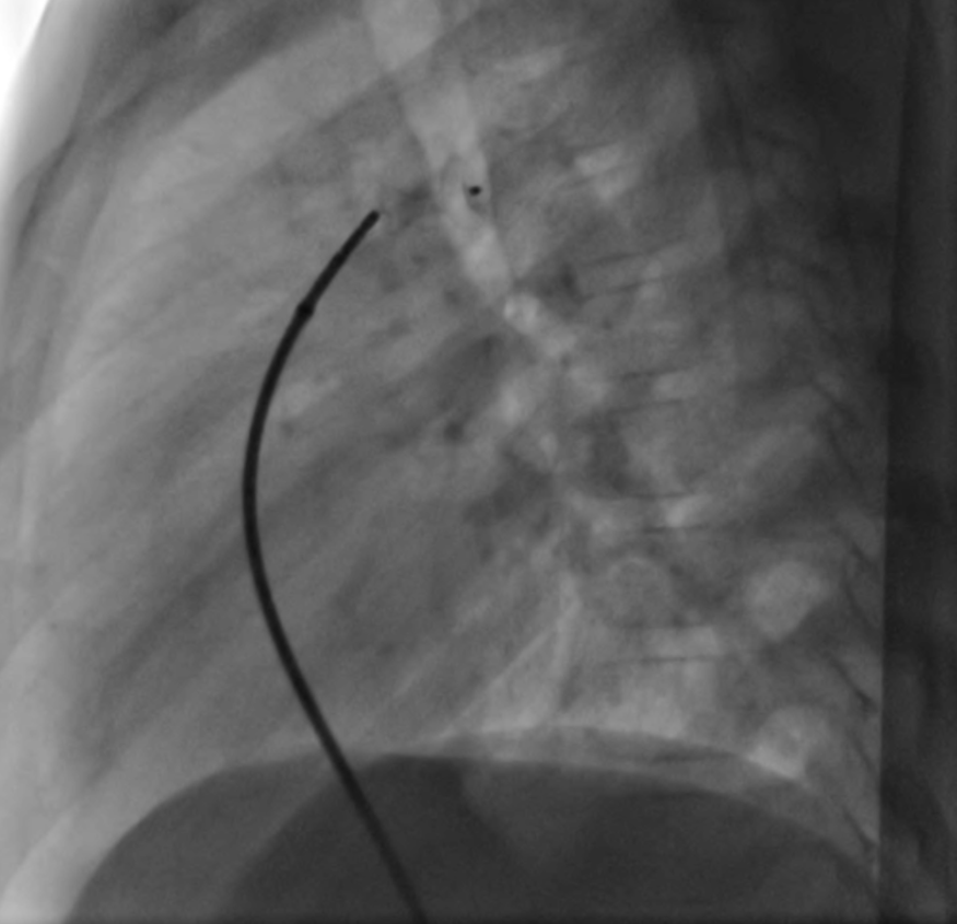
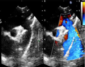
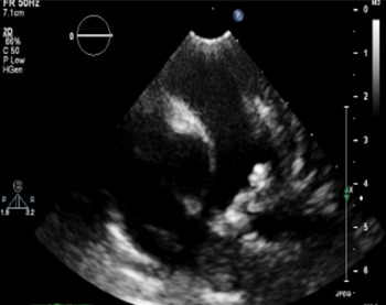
Before releasing the device, the device was assessed by fluoroscopy making sure that there is good waist at the centre of the device. We noticed that the aortic disc of the device is usually at the posterior border of the trachea in the lateral view (Fig 4). Echocardiography was performed in the cardiac catheterisation laboratory before releasing the device. The device placement was assessed on two-dimensional echocardiography in standard parasternal short axis view, high parasternal view, and suprasternal view (Figs 5 and 6). The correct placement of the device was accepted when the retention skirt was seen opposing the ampulla in the descending aorta, and the conical portion of the device was seen in the pulmonary artery with a waist at the mouth of the duct (Fig 6). Colour flow mapping was done to check for residual flow through the duct as well as turbulence at the origin of the left pulmonary artery and in the descending aorta (Fig 5). Small residual color flow was accepted. Doppler gradients of up to 5 mmHg in the left pulmonary artery and up to 10 mmHg in the descending aorta was accepted. The device was deployed and the echocardiographic assessments were repeated. All patients underwent repeat echocardiography before discharge, where all the above-mentioned parameters were reassessed.
Lateral fluoroscopic view showing the PDA device opposite the posterior border of the trachea.

Suprasternal long axis showing the aortic disc of the device with no coarctation.

Parasternal short axis both the pulmonary and aortic end of Amplatzer duct occluder device, with the central narrowing of the device.

Statistical analysis
All the data were collected prospectively. Statistical Package for Social Sciences (SPSS Inc. version 16.0 for Windows) was used for the analysis of data. All quantitative or continuous variables were estimated using measures of central tendency (mean) and measures of central dispersion (standard deviation).
Results
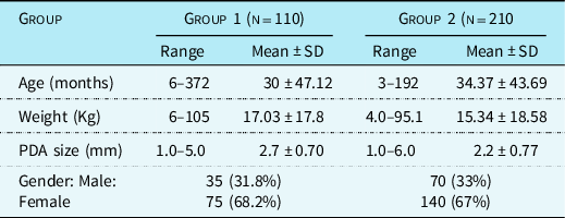
A total of 320 patients underwent patent ductus arteriosus device closure, 105 patients were males (32.8%) and 215 (67.2%) were females. The age in group 1 ranged from 6 to 372 months with the mean of 30 ± 47.12 months. The weight ranged from 6 to 105 kg with the mean of 17.03 ± 17.8 kg. In group 2, the age ranged from 3 to 192 months with the mean of 34.37 ± 43.69 months. The weight ranged from 4.0 to 95.1 kg with the mean of 15.34 ± 18.58 kg. The patent ductus arteriosus size at the pulmonary end ranged from 1 to 6 mm. Arterial access was achieved either alone or with concomitant venous as well in 210 (65.6%), while exclusive venous access was achieved in 110 (34.4%) patients (Table 1).
Demographics of the studied groups

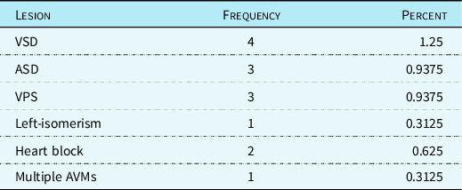
Concomitant findings were ventricular septal defect (n = 4), atrial septal defect (n = 3), valvular pulmonary stenosis (n = 3), left isomerism (n = 1), heart block (HB) (n = 2), and multiple arteriovenous malformations (n = 1) (Table 2).
Concomitant cardiac lesions

VSD: ventricular septal defect, ASD: atrial septal defect, VPS: valvular pulmonary stenosis, AVMs: arteriovenous malformations
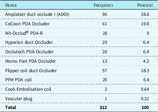
The majority of cases were closed by Amplatzer duct occlude I (28.8% of cases) or other similar devices according to the availability in the cardiac catheterisation laboratory or by coils if the patent ductus arteriosus is small (Table 3).
Devices used

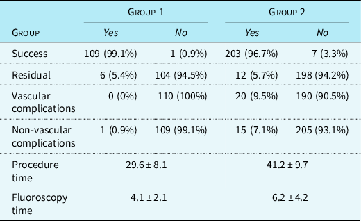
Successful closure of patent ductus arteriosus was achieved in 109 (99.1%) patients in group 1 and in 203 (96.7%) patients in group 2, while in 8 patients (one patient in group 1 and 7 patients in group 2), the patent ductus arteriosus was significantly large and the patients were referred for surgical closure. Residual patent ductus arteriosus was seen in 18 cases, 6 cases from group 1 (5.4%) and 12 patients from group 2 (5.7%). None of the cases in group 1 had vascular complications, while vascular complications were seen in 20 cases, all of them in group 2 (9.5%), of which 16 patients needed heparinisation, while the remaining 4 patients required streptokinase.
Nonvascular complications were seen in 16 patients, one patient only in group 1 (0.9%) and 15 patients in group 2 (7.1%). The nonvascular complications include one case had the device accidentally released (the device was not probably locked) with protrusion of the device into aorta causing coarcation of the aorta, the device was retrieved successfully and reinserted. Five cases had coil embolisation in the aorta, main pulmonary artery, left and right pulmonary arteries. All the embolised coils were retrieved successfully. One patient developed left pulmonary artery stenosis that was managed by surgical removal of the device with surgical patent ductus arteriosus closure. Some patients developed supra ventricular tachycardia, transient bradycardia that resolved spontaneously, others developed bleeding and haematoma without haemodynamic significance that needed any active intervention, (Table 4).
Procedure results

The procedure time was significantly less (29.6 ± 8.1 minutes) in patients with exclusive transvenous access (group 1), while it was 41.2 ± 9.7 minutes in patients of group 2. The fluoroscopy times was also significantly less (4.1 ± 2.1) in patients of exclusive venous access (group 1) than patients of group 2 (6.2 ± 4.2), (Table 4).
Discussion
In many studies using an antegrade procedure in the transcatheter closure of patent ductus arteriosus (from venous side only), an additional arterial approach was used for pressure monitoring and contrast substance injection. In these studies, transcatheter closure was carried out only via the venous approach in a few patients with occluded femoral arteries or in cases of failure to use the femoral artery route. Reference Hildebrandt, Schneider, Schweigl and Schneider8-Reference Grabitz, Schrader and Sigler10
Over the past two decades, transcatheter occlusion of patent ductus arteriosus has evolved to be the procedure of choice. Transcatheter closure has become the first line of treatment in many centres except in the newborn period. With the development of new devices, the success rate of the procedure is above 95% even in difficult patient groups such as premature and adult patients. Although complete closure of the patent ductus arteriosus can change in some studies depending on patients age, patent ductus arteriosus size and the device used, the highest closure rates have been observed with Amplatzer devices and the similar devices. Reference Baykan, Narin and Özyurt11
When the literature was reviewed, few studies were done for patent ductus arteriosus closure from the venous side only without arterial access, under echocardiographic guidance; however, although there is no arterial access, all these studies were still doing angiographic aortogram from the venous side to profile the duct. Reference Baykan, Narin and Özyurt11 To our knowledge to may be the first study for trans-catheter patent ductus arteriosus device closure form the venous side only under complete echocardiographic guidance without the use of angiographic dye.
Routine practice involves arterial access for angiography before, during, and after deployment of the device. The reasons for using arterial access are that it allows initial aortography to be performed without crossing the duct, supposedly minimising the risk of ductal spasm and consequent underestimation of the size of the duct. Also, it permits subsequent aortography to confirm a good position of the device prior to release and to exclude iatrogenic coarctation. Reference Garg, Srivastava, Tyagi, Reddy and Radha12
However, our experience at Aswan Heart Centre, Egypt, proved that the shape and the size of the duct can be evaluated clearly by echocardiography without any risk of ductal spasm and therefore no risk for any underestimation of the ductal size. Echocardiography also will let the operator to release the device with confidence. The correct placement of the device is accepted when the retention skirt is seen opposing the ampulla in the descending aorta and the conical portion of the device is seen in the pulmonary artery with a waist at the mouth of the duct. Color flow mapping is also done to check for residual flow through the duct as well as turbulence at the origin of the left pulmonary artery and in the descending aorta to rule out any coarctation or left pulmonary artery stenosis. For more confidence before the device release, the device is assessed by fluoroscopy making sure that there is good waist at the centre of the device.
Patients who underwent venous access (group 1) tended to have a slightly higher success rate and much lower incidence of vascular as well as non-vascular complications. Patient who underwent arterial access (group 2) tended to have slightly lower success rate s (96.7%) with a significant higher incidence of vascular as well as non-vascular complications.
In our study, vascular complications were more often seen in concomitant arterial and venous access. No correlations were seen between age or weight or patent ductus arteriosus size and the incidence of complications. Patent ductus arteriosus size was not related to vascular complications or residual defects. Weight was not related to vascular complications, however, the smaller the weight the higher chances of residual defect.
This study demonstrates the feasibility of closure of the patent ductus arteriosus using venous access alone. In this study, 320 patients evaluated at our institute with PDA, we were able to close patent ductus arteriosus in 110 patients with venous access alone. The key steps in our opinion, included, careful patient selection by thorough 2D and color flow imaging, echocardiographic definition of the duct and ampulla at the parasternal short axis, ducal view and suprasternal long axis view with carful measurement of the ductal ends.
The advantages of avoiding arterial access include, avoiding heparinisation thereby potentially accelerating flow elimination across the duct and elimination of inherent risks of arterial puncture (bleeding, femoral artery thrombosis). The procedure is considerably short as indicated by the short fluoroscopy time and many patients can potentially be discharged early. At the same time, we did not use any contrast media, thus, although it is not common, we avoided contrast-induced acute kidney injury.
Conclusion
Patent ductus arteriosus device closure without arterial access with echocardiographic guidance can be accomplished safely and effectively in vast majority of patients that prevents the arterial complications of the standard approach in small children. But for the reliability and validity of this method, randomised multicentre clinical studies are necessary.
Acknowledgements
None.
Financial support
This research received no specific grant from any funding agency, commercial or not-for-profit sectors.
Conflicts of interest
None.
Ethical standards
The authors declare review and approval of the study by Aswan Heart Centre research ethical committees.
Consent to participate
Freely given, informed consent to participate in the study was obtained from patients or parents. Patients were informed about objectives of the study.
Availability of data
Authors declare availability of data used in the study and declare no conflict of interest.
List of abbreviations
- PDA:
-
patent ductus arteriosus
- ADO-I:
-
Amplatzer ductal occlude -I
- VSD:
-
ventricular septal defect
- ASD:
-
atrial septal defect
- VPS:
-
valvular pulmonary stenosis
- HB:
-
heart block
- AVMs:
-
arteriovenous malformations