Implications
Heat stress (HS) is related to poorer performance of birds. A comparison of animals at different production stages revealed that older animals (42 days old) are more affected by HS than younger animals (21 days old). In our study, the effects of HS manifested as a greater reduction in weight gain and feed intake in older animals subjected to HS. Analysis of the expression of genes related to antioxidant capacity, antioxidant enzyme activity and biological markers of stress showed that 21-day-old broilers have a greater antioxidant capacity than 42-day-old broilers. Future studies should be conducted to expand the knowledge of how the environment affects animal physiology and metabolism at different ages. A better understanding of how metabolism effectively fights stress in birds could be useful for the development of new approaches to improving poultry welfare and production by making both younger and older birds less susceptible to damage caused by HS.
Introduction
High temperatures can lead to HS, followed by a reduction in food intake (Song et al., Reference Song, Liu, Sheikhahmadi, Jiao and Lin2012), lower IGF-I messenger RNA (mRNA) expression (Gomes-Marcondes and Tisdale, Reference Gomes-Marcondes and Tisdale2002) and increased expression of genes related to protein degradation (Lee et al., Reference Lee, Giordano and Zhang2012). In addition to losses related to productive performance characteristics, HS is also related to several metabolic disorders, such as disorders in the central nervous system (Ahmed, Reference Ahmed2005), the endocrine system (Das et al., Reference Das, Palai, Mishra, Das and Jena2011), the acid/base balance, membrane integrity and muscle (such as myopathy) (Sandercock et al., Reference Sandercock, Hunter, Nute, Mitchell and Hocking2001).
A comparison of animals at different stages of production revealed that older animals are more affected by HS compared with younger animals (Sandercock et al., Reference Sandercock, Hunter, Nute, Mitchell and Hocking2001). Independent of the above related disorders, the differential effect of age may be due in part to oxidative stress that occurs in birds subjected to HS (Yang et al., Reference Yang, Tan, Fu, Feng and Zhang2010); studies have shown that older animals may have increased production of reactive oxygen species (ROS) and thus are more susceptible to the effects of oxidative stress compared with younger animals (Cui et al., Reference Cui, Kong and Zhang2012).
Thus, the hypothesis of this study was that HS and advanced age induce increased ROS production and that these factors cause failures in the main antioxidant mechanisms, leading to oxidative stress in these animals. To verify our hypothesis, we aimed to evaluate the effects of acute HS and age on the expression of genes related to antioxidant capacity; the activity of superoxide dismutase (SOD), catalase (CAT) and glutathione peroxidase (GPx) in the liver; and oxidative stress markers in the liver and plasma of broilers aged 21 and 42 days.
Material and methods
The Committee on Animal Care of the Universidade Estadual de Maringá – Brazil approved this study.
Experimental design and animals
We used 200 male broilers (Cobb 500) (Gallus gallus) beginning at 1-day-old in this experiment. The animals were separated in collective cages (10 animals/cage; cage is the experimental unit for performance parameters) and raised in an acclimatized room at thermoneutral: temperature was maintained at 34°C with 50% relative humidity for the first 7 days, decreased gradually to 27°C with 60% relative humidity until day 20, and then decreased gradually to 18°C and 60% relative humidity until day 42.
To evaluate the effect of HS, 50 animals (at each experimental age) were subjected to acute HS (38°C) for 24 h at days 20 and 41. Heat stress treatment was applied by increasing the room temperature gradually (the temperature was increased by 3° per hour); the evaluation period started after the room reached 38°C.
In total, 50 animals from each group (thermoneutral and HS) were slaughtered by cervical dislocation at 21 or 42 days. Rectal temperature was measured in the thermoneutral and HS birds.
Throughout the experimental period, the animals had free access to water and a balanced diet to meet their nutritional demands (Rostagno et al., Reference Rostagno, Albino, Donzele, Gomes, Oliveira, Lopes, Ferreira and Barreto2011); the diet consisted of a corn- and soy-based feed with 21.60% CP and 3052 kcal/kg and 19.70% CP and 3170 kcal/kg in the starter and grower periods, respectively.
The birds in the thermoneutral and HS groups were weighed at days 20, 21, 41 and 42. Feed intake was calculated as the difference between the amount of feed offered at day 20 and the feed residue at day 21 and between the amount of feed offered at day 41 and the feed residue at day 42 in both environments. Water intake and mortality were measured during the 24-h thermal comfort period and the 24-h stress period at both ages (21 and 42 days).
Gene expression
For the gene expression analysis (bird was the experimental unit, n=5), liver samples were collected from five animals per treatment and stored in RNA Holder (BioAgency Biotecnologia, São Paulo, Brazil) at −20°C until total RNA was extracted.
Total RNA was extracted using Trizol® (Invitrogen, Carlsbad CA, USA) according to the manufacturer’s instructions (1 ml/100 mg of tissue). All of the materials used were pre-treated with the RNase inhibitor RNase AWAY® (Invitrogen). The total RNA concentration was measured using a spectrophotometer at a wavelength of 260 nm. Ribonucleic acid integrity was analyzed by 1% agarose gel electrophoresis and 10% ethidium bromide stainingand visualized under UV light. The RNA samples were treated with DNase I (Invitrogen) according to the manufacturer’s instructions to remove potential genomic DNA contamination.
A SuperScriptTM III First-Strand Synthesis Super Mix (Invitrogen Corporation, Brasil) kit was used for cDNA synthesis from 1 µg of DNase-treated total RNA, according to the manufacturer’s instructions. The cDNA concentration was measured using a spectrophotometer at a wavelength of 260 nm. The cDNA samples were diluted to 40 ng/µl and stored at −20°C until further use as template in the amplification reaction. Real-time PCR reactions were performed using the fluorescent dye SYBR GREEN (SYBR® GREEN PCR Master Mix; Applied Biosystems, Carlsbad, CA, USA). The amplification reaction consisted of 5 µl of diluted cDNA, 0.5 µl of each primer (forward and reverse) at 10 µM (final concentration: 200 nM), 12.5 µl of SYBR® GREEN PCR Master Mix, and water to a total volume of 25 µl. To measure the efficiency of each primer/gene set, a series of 25-µl reactions was analyzed as described above using 5 µl of a serial dilution of pooled cDNA as the template. The thermal cycling parameters for all genes were as follows: hot-start at 95°C for 10 min, followed by 40 cycles of denaturation at 95°C for 15 s and annealing/extension at 60°C for 1 min, and ending with a melt curve from 65°C to 95°C.
The primers used for the thioredoxin (Trx), thioredoxin reductase 1 (TrxR1), SOD, glutathione peroxidase 7 (GPx7), glutathione synthetase (GSS) and glutathione reductase (GSR) amplification reactions were designed based on the gene sequences deposited at www.ncbi.nlm.nih.gov (accession numbers, NM_205453, NM_001030762.2, NM_205064.1, NM_001163245.1, XM_425692.3 and XM_001235016.2, respectively) using the site www.idtdna.com. Two endogenous controls, ß-actin and glyceraldehyde-3-phosphate dehydrogenase, were tested, and ß-actin (accession number L08165) was selected because its amplification efficiency was higher and similar to the amplification efficiency of the target genes (Table 1). All of the analyses were performed in duplicate.
Primer sequences used for quantitative real-time PCR

Trx=thioredoxin; TrxR1=thioredoxin reductase 1; SOD=superoxide dismutase; GSS=glutathione synthetase; GPx7=glutathione peroxidase 7; GSR=glutathione reductase.
1 Amplicon size in base pairs (bp).
2 Amplification efficiency: E=10−1/slope−1. The slope is of the curves plotted after linear regression of the threshold cycle (Ct) value v. log10 dilution.
Gallus gallus-specific primers were used to amplify all genes. The amplification efficiencies (90% to 110%) were similar for the genes of interest (Table 1). Analysis of the dissociation curves did not reveal any non-specific PCR products, such as the formation of primer dimers, thus demonstrating the reliability of the data for estimating the mRNA expression of the evaluated genes. The endogenous control, β-actin, did not show any significant differences among treatments, which confirmed its suitability as a control.
Isolation of liver fractions
The following substrates and reagents were purchased from Sigma Chemical Co. (St. Louis, MO, USA): phenylmethylsulfonylfluoride (PMSF), reduced glutathione (GSH), 2ʹ,7ʹ-dichlorofluorescein diacetate (DCFH-DA) and o-phthalaldehyde (OPT). The other reagents were from Merck (Merck, Darmstadt, Germany), Carlo Erba (São Paulo, Brazil) and Reagen (Rio de Janeiro, Brazil).
To evaluate mitochondrial hydrogen peroxide (H2O2), liver mitochondria were isolated from six birds in each treatment group (n=6) by differential centrifugation in a mannitol–sucrose medium, as described by Bracht et al. (Reference Bracht, Ishii-Iwamoto and Salgueiro-Pagadigorria2003). The livers were chopped and homogenized using a Dounce homogenizer (Sigma-Aldrich, St Louis, MO, USA) in medium containing 200 mM mannitol, 76 mM sucrose, 0.2 mM ethylene glycol tetraacetic acid (EGTA), 0.1 mM PMSF, 1.0 mM Tris (pH 7.4) and 50 mg/100 ml fatty acid-free bovine serum albumin (w/v). The homogenate was fractionated via sequential centrifugation at 536×g and then 7080×g for 10 min each. The sediments containing mitochondria were washed twice by resuspension and centrifugation at 6392×g. The final mitochondrial pellet was suspended in a small volume of isolation medium to yield a protein concentration of 40 to 50 mg/ml.
Liver cytosolic fractions from six birds in each treatment group were used to measure soluble antioxidant enzyme activities (CAT, GPx and SOD, n=6). Liver samples were homogenized in ice-cold medium containing 0.1 M phosphate buffer (pH 7.4) (10 ml of homogenization buffer per 1 g of tissue). The homogenates were centrifuged at 10 000×g for 10 min, and enzyme activity was determined in the supernatant using classical methods, as briefly described below.
Homogenates (~50 mg/ml) obtained from freeze-clamped livers were used to measure GSH content and thiobarbituric acid-reactive substances (TBARS) (n=6). Pieces of ~1.0 g were clamped in liquid nitrogen and homogenized with a Potter-Elvehjem (Fisher Scientific, Waltham, MA, USA) in medium containing 250 mM sucrose, 1 mM EGTA and 10 mM 4-(2-hydroxyethyl)piperazine-1-ethanesulfonic acid (HEPES) at pH 7.2.
Antioxidant enzyme activity in liver cytosolic fractions
Catalase activity was measured based on the ability to break down H2O2. The tissue supernatant was added to 1 M Tris buffer containing 5 mM ethylenediaminetetraacetic acid (EDTA) and 50 mM H2O2 (pH 8.0). The rate of decomposition of H2O2 was monitored spectrophotometrically at 240 nm for 60 s. Catalase activity was expressed as H2O2 consumed/min×mg protein (ε, 33.33 M−1×cm−1) (Aebi, Reference Aebi1974).
The activity of the cytosolic isoform of GPx was assessed according to its ability to oxidize GSH in the presence of H2O2. The reaction was performed in a cuvette at 25°C, and the decrease in absorbance at 340 nm was recorded over a period of 90 s. The activity of this enzyme was expressed in nanomoles of NADPH oxidized/min×mg protein (ε, 6.22 M−1×cm−1).
Superoxide dismutase activity was measured according to its ability to inhibit the autoxidation of pyrogallol. The rate of increase in the absorbance at 420 nm was recorded for 180 s in a spectrophotometer. All experiments were performed at 25°C; liver homogenates containing the enzyme were added to 1 mM pyrogallol in 0.2 M Tris–HCl buffer containing 0.02 M EDTA (pH 8.2). One unit of SOD activity (U) was defined as the amount of the enzyme required to inhibit the rate of pyrogallol autoxidation by 50%; therefore, enzymatic activity was expressed as U of SOD/mg protein (Marklund and Marklund, Reference Marklund and Marklund1974).
Mitochondrial hydrogen peroxide generation
The generation of mitochondrial ROS was quantified by the oxidation of DCFH-DA into the fluorescent compound dichlorofluorescein (DCF) in the presence of H2O2. The reactions were conducted in cuvettes using an RF-5301PC Spectrofluorophotometer (Shimadzu; Kyoto, Japan). Mitochondria (1 mg protein/ml) were pre-incubated with 5 mM glutamate, 5 mM malate and 15 μM DCFH-DA. After 3 min, 0.6 mM ADP was added, and the fluorescence was recorded for 5 min (excitation, 495 nm; emission, 529 nm). Rates were then corrected for a substrate blank. Mitochondrial H2O2 generation was expressed as pmol DCF produced/min×mg mitochondrial protein (Berson et al., Reference Berson, De Beco, Lettéron, Robin, Moreau, El Kahwaji, Verthier, Feldmann, Fromenty and Pessayre1998).
Determination of Glutathione content in liver homogenates
Reduced GSH content was measured fluorometrically using OPT according to the method described by Hissin and Hilf (Reference Hissin and Hilf1976) with modifications. The homogenates were centrifuged at 10 000×g for 3 min after diluting 25 µl in 1.5 ml of a precipitating medium containing 125 mM sucrose, 65 mM KCl, 10 mM HEPES (pH 7.4) and 15% trichloroacetic acid. The reaction was started by adding 100 µl of OPT solution (1 mg/ml in methanol). o-phthalaldehyde reacts with GSH at pH 8.0 to form a fluorescent product (GSH-OPT), which was measured fluorometrically (350 nm excitation and 420 nm emission) after an incubation period of 15 min at room temperature. The results are expressed as µg GSH/mg protein in the supernatant.
Determination of lipid peroxidation levels in liver homogenates
Thiobarbituric acid-reactive substances were used as biomarkers of oxidative stress and lipid peroxidation (Ohkawa et al., Reference Ohkawa, Ohishi and Yagi1979) and were measured using direct spectrophotometry. The results are expressed as nanomole malondialdehyde (MDA)/mg protein using a molar extinction coefficient for MDA of 1.56×10⁵ M−1 cm−1.
Plasma analyses
Blood was collected from 10 animals per treatment group (21 or 42 days of age and thermoneutral or HS environment, n=10) to analyze creatinine content as well as plasma alanine aminotransferase (ALT) and aspartate aminotransferase (AST) activity. Blood was collected from the jugular vein into heparin tubes (Fisher Scientific) and was kept on ice. After centrifugation (1500×g, 10 min, 4°C), plasma was collected and stored at −20°C until further analysis.
Creatinine content and ALT and AST activity were analyzed using colorimetric methods with the following kits according to the manufacturer’s recommendations (Gold Analisa, Belo Horizonte, Minas Gerais, Brazil): creatinine-PP, MS 80022230066;ALT, MS 80022230086; and AST,MS80022230083. Alanine aminotransferase and AST enzymatic activity in the sample was calculated based on the decrease in absorbance at 340 nm when NADH is converted to NAD+.
Statistical analysis
The 2−∆CT method (Livak and Schmittgen, Reference Livak and Schmittgen2001) was used to analyze relative expression. The results are expressed as the average and SE. The Shapiro–Wilk’s test was applied to evaluate the normality of the data. The experiment had a completely randomized factorial design, with two thermal environments (thermoneutral or HS) and two ages (21 or 42 days). The data were submitted to two-way ANOVA, and when the effect was significant, the averages were compared using Tukey’s test (P<0.05) (SAS Institute Inc., Cary, NC, USA).
Results
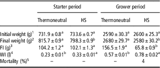
Birds in the HS group had a lower final weight and feed intake and a greater water intake than broilers in the thermoneutral group. Mortality was measured during the 24-h thermal comfort period and the 24-h stress period at each age (21 or 42 days). Death was observed in only older birds subjected to HS (Table 2).
Performance of broilers

HS=heat stress; FI=feed intake; WI=water intake.
1 Initial weight: initial weight at day 20 (starter period) or day 41 (grower period).
2 Final weight: final weight at day 20 (starter period) or day 41 (grower period).
3 FI: from day 20 to 21 (starter period) or from day 41 to 42 (grower period).
4 WI: from day 20 to 21 (starter period) or from day 41 to 42 (grower period).
5 There were no deaths from days 20 to 21 (starter period) or from days 41 to 42 (grower period) in the thermoneutral environment.
a,bMean values within a row with different superscript letters are significantly different by Tukey’s test (P<0.05).
Gene expression
The gene expression results in both environments and ages are shown in Figures 1 and 2.
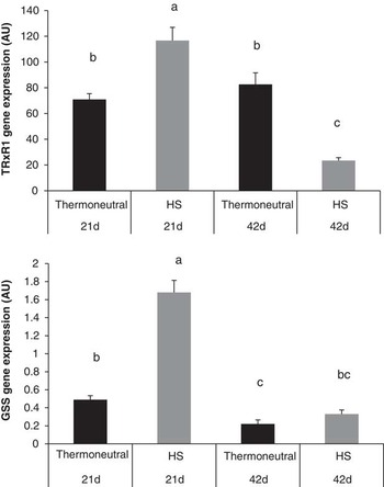
Thioredoxin reductase 1 (TrxR1) and glutathione synthetase (GSS) gene expression in the liver of broilers at 21 and 42 days of age in the thermoneutral and heat stress (HS) groups. The results are shown as the mean and SE. a,b,cDifferent letters between treatment groups represent a significant difference (P<0.05).

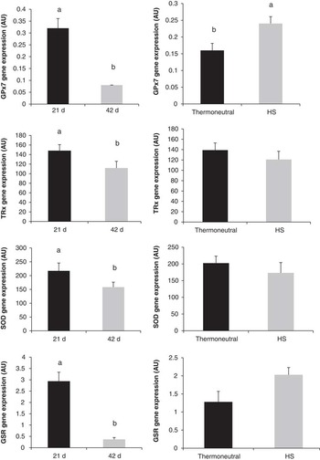
Glutathione peroxidase 7 (GPx7), thioredoxin (Trx), superoxide dismutase (SOD) and glutathione reductase (GSR) gene expression in the liver of broilers at 21 and 42 days of age in the thermoneutral and heat stress (HS) groups. The results are shown as the mean and SE. a,bDifferent letters between treatment groups represent a significant difference (P<0.05).

We observed a significant effect for the environment×age interaction on TrxR1 (P<0.0001) and GSS (P<0.0001) gene expression (Figure 1). Twenty-one-day-old animals exposed to HS showed the highest expression of these genes.
Age influenced Trx (P=0.0090), SOD (P=0.0194), GSR (P<0.0001) and GPx7 (P<0.0001) gene expression (Figure 2); we observed greater expression in 21-day-old birds than in 42-day-old birds. Regarding the environment, animals in the thermoneutral group had lower GPx7 gene expression than those exposed to HS.
Markers of oxidative stress and antioxidant enzyme activity
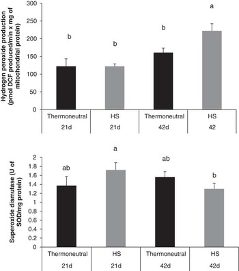
We observed an interaction effect for environment×age on H2O2 production (P=0.0380) and SOD activity (P=0.0391) (Figure 3). Forty-two-day-old birds subjected to HS showed the highest production of H2O2 (222.31 pmol DCF produced/min×mg mitochondrial protein). Twenty-one-day-old birds exposed to HS showed greater SOD activity than 42-day-old birds under the same environmental condition (1.72 v. 1.30 U SOD/mg protein).
Hydrogen peroxide (H2O2) production and superoxide dismutase (SOD) activity in the liver of broilers at 21 and 42 days of age in the thermoneutral and heat stress (HS) groups. The results are shown as the mean and SE. a,bDifferent letters between treatment groups represent a significant difference (P<0.05).

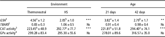
We also verified the effects of age and environment on GSH content (P<0.0001 and P=0.0039, respectively) and CAT enzyme activity (P=0.0007 and P=0.0004, respectively). Higher GSH content and lower CAT activity were observed in the animals in the thermoneutral group compared with those in the HS group and in animals at 21 days compared with those at 42 days. No effect of age or environment was observed on TBARS or GPx enzyme activity (Table 3).
Analysis of oxidative stress markers and antioxidant enzyme activity in the liver

HS=heat stress; GSH=glutathione; TBARS=thiobarbituric acid-reactive substances; CAT=catalase; GPx=glutathione peroxidase; H2O2=hydrogen peroxide.
a,bMean values within a row with different superscript letters are significantly different.
**P<0.01, ***P<0.001.
1 Expressed as µg GSH/mg protein.
2 Expressed as nmol TBARS/mg protein.
3 Expressed as H2O2 consumed/min×mg protein.
4 Expressed as NADPH oxidized/min×mg protein.
Plasma analyses
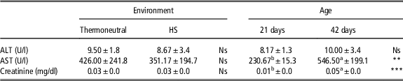
The effects of age and environment on particular factors in plasma from broilers are shown in Table 4. Among the variables analyzed in this study, only age (P<0.0001) influenced creatinine levels and AST enzyme activity. Forty-two-day-old chickens had higher creatinine content (0.05 v. 0.01 mg/dl) and higher AST activity (546.50 v. 230.67 U/l) than 21-day-old chickens. There was no effect of age or environment on ALT activity.
Plasma analysis of alanine aminotransferase (ALT), aspartate aminotransferase (AST) activity and creatinine content in broilers

HS=heat stress.
a,bMean values within a row with different superscript letters are significantly different.
**P<0.01 ***P<0.001.
Discussion
Increased production of mitochondrial ROS, which occurs with advanced age (Cui et al., Reference Cui, Kong and Zhang2012) and in HS-exposed animals (Mujahid et al., Reference Mujahid, Sato, Akiba and Toyomizu2006), is related to greater oxidative damage of macromolecules and to depletion of energy production machinery. Hydrogen peroxide, a type of ROS, is synthesized via a dismutation reaction catalyzed by SOD (Gutteridge and Mitchell, Reference Gutteridge and Mitchell1999); in the presence of reactive metals, H2O2 can participate in OH synthesis, and H2O2 can be converted into water by CAT and GPx (Oktyabrsky and Smirnova, Reference Oktyabrsky and Smirnova2007; Nishikawa et al., Reference Nishikawa, Hashida and Takakura2009).
Increased ROS production has been consistently observed in broilers subjected to acute (Lin et al., Reference Lin, Decuypere and Buyse2006) or chronic HS (Yang et al., Reference Yang, Tan, Fu, Feng and Zhang2010). Consistent with data from the literature, we observed in this study that 42-day-old animals exposed to HS had the highest H2O2 production. Cellular ROS production occurs normally during ATP production; however, this production increases with advanced age, thus creating a vicious cycle (Tengan et al., Reference Tengan, Gabbai and Moraes1998). Younger animals exposed to HS showed H2O2 production similar to that in animals that remained in a thermoneutral environment. This result may be due to the greater antioxidant capacity of younger animals, as evidenced by the expression of the evaluated genes and the SOD enzyme activity results: 21-day-old animals exposed to HS showed the highest gene expression and the highest activity of this antioxidant enzyme.
In addition to SOD (Aluwong et al., Reference Aluwong, Kawu, Raji, Dzenda, Govwang, Sinkalu and Ayo2013), an organism utilizes several antioxidant defense mechanisms, including non-enzymatic and enzymatic mechanisms such as those based on the actions of CAT and the GSH systems (Mahmoud and Edens, Reference Mahmoud and Edens2003), to avoid further damage. Glutathione is involved in a variety of biological actions and has a key role in defense against free radicals (Morand et al., Reference Morand, Rios, Moundras, Besson, Remesy and Demigne1997). Glutathione biosynthesis occurs in most tissues and is dependent on the consecutive action of two enzymes, γ-glutamylcysteine synthetase and GSS (Huber et al., Reference Huber, Almeida and Fátima2008). In our work, we observed that animals exposed to HS, which induced greater H2O2 production, also showed higher expression of the GSS gene. However, unlike the results observed for H2O2 production, younger animals showed the highest GSS expression, suggesting that younger animals may have an increased ability to synthesize GSH when needed, as in the case of HS.
The GSH system is composed of GSH, GPx and GSR (Huber et al., Reference Huber, Almeida and Fátima2008). Heat stress has been shown to be one of several factors that affect the activity of these enzymes (Pamok et al., Reference Pamok, Aengwanich and Komutrin2009). This previous study found increased activity in animals exposed to high temperature and suggested that this may be an attempt to eliminate ROS, which were at higher levels under this condition. Accordingly, we observed effects of not only environment (animals exposed to HS had higher GPx7 mRNA expression) but also age on GSR and GPx7 gene expression. We observed increased expression of these genes in animals at 21 days of age, potentially suggesting higher antioxidant capacity in younger animals.
A deficiency in antioxidant mechanisms is linked to cellular damage, including DNA damage, greater lipid peroxidation and increased protein oxidation, such as the formation of disulfides (Willemsen et al., Reference Willemsen, Swennen, Everaert, Geraert, Mercier, Stinckens, Decuypere and Buyse2011). Glutathione, besides directly combatting H2O2 through the action of GPx enzymes, is also related to the protection of proteins by a mechanism known as glutathionylation. This mechanism describes the formation of a disulfide bond between a thiol group of a protein and a thiol group of GSH, resulting in protein-S-S-glutathione. This disulfide can be reduced by GSH-dependent thioltransferase enzymes known as glutaredoxins. Thus, glutathionylation protects proteins by preventing the formation of protein–protein disulfides and protects against other types of protein oxidation that are irreversible (Lou, Reference Lou2003; Ghezzi, Reference Ghezzi2013).
The cellular ratio of reduced to oxidized GSH (GSH/glutathione disulfide (GSSG)) is an important parameter that provides information regarding the redox state of an organism and should be >10 (Wu, Reference Wu2013). In our work, to verify the redox condition of the birds, we determined that animals that remained in the thermoneutral environment had a higher hepatic GSH concentration than those that were exposed to HS. Younger animals had a higher GSH concentration than older animals. This suggests that under these conditions, an increased amount of GSH is in the oxidized form, possibly due to failures in mechanisms for generating GSSG.
Another enzymatic complex involved in protecting proteins is the Trx complex (Koháryová and Kollárová, Reference Koháryová and Kollárová2008). Thioredoxin acts specifically in the reduction of protein–protein disulfides and protects against the formation of sulfonic acid from cysteine residues; both processes are responsible for changes in protein activity (Lou, Reference Lou2003). In the protein reduction reaction, Trx is oxidized, and its reduction is dependent on TrxR at the expense of NADPH (Tan et al., Reference Tan, Greetham, Raeth, Grant, Dawes and Perrone2010). In our work, we observed increased TRx and TRxR1 gene expression when animals were exposed to HS and had H2O2 production increased. However, we must emphasize that the highest TRxR1 gene expression was observed in HS-exposed broilers at 21 days of age. This result suggests that younger animals may have greater antioxidant GSH complex activity, in addition to greater Trx complex activity, to eliminate H2O2. This result was not observed in 42-day-old animals.
We assessed plasma AST enzyme activity as a marker of oxidative stress-induced damage because studies have shown that AST has increased activity in serum under conditions of injury (Khan et al., Reference Khan, Alhomida, Sobki, Habib, Aseri, Khan and Moghairi2013); creatinine concentration was also assessed as a parameter related to the failure of some organs and to increased protein degradation (Fernandes and Larsson, Reference Fernandes and Larsson2000). The analysis of these markers corroborated our gene expression and enzymatic activity findings; increased AST activity and increased creatinine content were observed in 42-day-old animals, an age at which we noted lower performance in the evaluated antioxidant mechanisms.
Another interesting finding from our work that is contrary to the data presented above relates to CAT enzyme activity, which, in addition to the complex formed by GSH, GPx and GSR, eliminates H2O2 from the cell (Nishikawa et al., Reference Nishikawa, Hashida and Takakura2009). Unlike the results from previous studies, we observed increased CAT activity in animals that were exposed to stress and in older birds but not in 21-day-old birds. Our results suggest that under HS conditions, in which there is more H2O2 production, 21-day-old broilers have a greater antioxidant capacity than 42-day-old broilers. Younger animals showed greater expression of genes related to antioxidant activity, greater SOD activity, and more robust activity of enzymes used as stress markers, all of which could be due to greater antioxidant capacity. These findings highlight some of the differences between birds of different ages when they are exposed to thermal stress. Thus, these results may help reduce losses in poultry production due to heat because they provide new possibilities for improving broiler chicken production; a better understanding of the mechanisms involved in combating stress will potentially enable the determination of strategies for preventing and eliminating damage.
Acknowledgments
This study was supported by the National Council for Scientific and Technological Development – CNPq.