Introduction
The common vole, Microtus arvalis (Pallas, 1778) is a pest species of the agroecosystems of Central Europe. In this region and habitat, there are generally fluctuating populations. As the dominant rodent found in fields, it is also an important host and reservoir species of various pathogens and an obvious target for parasitological research (Erhardova, Reference Erhardova1958; Rosicky, Reference Rosicky and Kratochvil1959; Tenora, Reference Tenora1972, Reference Tenora1976; Prokopic, Reference Prokopic1973). Heligmosomum costellatum (Heligmosomidae: Trichostrongyloidea; Dujardin, 1845) is a nematode parasite with a direct life cycle (without intermediate hosts) with three free-living larval stages and the parasitic fourth stage to adult form living in the lumen of the small intestine (Brown et al., Reference Brown, Macdonald, Tew and Todd1994; N'Zobadila et al., Reference N'Zobadila, Boyer and Durette-Dess1996). Some previous studies of H. costellatum have not only revealed its frequent occurrence in common voles (Holisova & Kocis, Reference Holisova and Kocis1955; Rosicky, Reference Rosicky and Kratochvil1959; Gabriel, Reference Gabriel1972; Prokopic, Reference Prokopic1972), but also that Heligmosomum is the only genus observed so far in the common vole population in Central Europe (Tenora et al., Reference Tenora, Barus, Hildebrand and Prokes2002). Some studies have also dealt with the dynamics of parasitaemia and examined its dependency on host age (Tenora, Reference Tenora1972; Kisielewska & Zubczewska, Reference Kisielewska and Zubczewska1973).
In our research, we have concentrated on one fluctuating common vole population and the dynamics of the H. costellatum prevalence over the course of 2 years. The aim of the study was to evaluate the dependence of H. costellatum prevalence on common vole population dynamics as well as the weight, age, gender and reproduction condition of the common voles.
Materials and methods
The study was conducted on an alfalfa field near the village Drnholec in the Breclav region, Czech Republic (48°53′30″N, 16°27′30″E). A study plot 200 × 900 m in size was chosen in a typical agricultural region of southern Moravia. The landscape was composed of 91% arable land, while woodland represented less than 4% of the area.
The community of small terrestrial mammals (rodents and insectivores) was sampled by snap-trapping from January 2002 until November 2003. Small mammals were captured monthly. Snap-traps were baited with fried pieces of wick for a kerosene lamp (soaked in fat and flour) and exposed in lines of 100 traps 3 m apart for one night. The species of every captured animal was first determined, after which all trapped mammals were weighed, measured, sexed and dissected to obtain information on reproductive status, as well as examined for the presence of parasites and pathological changes. The reproductive state was specified as immature or adult. Animals that had not yet entered into reproduction were designated as immature. Immature females were without placental scars (maculae cyanae; MC) or embryos, and the testes of immature males were less than 8 mm long (usually 3–4 mm long). Adults that had embryos or MC were classified as sexually active females. If the eye lenses were not damaged, the age of animals was determined based on eye lens mass (Janova et al., Reference Janova, Nesvadbova and Tkadlec2007a). The dominant species, common vole (Microtus arvalis), was subjected to helminthological examination and H. costellatum prevalence and dynamics were investigated.
The abundance of voles was expressed as the number of captured animals per 100 trap-nights (relative abundance, rA). In order to restore the original variation in the data that was presumably lost due to the ‘saturation effect of the traps’ (i.e. occupied traps cannot capture other animals), the index was transformed by assuming a random (Poisson) distribution of voles, i.e. transformed index = [ − ln(1 − index/100)] × 100 (Caughley, Reference Caughley1977).
During the necropsy, whitish miliary nodular lesions were observed in the stomach wall. The occurrence of parts of parasite (H. costellatum) bodies was observed (fig. 1). Only larvae encapsulated in the stomach wall were monitored and examined. As these larvae can also occur in the wall of the small intestine, our data are relative indicators of parasitic prevalence. Encapsulated larvae were examined randomly (at least one in each group) and identified microscopically. All the individuals were members of H. costellatum species as identified by the key published by Tenora et al. (Reference Tenora, Barus, Hildebrand and Prokes2002) and N'Zobadila et al. (Reference N'Zobadila, Boyer and Durette-Dess1996). Other larvae were determined without extraction from cysts and it was assumed that they were conspecific. The intensity of infection in the stomach wall ranged from one to six nodules containing parasites. Further analysis was focused on the prevalence of H. costellatum in common voles, although the number of parasites in the host animal was not evaluated.
Larvae of H. costellatum localized in deep parts of the gastric mucosa with a predominantly lymphoplasmocytic inflammatory reaction surrounding the parasites. Haematoxylin and eosin; original magnification × 100.

Analysis of seasonal influence and individual vole variables on the occurrence of H. costellatum was carried out using the binomial multifactor module Generalized linear/nonlinear models (GLZ) in the program Statistica version 5 (StatSoft Inc., Tulsa, Oklahoma, USA). The following were analysed as continuous predictors: date of trapping, rA, weight of individual animals, age according to eye lens; and categorical factors: year, status of reproduction and gender. The level of statistical significance was stated as α = 0.05 and α = 0.1 to allow us to determine the slightest of tendencies. The date of trapping was recorded as the number of days since the first day of the year.
Results
Nodules containing distinct parts of the parasite body (H. costellatum) were commonly found in the gastric mucosa, in the upper parts as well as in deeper layers. The surrounding tissue was evidently compressed by encapsulated larvae. The presence of H. costellatum in stomach tissue was restricted to the mucosa, resulting in local disruptions and slight hyperplasia of the mucous cells, with a limited inflammatory response represented by lymphocytes and eosinophilic granulocytes surrounding the parasite. There was no evidence of haemorrhaging. In a few cases, the nematodes elicited a chronic granulomatous response with focal destruction of gastric glands. Worms were not observed to penetrate the muscularis mucosae.
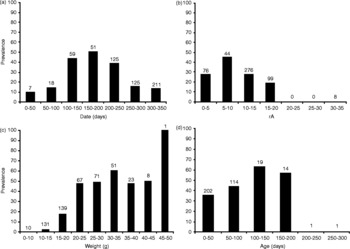
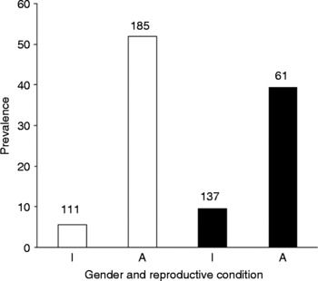
Of the 503 voles examined, 139 (27.6%) were detected as being H. costellatum positive. Analysis of the influence of age was carried out on 351 animals, where undamaged eye lenses were available. The date of capture, weight, year, reproduction and interactions year × gender and year × reproduction influenced the prevalence of H. costellatum in the common vole population (figs 2 and 3). Gender, rA and interactions of other factors (except the former) did not affect the presence of H. costellatum.
Influence of (a) the trapping date; (b) relative abundance (rA); (c) body weight and (d) host age on the prevalence (%) of H. costellatum. The sample size is shown above each column.

Influence of gender and reproductive condition on the prevalence (%) of H. costellatum: I, immature; A, adult; white bars, female; black bars, male. The sample size is shown above each column.

A slightly higher prevalence of H. costellatum was found in older animals (fig. 2). A disproportionally higher parasite prevalence was observed in 2002, when 42.23% of animals (n = 251) were infected with H. costellatum, compared with 17.06% (n = 252) in 2003. Older animals and adults of higher weight were most often infected (fig. 2). Animals were mostly infected between April and September. Significant interactions between gender and year and and between reproductive state and year in 2002 are probably attributable to females (especially adult ones) which were mostly H. costellatum positive, while in 2003 the majority of adult females were H. costellatum negative.
Discussion
We found a significant relationship between the prevalence of H. costellatum and vole body size, age and reproductive status. Parasite prevalence was also affected by the year and trapping date of the animal.
The intestinal parasite H. costellatum did not penetrate the muscularis mucosae in our tissue samples. A very low level of mechanical damage to the gastric mucosa was observed, along with the absence of, or only a very mild, inflammatory reaction. Histopathologically, we observed focal erosions, hyperaemia, mild fibrosis of mucosa, and lymphocytic and eosinophilic inflammatory infiltrates surrounding the encapsulated parasites. Similar pathomorphological lesions were also found in the intestines. Thus, in the majority of the infected animals, H. costellatum was observed as an example of a parasite that causes temporary infection with a relatively low pathogenic impact on the host. In general, the absence of an inflammatory response or moderate reaction in a host tissue could also be explained by a short attachment time of the parasite to host cells. In the case of our samples, this explanation was unlikely because no strong correlation was noted in any of the examined animals.
Demographic parameters of the host vole population affected the prevalence of H. costellatum. A higher prevalence of H. costellatum was observed in adult animals of a larger body size, animals captured during the reproductive season (from April until the end of August) and in 2002. It should be mentioned that the reproductive state, age and date (time of the trapping) correlated well with the body weight data, due to the fact that reproductively active animals are heavier (Zejda, Reference Zejda1971, Reference Zejda1992; Markowski, Reference Markowski1980; Janova et al., Reference Janova, Nesvadbova and Tkadlec2007a, Reference Janova, Havelkova and Tkadlecb).
A significant effect of the interaction of gender and the year was demonstrated by intensive reproduction and consequently high abundances in 2002. Higher prevalence of females in 2002 than in 2003 indicated that the prevalence of different genders can also vary annually (Heroldova et al., Reference Heroldova, Janova, Bryja and Tkadlec2005; Janova et al., Reference Janova, Heroldova and Bryja2008). Reproducing females usually shared their burrows with daughters and other related females (Mackin-Rogalska, Reference Mackin-Rogalska1979; Boyce & Boyce, Reference Boyce and Boyce1988a). As a consequence, the probability of transmission of infection is higher. In contrast, the home ranges of males are larger and their mobility is greater in comparison with that of females (Boyce & Boyce, Reference Boyce and Boyce1988b; Zejda et al., Reference Zejda, Zapletal, Pikula, Obdrzalkova, Heroldova and Hubalek2002). This can lead to a higher infection prevalence in female residents than in migrating males. Although the effect of host density might operate with a time-lag, we should mention that the infection with H. costellatum was affected more by the year than by immediate rA. It is probable that other environmental factors (biotic or abiotic) may have had some influence.
Even though high host density leads to a higher prevalence of various diseases (Zejda et al., Reference Zejda, Zapletal, Pikula, Obdrzalkova, Heroldova and Hubalek2002), we found a lower prevalence of H. costellatum in autumn when the highest density of vole populations occurs. Previous studies have stipulated that the higher parasite prevalence is common during the reproductive period, which ends in September in Central Europe (Tenora, Reference Tenora1972; Kisielewska & Zubczewska, Reference Kisielewska and Zubczewska1973). An alternate explanation is also possible. In autumn, vole populations consist mostly of young animals. On the other hand, adults, which are sexually active in the first half of the reproductive season, mostly comprise the over-wintered animals.
It is known that during the winter months voles have a tendency to share nests. Therefore, H. costellatum infection can be spread in this cohort easily (Prokopic, Reference Prokopic1972; Tenora, Reference Tenora1972; Tenora & Zejda, Reference Tenora and Zejda1974). This reflects the higher prevalence of animals caught until August, the time when the last hibernating animals die. The ascertainment of the true age of animals (based on the eye lens mass) revealed that age had a marginal effect on the prevalence of H. costellatum infection. However, this finding is not conclusive as only undamaged eye lenses from a relatively low number of age-determined animals were examined. Kisielewska & Zubczewska (Reference Kisielewska and Zubczewska1973) observed that H. costellatum was more prevalent among older (heavier) animals.
Acknowledgements
This study was supported by projects nos. GA CR 521/08/P529 and MZE 0002716202 (Ministry of Agriculture of the Czech Republic) and AdmireVet CZ 1.05/2.1.00/01.0006 (Ministry of Education, Youth and Sports of the Czech Republic). The work complied with Council directive 86/609/EEC regulations on the experimental use of animals. We would like to thank Neysan Donnelly (University of Aberdeen, UK) and Maria Vass (Scientific Editing Services, m_vass@hotmail.com) for critical grammatical checking of the article.