




No CrossRef data available.
Article contents
Characterization of excretory–secretory products from protoscoleces of Echinococcus granulosus and evaluation of their potential for immunodiagnosis of human cystic echinococcosis
Published online by Cambridge University Press: 23 August 2004
Abstract
This study describes, for the first time, the characterization of excretory–secretory antigens (ES-Ag) from Echinococcus granulosus protoscoleces, evaluating their usefulness in the immunodiagnosis of human cystic echinococcosis. ES-Ag were obtained from the first 50 h maintenance of protoscoleces in vitro. This preparation contained over 20 major protein components which could be distinguished by 1-dimensional SDS–PAGE with apparent masses between 9 and 300 kDa. The culture of of protoscoleces from liver produced a greater variety of excretory–secretory protein components than those from lung. Determination of enzymatic activities of secreted proteins revealed the presence of phosphatases, lipases and glucosidases, but no proteases. These findings were compared to those obtained from somatic extracts of protoscoleces and hydatid cyst fluid products. Immunochemical characterization was performed by immunoblotting with sera from individuals infected by cystic echinococcosis (n=15), non-hydatidic parasitoses (n=19), various liver diseases (n=24), lung neoplasia (n=16), and healthy donors (n=18). Antigens with apparent masses of 89, 74, 47/50, 32, and 20 kDa showed specificity for immunodiagnosis of human hydatidosis. The 89 and 74 kDa components corresponded to antigens not yet described in E. granulosus, whereas proteins of 41–43 kDa and 91–95 kDa were recognized by the majority of the non-hydatid sera studied.
Keywords
Information
- Type
- Research Article
- Information
- Copyright
- © 2004 Cambridge University Press
INTRODUCTION
Unilocular hydatidosis, caused by the metacestode Echinococcus granulosus, is an important human and animal health concern worldwide, as well as the cause of major economic losses. In humans, the early detection of the disease is very important, since it allows initiation of chemotherapy or surgical removal of the cyst. Currently, diagnosis is based on a combination of imaging techniques (ultrasonography, computerized axial tomography, X-rays) and immunodiagnostic methods such as ELISA and immunoblotting (WHO, 2001; Ortona et al. 2003; Zhang, Li & McManus, 2003). Historically, hydatid cyst fluid (HCF) has been used as a main antigenic source for the serodiagnosis of the disease, with the two components of major diagnostic relevance identified as antigen 5 (Capron, Vernes & Biguet, 1967) and antigen B (Oriol et al. 1971). Although, initially, both antigens were considered specific to E. granulosus, later studies demonstrated cross-reactivity with sera from individuals infected by other helminths, mainly E. multilocularis and Taenia solium (Lightowlers et al. 1989; Legatt, Yang & McManus, 1992). These observations, together with the variability of sources of HCF and the inclusion of serological components from the host, represent complicating factors for immunodiagnosis of the disease.
In order to improve the specificity and the sensitivity of diagnosis of human cystic echinococcosis, several strategies have been followed, such as purification of antigens 5 and B or utilization of other HCF antigens. Additionally, protoscolex-derived crude antigen preparations (Craig, Zeyhle & Romig, 1986; Rafiei & Craig, 2002) as well as recombinant antigens and synthetic peptides (Zhang et al. 2003) have been used in the sero-diagnosis of the disease. Nevertheless, results are highly variable dependent on the sera, antigen, and laboratory (Ortona et al. 2003). It is, therefore, important to consider the characterization of new antigens with greater specificity and enhanced diagnostic sensitivity. In this paper, we describe for the first time the biochemical characterization of excretory–secretory antigens from protoscoleces of E. granulosus (ES-Ag), and evaluate their potential for immunodiagnosis of human cystic echinococcosis.
MATERIALS AND METHODS
Hydatid cyst fluid antigens (HCF)
HCF was obtained as described by Varela-Díaz et al. (1974) from liver and lung cysts of sheep origin. The HCF was centrifuged at 2000 g for 45 min, then passed through a Millipore AP20 filter (Bedford, USA) and dialysed against distilled water, using dialysis tubing with a cut-off of 5 kDa (Medicell, London, UK). Finally, it was centrifuged at 6500 g for 30 min, lyophilized and stored at 4 °C. All batches were analysed by SDS–PAGE and stained with Coomassie Brilliant Blue R-250. To perform this study a batch from sheep lung hydatid fluid was used.
Somatic antigens (S-Ag)
Somatic antigen was also prepared from protoscoleces obtained by aseptic puncture from hydatid cysts of ovine origin. The protoscoleces were washed with phosphate-buffered saline (PBS) and stored at −25 °C with proteolytic enzyme inhibitors (2 mM phenyl methyl sulfonyl fluoride (PMSF) and 5 mM EDTA). Protoscoleces were thawed and sonicated (10 cycles of 12 s at 60 Hz frequency), freeze-thawed once more and centrifuged for 35 min at 2300 g. Supernatant fractions were aliquotted and stored at −25 °C. To perform this study a sheep liver batch was selected, with protoscoleces of 97·4% viability at the time of their extraction from the cyst.
Excretory–secretory antigens (ES-Ag)
To obtain excretory–secretory products, protoscoleces with viability higher than 90% were selected. Viability was assessed by morphological appearance, flame cell motility and general contractile movements (Smyth & Davies, 1974; Howell, 1986). Protoscoleces were cultured in PBS complemented with 10% glucose, 100 U/ml penicillin and 100 μg/ml streptomycin at 37 °C in 5% CO2, which promoted parasite survival for several days (Carmena et al. 2002). The medium was renewed every 8 h and concentrated by Ultrafree 15 filters with a 5 kDa pore diameter membrane (Millipore). EDTA (5 mM) and PMSF (2 mM) were added, and the ES products aliquotted and stored at −20 ° C. Protein concentration was determined by the bicinchoninic acid method (Sigma). In total, 10 cultures of protoscoleces from sheep were carried out, 8 from liver and 2 from lung. Medium corresponding to the first 48–64 h of culture was subsequently used. The concentration of proteins obtained for each preparation was: ES-Ag: 0·2 mg/ml; S-Ag: 2·95 mg/ml; HCF: 736 mg/g dry weight.
Enzymatic activities
Parasite preparations (HCF, S-Ag and ES-Ag) were screened for selected enzymatic activities by means of the Api-Zym System (Bio Mérieux, Marcy-l'Etoile, France), in accordance with manufacturer's instructions. Nineteen enzymes were assayed: acid phosphatase, esterase, esterase lipase, lipase, leucine aminopeptidase, valine aminopeptidase, cysteine aminopeptidase, trypsin, chymotrypsin, phosphoamidase, α-galactosidase, β-galactosidase, β-glucuronidase, α-glucosidase, β-glucosidase, β-N- acetylglucosaminidase, α-mannosidase, α-fucosidase, and alkaline phosphatase. Either 65 μl of each preparation (ES-Ag at 0·2 mg/ml; S-Ag and HCF at 1 mg/ml) were added to each well of the Api-Zym tray, containing the appropriate freeze-dried buffer and naphthol-coupled substrate. The tray was incubated at 37 °C for 4 h. After incubation, the free naphthol released from the substrate was detected by coupling with equal volumes (33 μl) of fast blue BB (0·35% wt[ratio ]vol in 2-methoxyethanol) and 2 M Tris buffer, pH 7·6, containing 10% wt[ratio ]vol of sodium dodecyl sulphate. The amount of product formed was semi-quantitatively assessed in accordance with the colour chart supplied with the system.
SDS–PAGE and immunoblotting
Proteins were separated by SDS–PAGE (8, 12·5 and 16% acrylamide) under reducing conditions according to the method of Laemmli (1970) and silver stained. Resulting protein patterns were studied by image analysis with the Bio-Image System (Millipore). For immunoblotting, proteins were transferred to polyvinylidene difluoride (PVDF) membranes (Immobilon-P, Millipore), essentially according to the method described by Towbin, Staehelin & Gordon (1979). Primary sera were used at a dilution of 1[ratio ]800 in 20 mM Tris-buffered saline (pH 7·4), 8% skimmed milk (TBS-M) and the membranes incubated overnight at 4 °C. Peroxidase-conjugated rabbit anti-human IgG (Dako, Copenhagen, Denmark) was used at a dilution of 1[ratio ]1000 in TBS-M buffer for 4 h at room temperature, and binding was visualized with 4-chloro-1-naphthol. A peroxidase-conjugated swine anti-rabbit total Ig (Dako) was used as secondary antibody in experiments with the hyperimmune rabbit sera under the same conditions.
Human sera
Fifteen pre- and post-surgery sera from 11 individuals with confirmed liver hydatidosis were assayed. Another 19 sera were used from individuals with other parasitoses confirmed by coprological examination. Of these, 8 were infected with a single parasite, whereas the other 11 were parasitized by 2 different species, namely Endolimax nana, Entamoeba coli, Entamoeba histolytica, Blastocystis hominis, Giardia lamblia, Ascaris lumbricoides, Toxocara spp. and Taenia spp. In addition, 40 sera from individuals with non-parasitological pathologies were also used. Of these, 24 corresponded to individuals suffering from various liver diseases (abscess, hepatitis, neoplasias, carcinomas, cholangitis, cholestasis and jaundice), while the other 16 belonged to individuals with lung neoplasia. As has previously been established, some of these diseases may provide false positive results in hydatid serology (Guisantes, 1979). Finally, we used 18 sera from individuals with no evidence of bacterial, viral or parasitic infections, obtained from blood banks and voluntary donors from our laboratory.
Hyperimmune rabbit sera anti-ES-Ag
A polyclonal immunoserum anti-ES-Ag was obtained according to the method described by Gallart et al. (1985). Titration of rabbit antiserum was performed by ELISA, as described by Nieto & Carbonetto (1989), using ES-Ag as solid phase.
RESULTS
Comparison of ES-Ag protein patterns of liver and lung cysts
Two cultures of protoscoleces from liver cysts (EH1 and EH2) and 2 from lung cysts (EP1 and EP2) were prepared simultaneously. Comparison of protein profiles by 12·5% SDS–PAGE corresponding to the first 50 h of in vitro maintenance indicated that culture of protoscoleces from liver produced a greater variety of excretory-secretory protein components than those from lung (Fig. 1). Analysis by the Bio-Image System (Millipore) showed that liver protoscoleces released detectable proteins with apparent molecular masses between 10 and 98 kDa, whereas lung protoscoleces released proteins within a range of 10 and 111 kDa. In both cases the profile was consistent, with major products including a protein triplet of 55/65 kDa and 3 polypeptides of 32–34, 21 and 14–16 kDa. In protoscoleces isolated from lung cysts an additional major component was a doublet of 49/51 kDa. In addition, ES products from the other two cultures of protoscoleces (FH1 and GN2) from hepatic cysts were also analysed by 12·5% SDS–PAGE, showing major products of 90, 69, 21, 16 and 13 kDa, including doublets of 43/47, 35/38 and 29/31 kDa. The excretory–secretory products of 4 cultures of protoscoleces from hepatic cysts (GN1, GN2, GP1 and GP2) were analysed by 8% SDS–PAGE to identify the high molecular mass proteins. The protein profiles were very similar, with prominent bands at approximately 325, 230, 170, 137, 100, 81 and 50 kDa. Use of 16% gels (with the same cultures) allowed resolution of smaller proteins. Again, the profile was consistent through the duration of culture, with major products of 16·5, 12 and 10 kDa (data not shown).

Fig. 1. SDS–PAGE (12·5%) of the excretory–secretory products obtained during the first 50 h of culture of protoscoleces. (A) Culture EH2 (liver). (B) Culture EP1 (lung). M, Molecular mass marker, expressed in kDa. Lane 1, 13 h; Lane 2, 25 h; Lane 3, 37 h; Lane 4, 43 h; Lane 5, 50 h.
Enzymatic activities
Parasite preparations were screened for a variety of enzymatic activities using an assay system which allowed a semi-quantitative analysis. This is shown in Table 1, demonstrating that a range of phosphatases and lipases were present in the ES-Ag, S-Ag and HCF. Of the 5 proteases screened, only leucine aminopeptidase could be detected in the S-Ag and HCF. Of the 8 glucosidases, only β-galactosidase was present in all 3 preparations, with highest activity in HCF. ES-Ag was also positive for α-galactosidase and N-acetyl-β-glucosaminidase activity. In general, however, phosphatases were most prominent in all preparations, with average values of 22, 37, and 40 nanomoles of hydrolysed substrate in ES-Ag, S-Ag and HCF, respectively.
Table 1. Semi-quantitative assessment of enzymatic activities in different parasite extracts, expressed in nanomoles of hydrolysed substrate (ES-Ag: excretory–secretory products of protoscoleces; S-Ag: somatic extract of protoscoleces; HCF: hydatid cyst fluid.)

Immunoblotting
Figure 2 shows an analysis of recognition of individual components of ES-Ag with the sera from hydatid patients. The human sera identify 9 major components, those of 113, 89 and 67 kDa and the doublet 47/50 kDa standing out. Fig. 3 shows the antigenic profile of the ES-Ag recognized with sera from individuals with non-hydatid parasitoses, non-parasitic pathologies that can be confused with hydatidosis in the immunodiagnosis and healthy donors. Proteins of 91–94 kDa and 41–43 kDa were identified by many of these sera, indicating their lack of appropriate specificity for diagnostic purposes. Table 2 summarizes the data obtained by immunoblotting.

Fig. 2. Immunoblot analysis of 15 serum samples from patients with cystic echinococcosis with ES-Ag. M, Molecular mass marker, expressed in kDa.

Fig. 3. Immunoblot analysis with ES-Ag. (A) Human sera corresponding to individuals with non-hydatid parasitoses. M, Molecular mass marker, expressed in kDa. Lane 1, E. nana+E. coli; Lane 2, E. nana+E. histolytica; Lane 3, A. lumbricoides+E. histolytica+E. coli; Lane 4, E. coli+A. lumbricoides; Lane 5, B. hominis; Lane 6, E. histolytica+E. coli; Lane 7, G. lamblia; Lane 8, G. lamblia+E. coli; Lanes 9 and 10, E. nana; Lane 11, E. histolytica+E. coli; Lane 12, E. histolytica; Lane 13, E. histolytica+G. lamblia; Lane 14, E. histolytica+E. coli; Lane 15, E. histolytica+G. lamblia; Lane 16, Taenia spp; Lane 17, G. lamblia; Lane 18, E. histolytica+E. coli; Lane 19, A. lumbricoides; Lane 20, Taenia spp; Lane 21, Toxocara spp. (B) Human sera corresponding to healthy donors. (C) Human sera corresponding to individuals with liver diseases. (D) Human sera corresponding to individuals with lung neoplasia. M, Molecular mass marker, expressed in kDa.
Table 2. Components of excretory–secretory products of protoscoleces recognized by different sera by immunoblotting (The percentage of sera which bound each antigenic component is expressed in parentheses. Molecular masses are expressed in kDa. D, doublet.)

DISCUSSION
For immunodiagnosis of human cystic echinococcosis, the major antigenic source used thus far has been hydatid fluid, specifically antigen 5 and antigen B. The diagnostic relevance of subunits of these antigens has been reviewed by several authors (Lightowlers & Gottstein, 1995; Zhang & McManus, 2003). On the other hand, protoscolex somatic extracts have been used for immunodiagnosis of canine echinococcosis (Gasser, Lightowlers & Rickard, 1989, 1991; Gasser et al. 1992; Benito et al. 2001) and for identification of sheep infected with E. granulosus (Craig et al. 1981; Kittelberger et al. 2002). In order to improve diagnostic sensitivity and specificity, a third preparation constituted by ES products of protoscoleces has been assayed for the diagnosis of human alveolar echinococcosis (Auer, Hermentin & Aspöck, 1988) as well as canine echinococcosis (Gasser et al. 1992; Benito et al. 2001). Nevertheless, as this preparation has been barely studied for E. granulosus, we present a detailed characterization of ES-Ag from protoscoleces, highlighting its potential for immunodiagnosis of human cystic echinococcosis.
The ES-Ag used for this study was obtained from the first 50 h of culture of protoscoleces, as parasites maintained for this time exhibit a high survival rate (>85%), assuring minimal contamination with somatic products (Carmena et al. 2002). Analysis by SDS–PAGE revealed very similar protein profiles between different cultures, with major protein components with apparent masses of 90, 69, 34, 21, 16 and 13 kDa, as well as doublets of 43/47, 35/38 and 29/31 kDa, and a triplet of 55/65 kDa. ES-Ag from liver cyst protoscoleces had a greater variety of protein components than those of lung origin. It is unclear why this should be the case, but we assume that different environmental pressures stimulate parasites to secrete different products. Interestingly, this phenomenon has also been observed for hydatid fluid from liver and lung cysts (Varela-Díaz et al. 1974; Guisantes, 1979).
With regard to the current study, polypeptides of 38 and 22 kDa may correspond to subunits of antigen 5, reinforced by suggestions that antigen 5 is produced in the parenchymatous tissue of protoscoleces and excreted through the collecting ducts of the osmoregulation system (Yarzábal et al. 1976; Rickard et al. 1977; Sánchez et al. 1993). Similar protein profiles were observed from secreted products of protoscoleces at different time-periods of culture, with major proteins of 325, 230, 170, 137, 100, 81 and 49, 16·5, 12 and 10 kDa. The 8 kDa subunit corresponding to antigen B was not observed, suggesting that it is synthesized later by protoscoleces or from the metacestode germinal membranes.
Characterization of enzymes from E. granulosus has been addressed by several research groups (Vercelli-Retta et al. 1975; McManus & Bryant, 1995; del Cacho et al. 1996). In the present work we have determined that protoscoleces secrete phosphatases, lipases and glucosidases, but no detectable proteases. Both alkaline and acid phosphatases had already been described in the integument of adult protoscoleces (McManus & Bryant, 1995). Alkaline phosphatase activity has also been found on the surface of the germinal membrane of the hydatid cyst (del Cacho et al. 1996) and as a component of the hydatid cyst fluid (Vatankhan et al. 2003). The only secreted protease detected in the current study was leucine aminopeptidase in HCF, which was undetectable in secreted products of protoscoleces. This fact apparently disagrees with the proposal of Tort et al. (1999) which suggests that proteolytic enzymes excreted by the metacestode may play a role in the growth of cysts in the host tissues.
Secreted products of E. multilocularis protoscoleces have been characterized by immunoblotting using sera from patients with alveolar echinococcosis (Auer et al. 1988). These authors identified antigenic components of 65, 62, 60, 57, 55, 52 and 47 kDa. In our study we have identified at least 8 secreted components from E. granulosus protoscoleces ranging from 20 to 135 kDa, some of which (proteins of 67, 50 and 47 kDa) have similar molecular masses. In order to determine possible cross-reactivity, these products were analysed by immunoblotting with sera from patients with other parasitoses, lung neoplasias and hepatic diseases, as well as healthy donors. The obtained results revealed that there is no cross-reactivity between the human sera positive to E. granulosus and the sera from patients with other parasitoses, neither with the sera from healthy donors.
Sera from patients with other pathologies reacted specifically with a 67 kDa protein which was also recognized by the hydatid sera. This polypeptide was detected by 19% of sera from patients with lung neoplasia and by 67% of sera from patients with various hepatic pathologies. Additional cross-reacting proteins of 41–43 kDa and 91–95 kDa were recognized by 100 and 67% of human sera with other parasitoses, by 84 and 43% of human sera with other pathologies, and by 72 and 44% of human sera from healthy donors. It was difficult to highlight specific recognition patterns attributable to individual infections, possibly due to multiple infections not determined by coproparasitogical examination.
In summary, the ES-Ags from protoscoleces with diagnostic potential have molecular masses of 89, 74, 47/50, 32 and 20 kDa, although the 47 kDa component has been also described for E. multilocularis (Auer et al. 1988). Having reviewed existing data, the 89 and 74 kDa proteins appear to correspond to antigens not described thus far in any E. granulosus extract. Nevertheless, the low number of sera used in this study from patients infected by other taeniids suggests that a more comprehensive analysis should be carried out in order to determine possible cross-reactivity with related parasitic infections. We are, therefore, carrying out these studies, and have initiated a programme to clone and express individual protein components from secreted products of protoscoleces which display both high immunoreactivity and specificity to improve the sensitivity and specificity of immunodiagnosis of human hydatid disease.
The authors are very grateful to Professor Murray E. Selkirk (Department of Biological Sciences, Imperial College, London, UK) for his critical revision of this manuscript. We also thank Dr José Errasti-Alustiza (Surgery Service of Hospital de Txagorritxu, Vitoria, Spain), Dr Luz Estela Alzate-Palacio (Health Service of Risaralda, Colombia), and Dr José Luis Sánchez-Quesada (Biochemistry Department of Hospital Santa Creu I Sant Pau, Barcelona, Spain) for the human sera provided for this study.
References
REFERENCES
AUER, H.,
HERMENTIN, K. &
ASPÖCK, H. (
1988).
Demonstration of a specific Echinococcus multilocularis antigen in the supernatant of in vitro maintained protoscoleces.
Zentralblatt für Bacteriologie, Microbiologie und Hygiene
268,
416–423.Google Scholar
BENITO, A.,
CARMENA, D.,
SPINELLI, P.,
POSTIGO, I.,
MARTÍNEZ, J.,
ESTIBALEZ, J. J.,
MARTÍN DE LA CUESTA, F. &
GUISANTES, J. A. (
2001).
The serological diagnosis of canine echinococcosis by an enzyme immunoassay useful for epidemiological surveys.
Reviews in Parasitology
61,
17–23.Google Scholar
CAPRON, A.,
VERNES, A. &
BIGUET, J. (
1967).
Le diagnostic immuno-électrophorétique de l'hydatidose. In
Le Kyste Hydatique du Foie, pp.
27–40.
SIMEP,
Lyon.
CARMENA, D.,
BENITO, A.,
POSTIGO, I.,
ARTEAGA, J.,
MARTINEZ, J. &
GUISANTES, J. A. (
2002).
Short term culture of protoscoleces to obtain excretory-secretory proteins of Echinococcus granulosus.
Research and Reviews in Parasitology
62,
84–88.Google Scholar
CRAIG, P. S.,
HOCKING, R. E.,
MITCHELL, G. F. &
RICKARD, M. D. (
1981).
Murine hybridoma-derived antibodies in the processing of antigens for the immunodiagnosis of hydatid (Echinococcus granulosus) infection in sheep.
Parasitology
83,
303–317.CrossRefGoogle Scholar
CRAIG, P. S.,
ZEYHLE, E. &
ROMIG, T. (
1986).
Hydatid disease: research and control in Turkana. II. The role of immunological techniques for the diagnosis of hydatid disease.
Transactions of the Royal Society of Tropical Medicine and Hygiene
80,
183–192.Google Scholar
DEL CACHO, E.,
CAUSAPÉ, C.,
SÁNCHEZ-ACEDO, C. &
QUILEZ, J. (
1996).
Cytochemical study of the germinal membrane of the Echinococcus granulosus cyst.
Veterinary Parasitology
62,
101–106.CrossRefGoogle Scholar
GALLART, M. T.,
BLADE, J.,
MARTINEZ, J.,
SIERRA, J.,
ROZMAN, C. &
VIVES, J. (
1985).
Multiple myeloma with monoclonal IgG and IgD of Lambda type exhibiting under treatment, a shift from mainly IgG to mainly IgD.
Immunology
55,
45–47.Google Scholar
GASSER, R. B.,
LIGHTOWLERS, M. W. &
RICKARD, M. D. (
1989).
Identification of protein components of Echinococcus granulosus protoscolex antigens for specific serodiagnosis of Echinococcus granulosus in dogs.
Parasite Immunology
11,
279–291.CrossRefGoogle Scholar
GASSER, R. B.,
LIGHTOWLERS, M. W. &
RICKARD, M. D. (
1991).
Echinococcus granulosus: antigenic proteins in oncospheres and on the surface of protoscoleces identified by serum antibodies from infected dogs.
Research in Veterinary Science
50,
340–345.CrossRefGoogle Scholar
GASSER, R. B.,
JENKINS, D. J.,
HEATH, D. D. &
LAWRENCE, S. B. (
1992).
Use of Echinococcus granulosus worm antigens for immunodiagnosis of Echinococcus granulosus infection in dogs.
Veterinary Parasitology
45,
89–100.CrossRefGoogle Scholar
GUISANTES, J. A. (
1979).
Estudio de diferentes antígenos hidatídicos en relación con el hospedador intermediario y la localización de la larva. Ph.D. thesis. University of Navarra,
Pamplona, Spain.
HOWELL, M. J. (
1986).
Cultivation of Echinococcus species in vitro. In
The Biology of Echinococcus and Hydatid Disease
(ed. Thompson, R. C. A.), pp.
143–163.
George Allen & Unwin,
London.
KITTELBERGER, R.,
REICHEL, M. P.,
JENNER, J.,
HEATH, D. D.,
LIGHTOWLERS, M. W.,
MORO, P.,
IBRAHEM, M. M.,
CRAIG, P. S. &
O'KEEFE, J. S. (
2002).
Evaluation of three enzyme-linked immunosorbent assays for the detection of serum antibodies in sheep infected with Echinococcus granulosus.
Veterinary Parasitology
110,
57–76.CrossRefGoogle Scholar
LAEMMLI, U. K. (
1970).
Cleavage of structural proteins during the assembly of the head of bacteriophage T4.
Nature, London
227,
680–685.CrossRefGoogle Scholar
LIGHTOWLERS, M. W.,
LIU, D.,
HARALAMBOUS, A. &
RICKARD, M. D. (
1989).
Subunit composition and specificity of the major cyst fluid antigens of Echinococcus granulosus.
Molecular and Biochemical Parasitology
37,
171–182.CrossRefGoogle Scholar
LEGGATT, G. R.,
YANG, W. &
McMANUS, D. P. (
1992).
Serological evaluation of the 12 kDa subunit of antigen B in Echinococcus granulosus cyst fluid by immunoblot analysis.
Transactions of the Royal Society of Tropical Medicine and Hygiene
86,
189–192.CrossRefGoogle Scholar
LIGHTOWLERS, M. W. &
GOTTSTEIN, B. (
1995).
Echinococcosis/hydatidosis: antigens, immunological and molecular diagnosis. In
Echinococcus and Hydatid Disease
(ed. Thompson, R. C. A. & Lymbery, A. J.), pp.
355–410.
CAB International,
Oxon.
McMANUS, D. P. &
BRYANT, C. (
1995).
Biochemistry, physiology, and molecular biology of Echinococcus. In
Echinococcus and Hydatid Disease
(ed. Thompson, R. C. A. & Lymbery, A. J.), pp.
135–181.
CAB International,
Oxon.
NIETO, A. &
CARBONETTO, C. H. (
1989).
Enzimoinmunoensayo (ELISA).
In Inmunología e Inmunoquímica
(ed. Margni, R. A.), pp.
571–586.
Editorial Médica Panamericana,
Buenos Aires.
ORIOL, R.,
WILLIAMS, J. F.,
PÉREZ ESANDI, M. V. &
ORIOL, C. (
1971).
Purification of lipoprotein antigens of Echinococcus granulosus from sheep hydatid fluid.
American Journal of Tropical Medicine and Hygiene
20,
569–574.CrossRefGoogle Scholar
ORTONA, E.,
RIGANÒ, R.,
BUTTARI, B.,
DELUNARDO, F.,
IOPPOLO, S.,
MARGUTTI, P.,
PROFUMO, E.,
TEGGI, A.,
VACCARI, S. &
SIRACUSANO, A. (
2003).
An update on immunodiagnosis of cystic echinococcosis.
Acta Tropica
85,
165–171.CrossRefGoogle Scholar
RAFIEI, A. &
CRAIG, P. S. (
2002).
The immunodiagnostic potential of protoscolex antigens in human cystic echinococcosis and the possible influence of parasite strain.
Annals of Tropical Medicine and Parasitology
96,
383–389.CrossRefGoogle Scholar
RICKARD, M. D.,
DAVIES, C.,
BOUT, D. T. &
SMYTH, J. D. (
1977).
Immunohistological localization of two hydatid antigens (antigen 5 and antigen B) in the cyst wall, brood capsules and protoscoleces of Echinococcus granulosus (ovine and equine) and E. multilocularis using immunoperoxidase methods.
Journal of Helminthology
51,
359–364.Google Scholar
SÁNCHEZ, F.,
GARCÍA, J.,
MARCH, F.,
CARDEÑOSA, N.,
COLL, P.,
MUÑOZ, C.,
AULADELL, C. &
PRATS, G. (
1993).
Ultrastructural localization of major hydatid fluid antigens in brood capsules and protoscoleces of Echinococcus granulosus of human origin.
Parasite Immunology
15,
441–447.CrossRefGoogle Scholar
SMYTH, J. D. &
DAVIES, Z. (
1974).
In vitro culture of the strobilar stage of Echinococcus granulosus (sheep strain): a review of basic problems and results.
International Journal for Parasitology
4,
631–644.CrossRefGoogle Scholar
TORT, J.,
BRINDLEY, P. J.,
KNOX, D.,
WOLFE, K. H. &
DALTON, J. P. (
1999).
Proteinases and associated genes of parasitic helminths.
Advances in Parasitology
43,
161–166.CrossRefGoogle Scholar
TOWBIN, H.,
STAEHELIN, T. &
GORDON, J. (
1979).
Electrophoretic transfer of proteins from polyacrylamide gels to nitrocellulose sheets: procedure and some applications.
Proceedings of the National Academy of Sciences, USA
76,
4350–4354.CrossRefGoogle Scholar
VARELA-DÍAZ, V. M.,
COLTORTI, E. A.,
RICARDES, M. I.,
GUISANTES, J. A. &
YARZÁBAL, L. A. (
1974).
The immunoelectrophoretic characterization of sheep hydatid cyst fluid antigens.
American Journal of Tropical Medicine and Hygiene
26,
1092–1096.CrossRefGoogle Scholar
VATANKHAH, A.,
ASSMAR, M.,
VATANKHAH, G. R. &
SHOKRGOZAR, M. A. (
2003).
Immunochemical characterization of alkaline phosphatase from the fluid of sterile and fertile Echinococcus granulosus cysts.
Parasitology Research
90,
372–376.CrossRefGoogle Scholar
VERCELLI-RETTA, J.,
REISSENWEBER, N. J.,
LOZANO, W. &
SIRI, A. M. (
1975).
Histochemistry and histoenzymology of the hydatid cyst of Echinococcus granulosus. Part I. The germinal membrane.
Zeitschrift für Parasitenkunde
48,
15–23.Google Scholar
WORLD HEALTH ORGANIZATION (
2001).
WHO/OIE Manual on echinococcosis in humans and animals: a public health problem of global concern
(ed. Eckert, J., Gemmell, M. A., Meslin, F.-X. & Pawlowski, Z. S.).
WHO/OIE,
Paris.
YARZÁBAL, L. A.,
DUPAS, H.,
BOUT, D. T. &
CAPRON, A. (
1976).
Echinococcus granulosus: distribution of hydatid fluid antigens in tissues of the larval stage. I. Localization of the specific antigen of hydatid fluid (antigen 5).
Experimental Parasitology
40,
391–396.CrossRefGoogle Scholar
ZHANG, W.,
LI, J. &
McMANUS, D. P. (
2003).
Concepts in immunology and diagnosis of hydatid disease.
Clinical Microbiology Reviews
16,
18–36.CrossRefGoogle Scholar

Fig. 1. SDS–PAGE (12·5%) of the excretory–secretory products obtained during the first 50 h of culture of protoscoleces. (A) Culture EH2 (liver). (B) Culture EP1 (lung). M, Molecular mass marker, expressed in kDa. Lane 1, 13 h; Lane 2, 25 h; Lane 3, 37 h; Lane 4, 43 h; Lane 5, 50 h.

Table 1. Semi-quantitative assessment of enzymatic activities in different parasite extracts, expressed in nanomoles of hydrolysed substrate
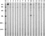
Fig. 2. Immunoblot analysis of 15 serum samples from patients with cystic echinococcosis with ES-Ag. M, Molecular mass marker, expressed in kDa.
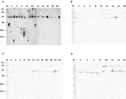
Fig. 3. Immunoblot analysis with ES-Ag. (A) Human sera corresponding to individuals with non-hydatid parasitoses. M, Molecular mass marker, expressed in kDa. Lane 1, E. nana+E. coli; Lane 2, E. nana+E. histolytica; Lane 3, A. lumbricoides+E. histolytica+E. coli; Lane 4, E. coli+A. lumbricoides; Lane 5, B. hominis; Lane 6, E. histolytica+E. coli; Lane 7, G. lamblia; Lane 8, G. lamblia+E. coli; Lanes 9 and 10, E. nana; Lane 11, E. histolytica+E. coli; Lane 12, E. histolytica; Lane 13, E. histolytica+G. lamblia; Lane 14, E. histolytica+E. coli; Lane 15, E. histolytica+G. lamblia; Lane 16, Taenia spp; Lane 17, G. lamblia; Lane 18, E. histolytica+E. coli; Lane 19, A. lumbricoides; Lane 20, Taenia spp; Lane 21, Toxocara spp. (B) Human sera corresponding to healthy donors. (C) Human sera corresponding to individuals with liver diseases. (D) Human sera corresponding to individuals with lung neoplasia. M, Molecular mass marker, expressed in kDa.
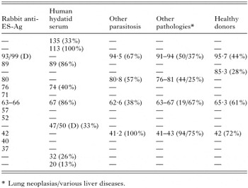
Table 2. Components of excretory–secretory products of protoscoleces recognized by different sera by immunoblotting
 You have
Access
- 26
- Cited by