Introduction
Tens of tons of interplanetary dust particles are believed to enter Earth's atmosphere daily, most of which vaporize (Rietmeijer Reference Rietmeijer2004). However, small particles with sizes below 40 μm remain largely intact and can be recovered from the stratosphere (Brownlee Reference Brownlee1985; Maurette et al. Reference Maurette, Olinger, Michel-Levy, Kurat, Pourchet, Braudstatter and Bourot-Denise1991). These have become known as interplanetary dust particles (IDPs) and have distinctive compositions (similar to carbonaceous and stony meteorites) and morphologies. The larger and denser particles show signs of melting and would be depleted in volatile components.
The theory of cometary panspermia proposes that grains in space consist of a mixture of complex organics and that their inclusion in many hundred billions of comets leads to the transition from prebiotic matter into primitive bacterial cells (Wickramasinghe Reference Wickramasinghe1974; Hoyle & Wickramasinghe Reference Hoyle and Wickramasinghe1976; Hoyle & Wickramasinghe Reference Hoyle and Wickramasinghe1977; Wickramasinghe et al. Reference Wickramasinghe, Brooks and Shaw1977). It has been suggested that pre-existing viable bacterial cells derived from interstellar space may have been present in comets in the primitive Solar System. This proposal is based on the following evidence: (1) the extinction properties of interstellar dust match precisely the expected behaviour of freeze-dried bacteria. (Hoyle & Wickramasinghe Reference Hoyle and Wickramasinghe1979); (2) the infrared (IR) absorption by dust in the 2.9–3.9 μm waveband for the galactic centre source GC-IRS7 has a distinctly bacterial signature (Hoyle et al. Reference Hoyle, Wickramasinghe and Al-Mufti1982; Rauf & Wickramasinghe Reference Rauf and Wickramasingh2010); and (3) the 217.5 nm (2175 Å) ultraviolet (UV) extinction is better explained by an ensemble of biological aromatics than by spherical graphite grains (Hoyle & Wickramasinghe Reference Hoyle and Wickramasinghe1991; Rauf & Wickramasinghe Reference Rauf and Wickramasingh2010). Other astronomical evidence, such as the diffuse IR bands, the complex organic composition of cometary dust and the extended red emission in the red rectangle, also serve to corroborate this proposal. However, the suggestion that the universe is replete with cosmic life is slow to gain wide acceptance.
In the early years of the space age, attempts were made to probe the upper atmosphere for the presence of microorganisms. The National Aeronautics and Space Administration (NASA)-supported balloon program (Bruch Reference Bruch, Gregory and Monteith1967) and Soviet rocket experiments (Lysenko Reference Lysenko1979) made attempts to show direct evidence for the presence of extraterrestrial microorganisms in the upper atmosphere, but the results were judged to be inconclusive due to the primitive nature of the sterilization procedures. In recent years, improved methodologies have been developed for the identification of microorganisms and stringent protocols applied to almost eliminate contamination from sampling and laboratory processes (Smibert & Kreig Reference Smibert, Kreis, Gerherdt, Murray, Wood and Kreig1994; Lopez-Amoros et al. Reference Lopez-Amoros, Mason and Lloyd1995; Lal et al. Reference Lal, Archarya, Patra, Rajaratnam, Subbarya and Venkataramani1996; Narlikar et al. Reference Narlikar, Ramadurai, Bhargava, Damle, Wickramasinghe, Lloyd, Hoyle and Wallis1998).
Claims to find viable microorganisms in the upper stratosphere have met with much scepticism. A common criticism concerns terrestrial contamination from sample handling and laboratory processes. In recognition of this criticism, stringent procedures have been used in the present investigation to prevent possible contamination during sample preparation. Experience at Cardiff with scanning electron microscopy (SEM) gives us high confidence in distinguishing contaminant particles from IDP-like cluster particles, while elemental composition identified via the energy dispersive spectroscopy (EDAX) facility adds confirmation (Miyake Reference Miyake2009). The current investigation uses high-resolution environmental SEM and ultraviolet-visible (UV-Vis) spectrophotometry to examine in detail some microdust samples collected at 38–41 km altitude.
Materials and methods
Collection of stratospheric air samples
The stratospheric air samples were collected at altitudes between 30 and 41 km using a cryosampler attached to a balloon according to the procedure as described in detail by Narlikar et al. (Reference Narlikar2003). The cryosampler was made of stainless steel and fitted with 16 probes. The whole assembly was electron-beam welded and electron plated. The cryosampler in pre-testing was found to be capable of withstanding temperatures between −246°C and 140°C. Before use, the interior components (probes and manifolds) were cleaned with acetone and thoroughly washed in de-mineralized water, steam baked and heated with an IR lamp to 140°C. During the balloon ascent, the probe inlet was closed via a metallic valve. The inlet was opened and closed via radio signal from ground. The air samples were passed through cellulose acetate filters (0.2 and 0.45 μm pore sizes) as described by Harris et al. (Reference Harris, Wickramasinghe, Lloyd, Narlikar, Rajaratnam, Turner, Al-Mufti, Wallis and Hoyle2001). Due to the limited availability of samples, the present study examined only the 0.45 μm pore membranes.
UV- Vis spectrophotometry
The filter containing stratospheric particles was placed in a glass vial containing de-mineralized water. The particles were removed by sonication for 15 min and centrifuged at 14 000 rpm for 30 min. The resulting pellet was resuspended in cyclohexane (1 ml) for 30 min, and the mixture centrifuged at 14 000 rpm for 10 min. The clear supernatant was transferred to the UV-Vis spectrophotometer (JASCO V-570 UV/VIS/NIR spectrophotometer) for analysis.
Scanning electron microscopy
Millipore filters (0.45 μm pore size) carrying the samples were cut into pieces (12 mm×12 mm) with a sharp razor blade, attached with sticky carbon tape onto metal stubs and gold coated. This sample preparation was conducted in a clean double-filtered air chamber. The samples were examined using a high-resolution environmental scanning electron microscope (Philips XL 30-ESEM) operated at 25–30 kV. Filter membranes containing no stratospheric samples were used as controls, being mounted and examined by SEM in exactly the same way.
Results
UV-Vis spectroscopy of stratospheric air samples
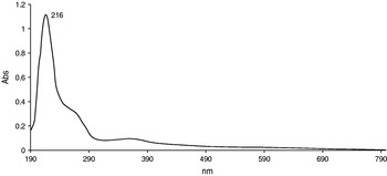
The UV-Vis spectrophotometric analysis of cyclohexane-dissolved samples produced a prominent peak centred at 216 nm, close to the 217.5 nm UV astronomical feature (Fig. 1).
UV-Vis spectrum of stratospheric air samples showing a prominent feature located at 216 nm and shoulder at 260 nm.

Scanning electron microscopy of stratospheric air samples
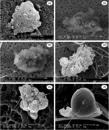
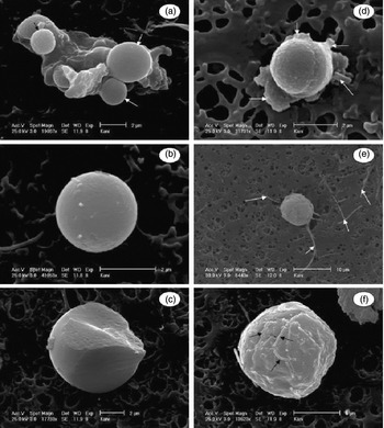
SEM of the membrane revealed numerous IDPs comprising clusters of submicron grains. These IDPs are sized between 7 and 32 μm across (Figs 2 and 3) and comprise coccoidal grains with some embedded rod or tubular objects (Fig. 2(a)). The sizes of the coccoids are between 0.4 and 1.3 μm in diameter, whereas the rods vary from 0.6 to 2.5 μm in length (Fig. 2(a)). Many globules with sizes ranging from 1.6 to 9 μm can also be observed in places (Figs 2(f), 3(a), (d), (e) and (f)), some of which show a surface covered with small granules (40–300 nm in diameter) (Fig. 2(f)) and filamentous features (Fig. 3(f)). None of the above features were found on the filter membranes used as controls.
Scanning electron micrographs of stratospheric air samples on 0.45 μm cellulose acetate filter. (a) A 20 μm sized clump formed by spherical particles (0.4–1.3 μm in diameter) as shown by short black arrows and rod-shaped particles (0.6–2.5 μm in length) as shown by long black arrows. (b) A 28 μm cluster of materials showing variable shapes from spherical (1.5–4.0 μm in diameter) as shown by short black arrows to irregular shaped objects (4.0–9.0 μm across) as shown by long black arrows; a few isolated filamentous or tubular objects can be occasionally seen (white arrow). (c) A 7.0 μm sized clump formed by small aggregates (0.6–3.0 μm) as shown by long black arrows; these aggregates are covered with granules (40–200 nm) as shown by short black arrows; the white arrows show filamentous or tubular objects. (d) A 32 μm clump formed by rod-shaped (2.0–5.0 μm in length) objects as shown by long black arrows and spherical features (0.4–2.0 μm) as shown by short black arrows. (e) A 16 μm clump formed by aggregates (2.5–4.5 μm across); these aggregates are composed of smaller granules (40–300 nm) and filamentous features of variable thickness (90–500 nm) as shown by long black arrows. (f) A 7.0 μm globule with a rough surface structure covered with tiny granules and rod-shaped features. The detached filaments may be contaminants (e), but not the embedded rods.

Scanning electron micrograph of stratospheric air particles. (a) A 12 μm sized clump showing globules of variable sizes arranging from 2.0 to 3.3 μm (as shown by white arrows); some globules showing a rough surface are sized between 1.6 and 1.8 μm (as shown by a black arrow); the white arrows show globules that have a smooth surface. (b) A 3 μm globule showing a rough surface structure. (c) A partially broken 9 μm globule showing its internal structure. (d) A 3 μm globule showing its coccoidal shape and rough surface structure; extracellular materials can be seen attached to the globule as shown by white arrows. (e) A 9 μm sized globule showing extra cellular filaments (arrows). (f) A 11 μm sized globule showing its rough surface covered with filamentous objects of variable lengths (as shown by arrows).

Discussion
Many particles of interplanetary origin were successfully collected on airborne plates coated with oil, flown at altitudes of 18–20 km (Hudson et al. Reference Hudson, Flynn, Fraundorf, Hohenberg and Shirck1981; McKeegan et al. Reference McKeegan, Walker and Zinner1985) and at higher altitudes in the stratosphere (Messenger Reference Messenger2000). These particles are largely fragments of comets and asteroids, but some components have been recently identified as pre-dating the Solar System (‘pre-solar grains’).
The present study using UV-Vis spectrophotometry shows the spectrum of the stratospheric samples containing only a single prominent peak centred at 216 nm, which is close to the 217.5 nm astronomical UV feature (Fig. 1). This astronomical UV feature was first observed in the spectrum of interstellar dust by Stecher (Reference Stecher1965). According to Hoyle & Wickramasinghe (Reference Hoyle and Wickramasinghe1962, Reference Hoyle and Wickramasinghe1969) and Stecher & Donn (Reference Stecher and Donn1965), the 217.5 nm extinction bump is attributable to small spheres of graphite. However, Hoyle & Wickramasinghe (Reference Hoyle and Wickramasinghe1977) later proposed the alternative molecular explanation of the 217.5 nm absorption; they argued that the requirement of graphite, which has a planar lattice structure to be in the form of small spheres in interstellar medium (ISM), appears scarcely relevant, and instead suggested the involvement of aromatic organic molecules. The feature has since been found along every galactic line of sight (Bradley et al. Reference Bradley, Humecki and Germani1992; Draine & Malhotra Reference Draine and Malhotra1993; Mathis Reference Mathis1993; Sandford Reference Sandford1996; Bradley et al. Reference Bradley, Keller, Snow, Hanner, Flynn, Gezo, Clemett, Brownlee and Bowey1999). Many astronomers attribute this feature to very small grains of carbon, possibly of aromatic and/or impure graphite (Draine & Malhotra Reference Draine and Malhotra1993; Mathis Reference Mathis1993; Sandford Reference Sandford1996). The carrier of the feature was proposed to be chondritic porous IDPs (CP-IDPs), since they have high carbon content (Thomas et al. Reference Thomas, Blanford, Keller, Klock and McKay1993) and contain inorganic carbon, including hydrogenated aliphatic and aromatic compounds (Clemett et al. Reference Clemett, Maechling, Zare, Swan and Walker1993; Flynn et al. Reference Flynn, Keller, Jacobsen, Wirick and Miller1999; Brownlee et al. Reference Brownlee, Joswiak, Bradley, Gezo and Hill2000).
Several laboratory models proposed for the carrier of the 217.5 nm feature are dehydrogenated and hydrogenated amorphous carbons, coals and polycyclic aromatic hydrocarbons (PAHs). However, none of these single compounds is generally accepted as the perfect model (Li & Greenberg Reference Li and Greenberg2003). It has been argued that the huge variation (±12%) with line of sight in the width and extinction bands is indicative of a large chemical dispersion in the Galaxy (Fitzpatick & Massa 2007). It has been suggested that the interstellar grain population also includes a substantial amount of ultra-small grains with PAH composition (Muthumariappan et al. Reference Muthumariappan, Maheswar, Eswaraiah and Pandey2008). Since PAHs have strong absorptions in the wide region (2000–2500 nm), the expected carrier of the 217.5 nm feature could be a mixture of PAHs, and the observed variation in the width of the feature may be attributed to differences in the PAH mixture (Muthumariappan et al. Reference Muthumariappan, Maheswar, Eswaraiah and Pandey2008). These observations seem to imply that in addition to the 217.5 nm extinction carrier, other dust components also exist in the ISM, including a population of amorphous silicate dust, aromatic hydrocarbons, PAH molecules and aliphatic hydrocarbons. Attempts to construct an average UV spectrum from 115 aromatic biomolecules also led to a synthetic 217.5 nm band (Wickramasinghe et al. Reference Wickramasinghe, Hoyle and Al-Jubory1989). Chen et al. (Reference Chen, Li, Li and Wang2008) have reported that buckyonions exhibit a peak close to the 217.5 nm extinction feature, but a mixture of silicate-graphite-PAH-buckyonions produces a feature that shows an almost perfect fit with the interstellar bump centred at 217.5 nm. Recently, Rauf & Wickramasinghe (Reference Rauf and Wickramasingh2010) have demonstrated that an ensemble of bio-degraded materials produced a feature centred precisely at 217.5 nm. Based on the early observations as mentioned above, the feature (216 nm) of the stratospheric air particles centred near 217.5 nm is likely to be organic in nature and interplanetary in origin.
The present study using a high-resolution environmental scanning electron microscope reveals the presence of 7–32 μm sized clusters of ovoids (0.4–1.3 μm in diameter) intermixed with elongated features (0.6–2.5 μm in length) (Fig. 2(a)). Other materials that were also found include globules (1.6–9 μm in diameter), some of which appear to have a granular surface structure (Figs 2(d) and (f)). Some of these globules display structural characteristics of bacteria, such as coccoids showing an apparent cell division with extracellular materials (Fig. 3(d)) and flagella (Fig. 3(e)). The ovoid features as shown in Fig. 2(a) appear similar in size and shape to nanobacteria found in travertine and limestone (Folk Reference Folk1993) and in Pleistocene ground water (McBride et al. Reference McBride, Picard and Folk1994). The above features may possibly be regarded as products of terrestrial contamination from sample handling and laboratory processing. However, none of these were observed on the membranes used as controls, thus suggesting an endogenous origin. The discovery of macrosized clusters of coccoids by the present study confirms the observations made by Harris et al. (Reference Harris, Wickramasinghe, Lloyd, Narlikar, Rajaratnam, Turner, Al-Mufti, Wallis and Hoyle2001) and Wainwright et al. (Reference Wainwright, Wickramasinghe, Narlikar and Rajaratnam2003, 2004a). Furthermore, the long filamentous features as shown in Figs 2(e) and 3(e) appear similar in structural characteristics to microfossils that have been found embedded in some carbonaceous meteorites, such as Orgueil and Murchison (Hoover Reference Hoover2009). Miyake (Reference Miyake2009) and Miyake et al. (Reference Miyake, Wallis and Wickramasinghe2009) ascribed these clusters to aggregates of IDPs rich in carbon and found that the rods are siliceous, suggesting that they are fragments of diatoms. Both diatoms and bacteria display an absorbance feature near 220 nm (2200 Å), similar to the observed UV spectral properties of interstellar grains, thus supporting the concept of a cosmic microbiological system in which these microorganisms might exist on comets (Hoover et al. Reference Hoover, Hoyle, Wickramasinghe, Hoover and Al-Mufti1999).
Although the present data obtained from the morphological and UV spectral analyses of stratospheric particles suggest that materials of biological origin are likely to be present in the samples, it may be argued that not all of the observed particles are of the same nature. The ovoid and elongated features could be the products of partial dissolution of the carbonate which is abundant in CP-IDPs, and have been reported to be similar to bacterially induced carbonate precipitation (Buczynsik & Chefetz Reference Buczynsik and Chafetz1991; McKay et al. Reference McKay, Gibson, Thomas-Keprta, Vali, Romanek, Clemett, Chillier, Maechling and Zare1996). A study made by Wainwright et al. (2004a) using secondary ion mass spectrometry (SIMS) has demonstrated that several clusters of bacteria-like particles found in the same stratospheric air samples are not biological, and thus pointed out that the SEM-based observation in itself cannot be used as evidence for the presence of bacteria in the stratosphere.
The early claim (Imshenetsky et al. Reference Imshenetsky, Lysenko and Kazakov1978) to have detected and isolated a wide range of common bacteria and fungi via rocket probes to heights of 48–77 km is beset with contamination objections. The recent studies using cryosamplers attached to balloons flying at altitudes of 20–41 km have largely overcome that problem (Harris et al. Reference Harris, Wickramasinghe, Lloyd, Narlikar, Rajaratnam, Turner, Al-Mufti, Wallis and Hoyle2001; Narlikar et al. Reference Narlikar2003; Wainwright et al. 2004b). The microorganisms recovered from the stratosphere include bacteria-like particles (Harris et al. Reference Harris, Wickramasinghe, Lloyd, Narlikar, Rajaratnam, Turner, Al-Mufti, Wallis and Hoyle2001; Narlikar et al. Reference Narlikar2003; Wainwright et al. 2004b), two bacteria (B. simplex and S. pasteuri) and one fungus (Engyodontium album) (Wainwright et al. Reference Wainwright, Wickramasinghe, Narlikar and Rajaratnam2003), P. Stutzeri (Narlikar et al. Reference Narlikar2003) and species of Penicillium and two bacteria (B. luciferensis and B. sphaericus (Griffin Reference Griffin2004). While most microorganisms appear unlikely to survive under harsh stratospheric conditions with low temperature and strong UV radiation, some microorganisms utilize protective strategies, such as clumping, forming thick cell walls, carbonizing outer layers and embedding in carbonaceous dust (Narlikar et al. Reference Narlikar2003; Wainwright et al. Reference Wainwright, Alharbi and Wickramasinghe2006). Furthermore, microorganisms can survive quite extreme conditions of temperature, pressure and even radiation (Hoyle & Wickramasinghe Reference Hoyle and Wickramasinghe2000). Secker et al. (Reference Secker, Wesson and Lepock1994) have shown that a carbonaceous coating of a few microns thickness of bacterial cells provides a protective shield against radiation. Microorganisms, such as spore-forming Bacilli (B. Simplex, Sarcina lutea) (Whisler Reference Whisler1940) and clump-forming bacteria (B. Subtilis and Staphylococcus aureus) (Horneck Reference Horneck1998; Darling Reference Darling2001), are relatively resistant to radiation. The pigmented microbes in the mesosphere discovered by Imshenetsky (Reference Imshenetsky1946) led him to propose that UV resistance is given by protective pigments (melanins and carotinoids) and suggests natural selection for these pigments.
Terrestrial dust particles are generally confined to the troposphere below 12–17 km, because the stratosphere lacks large-scale turbulence, being highly stratified. However, lofting mechanisms may exist (Wainwright et al. Reference Wainwright, Alharbi and Wickramasinghe2006) e.g. blue lightening strikes (Pasko et al. Reference Pasko, Stanley, Mathews, Inan and Wood2002) and gravito-photophoresis (Rohatschek Reference Rohatschek1996). The powerful volcanic eruption of Mt Pinatubo in the Philippines of 15 June, 1991 was reported to eject materials up to 32 km (Grant et al. Reference Grant1994). Imshenetsky et al. (Reference Imshenetsky, Lysenko and Kazakov1978) claimed that microorganisms were registered in a great number in the mesosphere during dust storms as compared to the absence of strong winds, but this is consistent with contaminants collected in flight through the troposphere. Terminal speeds for settling through the atmosphere are higher for larger sizes (and mean density). Moreover, lofting mechanisms are likely to discriminate in favour of smaller sizes, yet our recovered IDP-like particles show no statistical weighting to μm sizes. The ⩾10 μm clusters we found, with relatively few smaller than this, imply there is no stochastic mechanism kicking a spectrum of sizes upwards.
The absence of a plausible explanation for lofting our 7–32 μm clusters through the stratosphere to 40 km altitude adds weight to our identifying these materials as IDPs, and particularly carbonaceous IDPs (Miyake Reference Miyake2009). Although the detection in the samples of microfossil-like materials, such as coccoids, rods and filaments, suggests a biological origin, and the UV spectral data is consistent with this biological identification, more substantive evidence is needed to confirm this identification.