INTRODUCTION
Neospora caninum is an apicomplexan parasite, first described by Bjerkås et al. in 1984 and isolated and named by Dubey et al. (1988). Neospora caninum has a worldwide distribution and is a major cause of reproductive failure in cattle (Innes et al. 2001). Considerable economic losses are attributed to N. caninum in the farming industry, including the costs of still birth and neonatal mortality, increased calving interval resulting from early foetal death, increased culling, reduced milk production and reduced value of breeding stock (Trees et al. 1999; Dubey, 2003). There are currently no suitable chemotherapeutic agents to prevent transplacental transmission or to eliminate the parasite in cattle, making the development of an effective vaccine highly desirable. Epidemiological evidence indicates that persistently infected animals are less likely to abort following a point-source exposure than previously naïve animals, suggesting that cattle may develop some protective immunity against the disease (McAllister et al. 2000). Due to the intracellular nature of the parasite, it is expected that cell-mediated immunity (CMI), involving lymphoproliferative responses and the production of the cytokine interferon gamma (IFN-γ) will have a major protective role (Innes et al. 1995; Khan et al. 1997; Baszler et al. 1999). Eperon et al. (1999) demonstrated the importance of B-cells (and presumably antibodies) in controlling Neospora infections in mice. Although the role of antibodies in a protective immune response has still to be determined, a likely function would be in controlling the spread of extracellular stages of the parasite (Innes et al. 2002). Recent experimental studies in cattle have demonstrated that inoculation with N. caninum prior to pregnancy can protect against a second challenge administered at mid-gestation; transplacental transmission only occurred in the naïve control animals (Innes et al. 2001). In addition Williams et al. (2003) showed that seropositive cattle naturally infected with N. caninum were protected against abortion when experimentally challenged in early gestation. These two studies indicate vaccination may be a feasible option for the control of bovine neosporosis.
Neospora caninum infections can be established in mice, providing a convenient experimental model with which to test potential vaccination strategies. Live vaccines are more likely to induce appropriate CMI responses in host animals (Innes et al. 2002). Protective immunity against acute infection has been induced in mice using live attenuated, temperature-sensitive mutant strains of N. caninum (Lindsay et al. 1999) and sublethal doses of live N. caninum tachyzoites (Lundén et al. 2002). Virus-vectored vaccines including canine herpes virus (CHV) and vaccinia virus expressing the NcSRS2 N. caninum protein have also induced protective immune responses in mice (Nishikawa et al. 2000, 2001).
Attenuation of parasite virulence has been successfully applied in the development of live vaccines against other protozoan parasites. Leishmania major and Leishmania mexicana have been attenuated by repeated in vitro passage in the presence of gentamicin, and both attenuated strains of Leishmania induced significant protection in mice challenged with wild-type parasites (Daneshvar et al. 2003). Attenuation of virulence of Theileria annulata has been demonstrated following prolonged in vitro passage of schizont-infected cells (Hall et al. 1999). Babesia bovis and Babesia bigemina have both shown reduced virulence after repeated passage through splenectomized cattle (Pipano et al. 2002). Vaccination against Toxoplasma gondii-induced abortion in sheep has been achieved using a live incomplete (S48) strain of the parasite (O'Connell et al. 1988; Wilkins et al. 1988). The S48 strain of T. gondii was passaged over 3000 times in mice and has lost the ability to form both tissue cysts and oocysts (Buxton, 1993). A commercial vaccine based on this product is currently available in several countries worldwide and is licensed for veterinary use only.
The purpose of our study was to determine whether prolonged in vitro culture of N. caninum tachyzoites by repeatedly passaging parasites into fresh cell monolayers, would lead to attenuation of virulence in vivo. This study compared tachyzoites that were passaged successively between 33 and 39 times (low passage) and tachyzoites passaged between 70 and 84 times (high passage).
MATERIALS AND METHODS
Parasites and experimental inocula
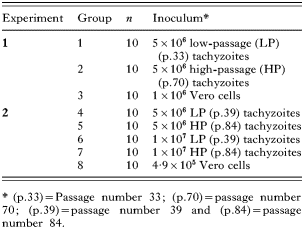
Neospora caninum tachyzoites (NC1 isolate) (Dubey et al. 1988) were successively propagated as previously described (Innes et al. 1995). Briefly, N. caninum parasites were cultured in Vero cell monolayers in 25 cm2 canted neck tissue-culture flasks (Corning, NY, USA). The monolayers were then disrupted using a sterile cell scraper (Corning, NY, USA), parasites were counted in a Neubauer haemocytometer and resuspended in phosphate-buffered saline (PBS) to produce inocula containing either 5×10⁷ or 1×10⁸ tachyzoites per ml. The control inoculum contained the same number of Vero cells as was present in the parasite inoculum and was prepared in the same manner. Both the parasites and Vero cells were inoculated into mice intraperitoneally (i.p.) within 1 h of their preparation in the laboratory, in a volume of 100 μl per mouse. During the routine passage of the parasites, aliquots were cryopreserved at regular intervals and stored in liquid nitrogen as described below.
The parasites described in this paper were isolated from a single source. The passage numbers refer to the continuous in vitro passage of the same stock of parasites for different lengths of time.
Cryopreservation and resuscitation of N. caninum tachyzoites
Infected monolayers were disrupted to release N. caninum tachyzoites as previously described (Innes et al. 2001). The tachyzoites were then counted and concentrated by centrifugation at 630 g for 5 min. The supernatant was discarded and the parasites resuspended at a concentration of 1×10⁷/ml in freezing medium (45% Iscoves modified Dulbecco's medium (IMDM) (Gibco BRL, Paisley, UK) 100 U/ml penicillin and 100 μg/ml streptomycin, (Northumbria Biologicals, Cramlington, UK), 45% foetal bovine serum (FBS) (Labtech, Austria) and 10% dimethyl sulphoxide (DMSO) (Sigma, Irvine, UK)). The parasites were aliquotted into 1 ml cryotube vials (Nunc, Roskilde, Denmark) and placed in 1 °C cryofreezing containers (Nalgene, USA) stored at −70 °C for 24 h, then transferred to vapour-phase liquid nitrogen storage. The parasites were resuscitated from liquid nitrogen storage by being rapidly warmed to 37 °C, then washed in 10 ml of IMDM supplemented with 5% FBS. Following centrifugation as above, parasites were resuspended in 2 ml of IMDM supplemented with 5% FBS, then inoculated into two 25 cm2 canted neck tissue culture flasks (Corning, NY, USA), each seeded 24 h previously with 1×10⁵ Vero cells.
Experimental design
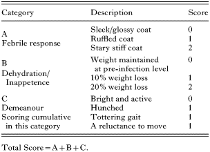
Female Balb/c mice, approximately 12 weeks old, were randomly assigned into groups of 10, identified by ear-marking, and fed rodent proprietary mix and fresh water ad libitum. The mice were inoculated intraperitoneally (i.p.) and observed daily. The morbidity of the animals was assessed according to a system agreed with the UK Home Office Inspectorate, (Table 1). A cumulative score of 5 on any day or a score of 4 for 2 consecutive days resulted in the mouse being culled. All surviving mice were euthanized on day 28 post-inoculation (p.i.) by CO2 inhalation. At post-mortem, samples of brain were removed and stored at −20 °C for analysis by N. caninum polymerase chain reaction (PCR). Brain, lung, liver and kidney samples were also removed and stored in 10% formal saline for histopathological examination, and blood was drawn from the heart to obtain serum.

Groups of mice were inoculated i.p. with different doses of low-passage (LP) and high-passage (HP) parasites or a control inoculum of Vero cells as indicated in Table 2.

ITS1 PCR
DNA was extracted from 25 mg samples of brain using the DNeasy Kit (Qiagen) as per manufacturer's instructions. Following extraction the DNA was stored at −20 °C prior to analysis by PCR. A nested PCR was used to detect N. caninum-specific internal transcribed spacer 1 (ITS1) DNA (Holmdahl and Mattsson, 1996) using the method previously described by Buxton et al. (1998). This produced a band of 297 bp when the products (5 μl each) were analysed by agarose gel electrophoresis (1·8%), stained with ethidium bromide and visualized under UV light.
Samples for histology and immunohistochemistry
Post-fixation, the brains were sliced coronally and, along with blocks of the other tissues, were processed to paraffin wax. Sections 5 μm thick were cut and stained with haematoxylin and eosin (H&E). To detect N. caninum, specific immunohistochemistry was used as previously described (Buxton et al. 1997). Briefly, tissue sections were incubated at 4 °C overnight with a primary polyclonal rabbit antiserum to N. caninum (diluted 1[ratio ]1000). The slides were washed in 0·5 M sodium chloride in 0·01 M phosphate buffer, a biotinylated goat-anti-rabbit IgG was used as a secondary antibody. Bound antibody was detected using the commercially available Vector-elite ABC system (Vector Laboratories, Peterborough, UK), with diaminobenzidine (DAB) being used as the chromogen. Sections were then counterstained with Mayer's haematoxylin and mounted under coverslips.
Serology
Blood taken at post-mortem was collected into sterile 1·5 ml tubes, allowed to clot overnight at 4 °C and then centrifuged at 5000 g for 2 min. The serum was decanted and stored at −20 °C prior to analysis. An indirect fluorescent antibody test (IFAT) for the detection of IgG was carried out as previously described (Buxton et al. 1997). Test sera were titrated in 2-fold dilutions from 1[ratio ]16 to a final concentration of 1[ratio ]4096. The endpoint was determined as the final concentration demonstrating distinct whole tachyzoite fluorescence (Conrad et al. 1993). The same procedure was used for the detection of IgM with the exception that fluorescein isothiocyanate (FITC)-conjugated goat anti-mouse IgM was used as the secondary antibody.
In vitro growth characteristics
The in vitro growth characteristics of the parasites were determined by quantifying their differential incorporation of tritiated [3H]uracil. Three replicate experiments were undertaken for low passage parasites (p.37, p.38 and p.39) and for high passage parasites using (p.74, p.75 and p.76). The procedure was carried out as previously described (Innes et al. 1995). In brief, Vero cells were cultured at a concentration of 5×10⁴ cells per well, in 96-well flat bottomed micro-titre plates (Nunc, Roskilde, Denmark) for 24 h prior to inoculation with the parasites. Parasites were added to quadruplicate wells containing Vero cells at a parasite to cell ratio of 4 tachyzoites: 1 cell. The cultures were labelled with 37 kBq of [3H]uracil per well (Amersham, Bucks, UK) and incubated at 37 °C in a humidified 5% CO2 atmosphere. Immediately prior to harvesting, parasite multiplication was stopped by the addition of 10 μl of 1 M sodium hydroxide. Cultures were harvested at 6, 12, 24, 48, 72 and 96 h post-inoculation onto glass-fibre filters and the parasite-associated radioactivity was quantified using a MATRIX 96™ gas proportional counter (Canberra Packard, Meriden CT, USA).
Statistical analysis
The mortality data from both Experiments 1 and 2 were analysed using the Kaplan-Meier procedure. In Exp. 2, mice were generally weighed on alternate days. A repeated measures model was fitted to data for days 2, 4 and 6 p.i. only as no mice in Group 6 (1×10⁷ LP) survived beyond day 7 p.i. The lack of independence between observations on the same mouse was modelled using an autoregressive type 1 correlation structure. To allow for the different mean starting weights for the groups, the day 0 reading was included in the model as a covariate. For ease of interpretation the results have been presented graphically as percentage changes from baseline. Analysis of variance (ANOVA) was used to analyse the in vitro growth experiment and IgG, IgM IFAT results. For the in vitro experiment a mixed model was used: counts per minute were analysed on a log scale, with low/high passage fitted as a fixed effect and the replicates within each type of passage fitted as random effects. A one-way ANOVA was used for the IgG and IgM data with the age of the animal at sampling being included in the model as a covariate. Differences in morbidity scores between two groups over successive times were assessed using Mann-Whitney tests. To account for multiple comparisons, the false discovery control method of Benjamini and Hochberg (1995) was used, as justified in this context by Benjamini and Yekutieli (2001). All statistical analyses were undertaken using Genstat 8th Edition apart from the false discovery rate procedure that was implemented within Microsoft Excel.
RESULTS
Exp. 1. Comparison of morbidity and mortality in mice inoculated with 5×10⁶ low- or high-passage N. caninum tachyzoites
Clinical observations
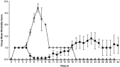
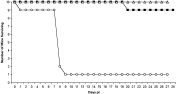
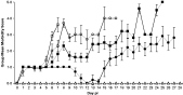
Low-passage parasites. Group 1 mice (5×10⁶ LP) developed ruffled coats from day 1 p.i., and from day 4 p.i. additional clinical symptoms were observed including hunching, reluctance to move, tottering gait and a stiff stary coat. A drop in group mean body weight of 6·6% (1·3 g) was observed by day 6 p.i. (data not shown). Animals were scored and culled in accordance with the scheme in Table 1 and results are illustrated in Fig. 1. By day 9 p.i. only 1 of the original 10 mice remained (Fig. 2). This animal became asymptomatic on day 17 p.i., and remained so until the end of the experimental period.
High-passage parasites
Group 2 mice (5×10⁶ HP) had less severe clinical symptoms of infection, when compared to the group 1 animals. Ruffled coats were seen on days 1–4 p.i., with the animals becoming asymptomatic on day 5 p.i. From day 11 p.i., symptoms including ruffled coats, hunching, a reluctance to move, and a tottering gait were observed in a few mice and were responsible for a group mean score of between 1 and 2 being maintained to the end of the experiment (Fig. 1). Only 1 of the 10 mice in group 2 died, and this occurred on day 23 p.i. (Fig. 2). Group 2 had a maximum group mean weight loss of 2·8% (0·6 g) on day 21 p.i. and by day 28 p.i. had returned to the group mean starting weight (data not shown).
Control animals
The control animals (group 3) showed no clinical symptoms and maintained a stable body weight throughout the experiment.

Fig. 1. Mean group morbidity score in Experiment 1. ○ – Group 1 (5×10⁶ LP). [squf ] – Group 2 (5×10⁶ HP). – Group 3 (1×10⁶ Vero cells). Error bars (S.E.M.).

Fig. 2. Mortality rates in Experiment 1. ○ – Group 1 (5×10⁶ LP). [squf ] – Group 2 (5×10⁶ HP). – Group 3 (1×10⁶ Vero cells).
Exp. 2. Comparison of morbidity and mortality in mice inoculated with different doses of low- or high-passage N. caninum tachyzoites
This experiment examined whether the difference in virulence demonstrated in Exp. 1 could be repeated following cryopreservation and resuscitation of parasites and using different doses of inoculum.
Low-passage parasites
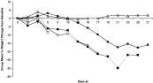
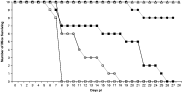
Group 4 mice (5×10⁶ LP) displayed symptoms from day 4 p.i., including hunching, reluctance to move, tottering gait and a stiff stary coat (Fig. 3). A decrease in group mean body weight of 20·9% (4·5 g) (Fig. 4) was also observed. All 10 animals in group 4 were culled or died by day 17 p.i. (Fig. 5).

Fig. 3. Mean group morbidity score in Experiment 2. ○ – Group 4 (5×10⁶ LP). [bull ] – Group 5 (5×10⁶ HP). □ – Group 6 (1×10⁷ LP). [squf ] – Group 7 (1×10⁷ HP). – Group 8 (4·9×105 Vero cells). Error bars (S.E.M.).

Fig. 4. Mean group % weight change from baseline in Experiment 2. ○ – Group 4 (5×10⁶ LP). [bull ] – Group 5 (5×10⁶ HP). □ – Group 6 (1×10⁷ LP). [squf ] – Group 7 (1×10⁷ HP). – Group 8 (4·9×105 Vero cells).

Fig. 5. Mortality rate in Experiment 2 ○ – Group 4 (5×10⁶ LP). [bull ] – Group 5 (5×10⁶ HP). □ – Group 6 (1×10⁷ LP). [squf ] – Group 7 (1×10⁷ HP). – Group 8 (4·9×105 Vero cells).
Group 6 mice (1×10⁷ LP) had more severe clinical symptoms by day 5 p.i. (Fig. 3), including an 8·3% (1·8 g) drop in group mean body weight between days 4 and 6 p.i. (see Fig. 4). All 10 mice were culled or died by day 8 p.i. (Fig. 5).
High-passage parasites
Group 5 mice (5×10⁶ HP) displayed milder symptoms of murine neosporosis than group 4, symptoms included a ruffled coat, weight loss and reluctance to move which were observed throughout the experiment (Fig. 3). The mean morbidity score for group 5 was significantly lower (P<0·05) (using Benjamini and Hochberg's false discovery control method) between 6 and 11 days p.i., compared to that seen in the animals in group 4 (5×10⁶ LP). A drop in group mean body weight was observed from day 12 p.i., reaching a maximum mean decrease of 17·6% (3·9 g) on day 25 p.i. (see Fig. 4). Only 2 mice in group 5 were culled or died prior to day 28 p.i. (Fig. 5). When the survival times from groups 4 and 5 were compared using the Kaplan-Meier procedure, group 5 was shown to have survived significantly longer (P<0·001) than group 4.
Group 7 animals (1×10⁷ HP) showed fewer symptoms than either group 4 or group 6 (Fig. 3). The mean morbidity score for group 7 was significantly lower (P<0·05) between 6 and 7 days p.i., compared to that seen in the animals in group 6 (1×10⁷ LP) (using Benjamini and Hochberg's false discovery control method). This dose of the high-passage parasites, however, resulted in a 29·9% (6·9 g) drop in group mean body weight (Fig. 4), and all 10 mice in this group died or were culled by day 25 p.i. (Fig. 5). When the survival times from groups 6 and 7 were compared using the Kaplan-Meier procedure, group 7 was shown to have survived significantly longer (P=0·006) than group 6.
Control animals
The control animals (group 8) displayed no clinical symptoms and maintained a stable body weight throughout the experiment.
Histopathology and PCR
Histological examination was performed on all samples collected and the extent of pathology, was based on the number and size of lesions observed in the brain. Lesions in the tissues were assessed histologically and the presence of parasites was demonstrated by immunohistochemistry. In all groups the severity of lesions was related to the time of survival. In general, mice culled before day 12 p.i. had mild or no lesions, whereas mice that survived for 15 days or longer had more severe pathology.
Low-passage parasites
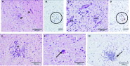
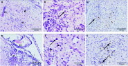
The mice from group 4 (5×10⁶ LP) that died before day 12 p.i. showed either mild, (Fig. 6A), or no pathology. The brains from the 2 mice that survived to days 15 and 18 p.i. had severe lesions, characterized by mononuclear perivascular cuffs and necrotic foci (Fig. 6C). Parasite tissue cysts or tachyzoites seen in H & E sections were confirmed by means of IHC (Fig. 6B and D).

Fig. 6. Group 4 (5×10⁶ LP) and Group 6 (1×10⁷ LP). (A). Brain from a mouse culled at day 7 p.i. showing small glial focus (gf) and a mild perivascular cuff (pc), (H&E). (B). Tissue cyst and tachyzoites within a small focus of gliosis in the brain of the same mouse (indicated within circle) (IHC). (C) Brain from a mouse that was culled at day 18 p.i. showing severe foci of necrosis (nf) and perivascular cuffing by mononuclear cells (pc) (H&E). (D) Small clumps of Neospora caninum parasites within lesions in the brain of the same mouse (indicated within circle) (IHC). (E). Brain from a mouse culled at day 7 p.i. showing a focus of necrosis. (F). N. caninum tissue cyst (arrow) (H&E). (G). Small aggregates of N. caninum parasites (arrow) within a focus of necrosis in the brain of the same mouse (IHC).
Group 6 mice (1×10⁷ LP) were all culled at day 7 p.i., 6 mice had mild lesions characterized by small to moderate numbers of foci of gliosis and necrosis (Fig 6E), whilst in 2 mice lesions and parasites were not detected. Parasite tissue cysts or tachyzoites were detected in 5 mice by histology (Fig. 6F) or IHC (Fig. 6G).
Parasite DNA was detected in 10 out of 10 brain samples from mice in group 4 (5×10⁶ LP) and in 7 out of 8 brain samples in group 6 mice (1×10⁷ LP).
High-passage parasites
Seven mice from group 5 (5×10⁶ HP), culled at the end of the experiment (day 28 p.i.) were severely affected (Fig. 7A), characteristically with large mononuclear perivascular cuffs, necrotic and glial foci and mononuclear meningitis. Many tissue cysts and tachyzoites were also observed (Fig. 7B and C), while in 1 mouse in group 5 only mild histological changes were detected.

Fig. 7. Group 5 (5×10⁶ HP) and Group 5 (1×10⁷ HP). (A). Brain from a mouse culled at day 28 p.i. showing severe lesions characterized by focal gliosis and vascular inflammation. (B) A small tissue cyst (arrow) associated with a focus of inflammation in the brain of the same mouse (H&E). (C). A small tissue cyst (arrow) and numerous tachyzoites within an area of necrosis and inflammation in the brain of the same mouse (IHC). (D). Brain from a mouse culled at day 24 p.i. showing a severe focus of necrosis in the molecular layer of the cerebellum, with associated meningeal inflammation. (E). One tissue cyst (arrow) and tachyzoites (arrowheads) in parasitophorous vacuoles, in the brain of a mouse culled on day 24 p.i. (H&E). (F) Neospora caninum tissue cysts (arrows) and tachyzoites within parasitophorous vacuoles in a focus of necrosis in the brain of a mouse culled at day 21 p.i. (IHC).
In group 7 (1×10⁷ HP), 7 mice had similar lesions to those observed in group 5. These mice were culled between days 18 and 25 p.i. and frequent foci of severe necrosis with gliosis and perivascular cuffing were recorded (Fig. 7D). Many tissue cysts and tachyzoites were present, mainly associated with lesions (Fig. 7E and F). In the 2 mice culled at day 7 p.i. only small foci of mild gliosis were seen.
Parasite DNA was detected in 7 out of 8 brain samples in group 5 mice (5×10⁶ HP) and in 9 out of 9 brain samples in group 7 animals (1×10⁷ HP).
Control animals
The control animals in group 8 had no clinical pathology, lesions, detectable parasites or parasite DNA.
Serology
Low-passage parasites
Group 4 mice (5×10⁶ LP) had IgM titres ranging from <1[ratio ]16 to 1[ratio ]128 and IgG titres ranging from <1[ratio ]16 to 1[ratio ]64. While group 6 (1×10⁷ LP) had IgM titres ranging from 1[ratio ]16 to 1[ratio ]256 and IgG titres of between <1[ratio ]16 and 1[ratio ]16. In general, low antibody titres were associated with early death or culling. The mouse in group 4 that survived to day 18 p.i. gave the highest IgG titre (1[ratio ]64) whilst also giving to lowest IgM titre (<1[ratio ]16).
High-passage parasites
Group 5 mice (5×10⁶ HP) mice showed no detectable IgM (titre of <1[ratio ]16) but had IgG titres ranging from 1[ratio ]256 to 1[ratio ]1024. Group 7 (1×10⁷ HP), had IgM titres ranging from <1[ratio ]16 to 1[ratio ]256 while the IgG titres in group 7 ranged from 1[ratio ]256 to 1[ratio ]2048.
When the IgG results were compared it was evident that group 5 produced greater levels of IgG than that produced by the animals in group 4, and group 7 produced greater levels of IgG than group 6. However, as IgG generally increased with time these differences are largely a reflection of the increased survival times in the groups receiving HP parasites.
Control animals
The control animals (group 8) had no detectable Neospora-specific IgM or IgG antibody titres.
In vitro multiplication rates of parasites
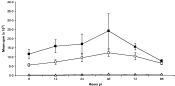
Parasite multiplication was measured by the differential incorporation of [3H]uracil by the multiplying parasites, and expressed as counts per min. Both the low-passage (p.37–p.39) and the high-passage (p.74–p.76) parasites multiplied steadily up to 48 h (Fig. 8), after which growth declined. There was strong evidence (P<0·001) of a difference in the pattern of growth rates over time between the high- and low-passage parasites with the high-passage parasites multiplying faster than the low-passage parasites at every time-point. The decrease in multiplication seen towards the end of the experiments is likely to be due to the tachyzoites lysing the Vero cell monolayers. The uninfected Vero cell controls did not incorporate [3H]uracil at any of the time-points tested.

Fig. 8. In vitro growth rates of Neospora caninum tachyzoites assessed by the differential uptake of [3H]uracil and expressed as counts per minute (cpm). □ – (low-passage tachyzoites p.37–p.39). [squf ] – (High-passage tachyzoites p.74–p.76). – (Vero cells) Error bars (S.E.M. from 3 separate experiments using quadruplicate samples).
DISCUSSION
Attenuation of virulence of N. caninum tachyzoites was achieved by continuous, passage of parasites in cell culture. In our study, differences were clearly demonstrated in the in vivo pathogenicity of N. caninum tachyzoites maintained for different lengths of time in tissue culture. The animals infected with the low-passage (virulent) parasites demonstrated a very rapid progression to severe clinical symptoms following inoculation with 5×10⁶ parasites, resulting in 95·0% (19/20) of the animals (groups 1 and 4) dying or being culled within 28 days p.i. By contrast, animals inoculated with the high-passage (attenuated) parasites, showed milder clinical symptoms and a 15·0% mortality rate with only 3 of 20 animals in groups 2 and 5 dying or being culled before day 28 p.i. The attenuation effect appears to be dose dependent as the mice that received 1×10⁷ high-passage parasites had all died by day 26 p.i. However, as a group they survived significantly longer than the mice that received 1×10⁷ low-passage parasites which all died or were culled by day 8 p.i. The attenuation phenotype observed in our study appears stable following cryopreservation and resuscitation, as similar results were obtained in 2 separate experiments. Other studies have shown that a subcutaneous (s.c.) inoculation with 2×10⁵ NC1 strain tachyzoites in Balb/c mice resulted in a mortality rate of 58% by day 70 p.i. (Lindsay et al. 1995). A subsequent paper reported a mortality rate of 70% in Balb/c mice within 41 days of an s.c. challenge with 5×10⁵ NC1 strain tachyzoites (Lindsay et al. 1999). These papers show that an s.c. inoculation of as few as 2×10⁵ NC1 strain parasites may be lethal in Balb/c mice, although no information is provided in these papers concerning the length of time the parasites had been maintained in culture prior to inoculation into the mice.
Symptoms of clinical neosporosis in mice include a rough coat, depressed appetite, dehydration, weight loss, tottering gait and reluctance to move (Lindsay and Dubey, 1989). Weight loss appears to be a characteristic symptom during a primary experimental infection with N. caninum tachyzoites in mice, as a 23% reduction in weight was observed by day 25 p.i. in Balb/c mice infected with NC Liverpool strain N. caninum parasites (Quinn et al. 2002). The initial weight loss that occurs during experimental N. caninum infections in mice is probably due to parasite multiplication causing tissue damage to the peritoneum and internal organs, leading to reduced appetite and dehydration. That this weight loss is due to tissue damage rather than the method of inoculation, is confirmed by the control animals, which received an i.p. inoculation of Vero cells but did not lose weight. Levels of weight loss may act as a useful quantifiable symptom of disease, following a primary experimental challenge with N. caninum parasites.
Detection of specific antibodies in mice inoculated with N. caninum was related to survival time, with higher antibody titres recorded in mice inoculated with HP parasites. The humoral immune response was characterized by an initial rise in levels of IgM for the first few days following infection. The antibody response then switched to IgG at about 12 days p.i. Those mice that succumbed to infection early still demonstrated detectable IgM titres (ranging from 1[ratio ]16 to 1[ratio ]256) and had either undetectable or very low titres of IgG (ranging from <1[ratio ]16 to 1[ratio ]64). Higher specific antibody responses (both IgM and IgG) were seen in the mice inoculated with 1×10⁷ HP N. caninum tachyzoites, compared to those inoculated with 5×10⁶ HP N. caninum tachyzoites, suggesting that the antibody titre produced was related to the challenge dose of parasites. The importance of the humoral immune response during murine Neospora infections was demonstrated by Eperon et al. (1999), using B-cell deficient μMT mice, which showed increased susceptibility to infection with the parasite, compared to wild-type mice. The anti-parasite function of antibodies is yet to be determined, although a likely role may involve controlling the spread of extracellular parasites (Innes et al. 2005).
The severity of the pathology seen in our study appears to be related to the survival time in the mice, with those that survived longest demonstrating the most severe lesions. Similar histological observations to those described in our study were seen by Long et al. (1998), Lindsay et al. (1995, 1999) and Lundén et al. (2002) when examining CNS tissue from infected mice. In these studies mice that received a primary challenge with NC1 N. caninum tachyzoites developed severe neuropathological lesions, including focal inflammation, focal necrosis and perivascular cuffing. The findings from our study are in agreement with those of Long et al. (1998), who observed a positive correlation between the dose of parasite challenge and levels of neuropathology, with mice that received larger challenge doses exhibiting more severe pathology than mice that received lower challenge doses.
Positive N. caninum specific ITS1 PCR results were seen as early as day 7 p.i. in samples of brain from animals inoculated with either low-passage or high-passage parasites, demonstrating that both are capable of disseminating to the brains of infected animals. Parasite DNA was detected in the brains of 17/18 mice that received the low-passage (virulent) parasites (groups 4 and 6), and 16/17 mice that received the high-passage (attenuated) parasites. Tissue cysts containing N. caninum parasites were seen in the brains of mice infected with either the low-passage (virulent) parasites or the high-passage (attenuated) parasites, demonstrating that both are capable of stage differentiation. Loss of life-cycle stage differentiation has been seen with T. gondii, where in 1 case following as few as 35–40 passages of tachyzoites in mice, the parasite lost its ability to form oocysts when the tissue cysts were fed to cats (the definitive host of T. gondii) (Frenkel et al. 1976). However, these parasites still retained the ability to form tissue cysts in mice. The S48 strain of T. gondii (O'Connell et al. 1988; Wilkins et al. 1988) was passaged over 3000 times in mice (Bos, 1993) and was found to have lost the ability to differentiate into either oocysts or bradyzoites. As the S48 strain tachyzoites undergo limited multiplication in vivo but do not persist, they proved to be a very effective means of immunizing animals against T. gondii (Buxton and Innes 1995). This attenuated (S48) strain of T. gondii is marketed as Toxovax™, a successful tissue culture-grown vaccine capable of controlling T. gondii induced abortion in sheep (O'Connell et al. 1988; Wilkins et al. 1988; Bos, 1993; Buxton, 1993).
The comparison of in vitro growth rates demonstrated that the high-passage (attenuated) parasites multiplied faster than the low-passage (virulent) parasites throughout. This may be because the attenuated parasites had become more adapted to tissue culture. A study by Schock et al. (2001) demonstrated no link between length of time in tissue culture and the in vitro growth rates of 6 different N. caninum isolates; although this study did not compare the growth rates of different passage numbers of the same isolate. The findings described in our paper suggest that the length of time in culture may have an effect on the pathogenicity of Neospora parasites in vivo. Long et al. (1998) noted that prolonged in vitro cultivation of N. caninum tachyzoites led to a reduction in virulence in vivo. However, Atkinson et al. (1999) demonstrated no attenuation effect in vivo of NC Liverpool strain N. caninum following 14 months of continuous in vitro culture, when compared to a culture of NC Liverpool stored in liquid nitrogen during this time. The same authors showed that there were considerable differences in the in vivo pathogenicity of NC Liverpool strain N. caninum in mice compared to the NC-SweB1 strain (Atkinson et al. 1999).
The mechanisms involved in the attenuation of other protozoan parasites are still not fully understood. Daneshvar et al. (2003) demonstrated attenuation of L. mexicana and L. major through repeated in vitro culture of promastigotes in the presence of gentamicin. Attenuation of T. annulata parasites has also been achieved through prolonged in vitro cell culture where the loss of virulence in vivo is thought to be a complex and gradual process caused by changes in the interaction between parasite and host cells (Preston et al. 2001). The less severe lesions observed following inoculation of the attenuated strain of T. annulata into cattle has also been linked with a reduced expression of matrix metalloproteases, which are required in cell adhesion and migration (Hall et al. 1999).
Lindsay et al. (1999) characterized 3 N. caninum temperature-sensitive mutants (NCts-4, NCts-8 and NCts-12) produced following exposure of parasites to N-methyl-N-nitro-N-nitrosoquanidine followed by 3–8 months continuous cell culture at 32·5 °C. These temperature-sensitive mutant parasites caused less severe lesions in Balb/c mice when compared to wild type NC1 strain parasites. However NCts-4, and NCts-12 reverted to the virulent wild type phenotype following culture at 37 °C, while NCts-8 showed a significantly reduced incidence of reversion to virulence following culture at 37 °C. This study by Lindsay et al. (1999) also demonstrated that vaccination of Balb/c mice with the NCts-8 parasites induced significant protection against a challenge with wild type NC1 strain N. caninum parasites. Live attenuated strains of intracellular protozoan parasites are attractive vaccine candidates as they are more likely to induce protective cell-mediated immune responses compared to immunization with killed vaccine preparations.
An interesting question arising from this study is whether the attenuation phenotype is due to the selection of a single genetic clone from a heterogeneous mix of parasites, or if the starting material was already clonal, then attenuation must have occurred through a more complex process such as loss or mutation of genes.
The results from our study show that it is possible to attenuate the virulence of N. caninum tachyzoites for mice through prolonged in vitro cell culture. The stability of the phenotype and the mechanisms involved in the attenuation process are still unknown and are the subject of continuing study.
The authors would like to acknowledge the Scottish Executive Environment and Rural Affairs Department for funding this study.