Introduction
Trematodes Haplosplanchnus pachysomus (Eysenhardt, 1829) and Provitellotrema crenimugilis Pan, Reference Pan1984 are members of the subfamily Haplosplanchninae Poche, 1926 (see Madhavi, Reference Madhavi, Jones, Bray and Gibson2005). Adult H. pachysomus are parasites of mugilid fishes in the Pacific, Indian and Atlantic Ocean basins (Skrjabin & Guschanskaja, Reference Skrjabin, Guschanskaja and Skrjabin1955; Bray, Reference Bray1984; Gajevskaja & Dmitrieva, Reference Gajevskaja and Dmitrieva1992). Characteristics of H. pachysomus, type-species of this genus, include the presence of a ventral sucker deeply immersed in the body, with a muscular sphincter around its opening. In addition to H. pachysomus, only five species from 25 listed in this genus have a similar ventral sucker – Haplosplanchnus caudatus (Srivastava, Reference Srivastava1939), H. straightum (Jehan, 1973), H. orientalis Gupta & Ahmad, Reference Gupta and Ahmad1979, H. stunkardi Gupta & Ahmad, Reference Gupta and Ahmad1979 and H. guptai Ahmad, Reference Ahmad1985. Bray (Reference Bray1984) and Nahhas et al. (Reference Nahhas, Rhodes and Seeto1997) considered these species junior synonyms of H. pachysomus. A single species of Provitellotrema, P. crenimugilis, was described by Pan (Reference Pan1984) in mullet from the coastal waters of China.
Species H. pachysomus was detected in Liza engeli (Bleeker) from the coastal waters of Cat Ba Island, Ha Long Bay, northern Vietnam, and P. crenimugilis in Liza haematocheila (Temminck & Schlegel) from Vostok Bay, Gulf of Peter the Great, southern Far-East Russia. In the present study, morphometric and molecular data are presented of these two digenean species, which are deposited in the Zoological Museum of the Institute of Biology and Soil Science, Far-Eastern Branch of the Russian Academy of Sciences, Vladivostok, Russia (e-mail: petrova@ibss.dvo.ru; deposited 29 July 2014).
Materials and methods
Collection of trematodes
Nine specimens of adult H. pachysomus were found in the intestine of L. engeli from coastal waters of Cat Ba Island, northern Vietnam. Seven specimens of adult P. crenimugilis were found in the intestine of L. haematocheila from Vostok Bay, Gulf of Peter the Great, Russian southern Far-East. Worms were rinsed in distilled water for a very short time, killed in hot distilled water, and preserved in 70% ethanol. Whole-mounts for adult descriptions were made by staining the specimens with alum carmine, dehydrating the worms in a graded ethanol series and clearing in xylene, followed by mounting in Canada balsam under a coverslip on a slide.
DNA extraction, amplification and sequencing
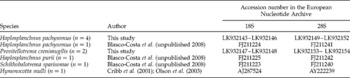
Adult specimens of H. pachysomus (n= 4) from Cat Ba Island and P. crenimugilis (n= 2) from the Primorsky Region, were used for molecular analysis (table 1). Total DNA was extracted from flukes, which were fixed in 96% ethanol, using a ‘hot shot’ technique (Truett, Reference Truett and Kieleczawa2006).
List of taxa incorporated in the molecular analysis of the family Haplosplanchnidae, with the number of DNA sequences given in parentheses.

Nuclear 18S rDNA and 28S rDNA fragments were successfully amplified using the polymerase chain reaction (PCR). 18S rDNA was amplified with the following primers: 18S-E (5′-CCG AAT TCG TCG ACA ACC TGG TTG ATC CTG CCA GT-3′), 18S-F (5′-CCA GCT TGA TCC TTC TGC AGG TTC ACC TAC-3′), described previously (Littlewood & Olson, Reference Littlewood, Olson, Littlewood and Bray2001). The initial PCR reaction was performed in a total volume of 20 μl containing 0.25 mm of each primer pair, 25 ng of total DNA in water, 10 × Taq buffer, 1.25 mm deoxynucleoside triphosphates (dNTPs), 1.5 mm magnesium and 1 unit of Taq polymerase. Amplification of a 2000-bp fragment of the 18S rRNA gene was performed in a GeneAmp 9700 (Applied Biosystems, USA), with a 5-min denaturation at 96°C; 35 cycles of 1 min at 96°C, 20 s at 58°C and 5 min at 72°C; and a 10-min extension at 72°C. Negative and positive controls using both primers were included.
28S rDNA was amplified with the primers DIG12 (5′-AAG CAT ATC ACT AAG CGG-3′) and 1500R (5′-GCT ATC CTG AGG GAA ACT TCG-3′), which were described previously (Tkach et al., Reference Tkach, Littlewood, Olson, Kinsella and Swiderski2003). The master mix for an initial PCR reaction for 28S rDNA was the same as for the amplification of the 18S rRNA gene. Amplification of a 1200-bp fragment of 28S rDNA was performed in a GeneAmp 9700 (Applied Biosystems), with a 3-min denaturation at 94°C; 40 cycles of 30 s at 94°C, 30 s at 52°C and 2 min at 72°C; and a 7-min extension at 72°C. Negative and positive controls using both primers were included.
PCR products were directly sequenced using an ABI Big Dye Terminator v.3.1 Cycle Sequencing Kit (Applied Biosystems), as recommended by the manufacturer, with the internal sequencing primers, described by Littlewood & Olson (Reference Littlewood, Olson, Littlewood and Bray2001) for 18S rDNA and Tkach et al. (Reference Tkach, Littlewood, Olson, Kinsella and Swiderski2003) for 28S rDNA. PCR products were analysed using an ABI 3130 genetic analyser at the Institute of Biology and Soil Science, Far-Eastern Branch of the Russian Academy of Sciences. Sequences were submitted to the European Nucleotide Archive (ENA) with the following accession numbers: LK932143–LK932154 (table 1).
Alignment and phylogenetic analysis
Ribosomal DNA sequences were assembled with SeqScape v. 2.6 software (Applied Biosystems). Alignments and estimation of the number of variable sites and sequence differences were performed using MEGA 6.0 (Tamura et al., Reference Tamura, Stecher, Peterson, Filipski and Kumar2013). Phylogenetic analyses of the nucleotide sequences were performed using the Maximum Likelihood (ML) algorithm with the MEGA 6.0 software, and the Bayesian algorithm with the MrBayes v. 3.1.2 software (Huelsenbeck, Reference Huelsenbeck2000). The best nucleotide substitution model, the general time reversible with estimates of invariant sites and gamma-distributed among-site variation (GTR+I+G), was estimated with Modeltest v. 3.7 software (Posada & Crandall, Reference Posada and Crandall1998). Bayesian analysis was performed using 5,000,000 generations and with two independent runs. Summary parameters and the phylogenetic tree were calculated with burning of 50,000 generations. Significance of the phylogenetic relationship was estimated by bootstrap (Felsenstein, Reference Felsenstein1985) for the ML algorithm, and posterior probabilities (Huelsenbeck et al., Reference Huelsenbeck, Ronquist, Nielsen and Bollback2001) for Bayesian inference. Phylogenetic relationships of the species of Haplosplanchnidae were inferred from our data and the nucleotide sequences of 18S rDNA and 28S rDNA from other trematode specimens obtained from the National Center for Biotechnology Information (NCBI) GenBank database, including sequences of 18S rDNA and 28S rDNA from the superfamily Haplosplanchnoidea, which were used as an outgroup (table 1).
Haplosplanchnus pachysomus (Eysenhardt, 1892)
Host. Liza engeli Bleeker.
Locality. Coastal water of Cat Ba Island, Ha Long Bay, northern Vietnam (20°84′N, 106°59′E).
Site. Intestine.
Intensity of infection. Nine specimens; one fish from 15 dissected
Description
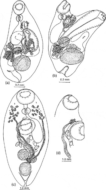
Based on five specimens (fig. 1a, b; table 2). Body sac-shaped in dorso-ventral position and Y-shaped in lateral position. Anterior and posterior extremities can invaginate inside body. Oral sucker subterminal. Pharynx round or oval, adjacent to oral sucker. Oesophagus short. Caecum single, reaching to mid-level of body posteriorly. Ventral sucker with thick muscular walls and deep cavity, and deeply immersed in body; opening surrounded by muscular sphincter. Testis single, in posterior third of body, round or oval. Seminal vesicle tubular, reaching to mid-line of ovary posteriorly. Prostatic part with thickened walls, surrounded by prostatic cells. Hermaphroditic duct short. Genital pore between pharynx and ventral sucker. Vitellaria in two fields between second and posterior third of body, between end of caecum and anterior margin of testis. Vitelline follicles elongated. Ovary spherical, dextral pre-testicular or contiguous with testis. Seminal receptacle round, contiguous to ovary. Uterus in middle third of body, with several loops, reaching mid-level of testis. Eggs operculated, in distal part of uterus only, containing miracidia, each with single eye-spot. Excretory bladder Y-shaped.
Adult worms: (a, b) Haplosplanchnus pachysomus; (c, d) Provitellotrema crenimugilis.

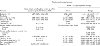
Morphometrics (measurements in mm) of Haplosplanchnus pachysomus after Gajevskaya & Dmitrieva (1992) and the present study (L, length; W, width).

18S rRNA and 28S rRNA gene sequence data
A total of 1796 and 1068 alignable characters were available for analysis in the 18S rRNA gene and 28S rRNA gene datasets for H. pachysomus, respectively. Of these, no variable and parsimony-informative sites for either 18S or 28S rDNA were presented.
Provitellotrema crenimugilis Pan, 1984
Host. Liza haematocheila (Temminck and Schlegel).
Locality. Vostok Bay, Gulf of Peter the Great, Russian southern Far-East (45°54′N, 132°44′E).
Site. Intestine.
Intensity of infection. Seven specimens, one fish from 12 investigated.
Description
Based on five specimens (fig. 1c, d; table 3). Body oval, with diffuse pigment at level of oesophagus and pharynx. Oral sucker subterminal. Prepharynx short. Pharynx transversely oval. Oesophagus short. Caecum single, sac-shaped, reaches to mid-length of body. Ventral sucker in second quarter of body; equal in diameter to oral sucker. Testis single, oval, located close to posterior extremity of body. Seminal vesicle tubular, long, reaches to level of ovary. Seminal receptacle large, round, sinistral. Uterine loops few, between ventral sucker and ovary. Seminal vesicle and uterus open into short hermaphroditic duct. Genital pore located just anterior to ventral sucker. Eggs operculated, in distal part of uterus only, contain miracidia, each with single eye-spot. Excretory bladder Y-shaped.
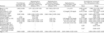
Morphometrics (measurements in mm) of Provitellotrema crenimugilis, Haplosplanchnus bivitellosus and H. purii after previous authors and the present study (L, length; W, width).

18S rRNA and 28S rRNA gene sequence data
A total of 1796 and 1068 alignable characters were available for analysis in the 18S rRNA gene and 28S rRNA gene datasets for P. crenimugilis. Of these, 3 (0.17%) and 1 (0.09%) variable sites were presented for 18S rDNA and 28S rDNA fragments, respectively.
Discussion
Adult H. pachysomus from our material have the greatest morphometric similarity with specimens of H. pachysomus found by Gajevskaja & Dmitrieva (Reference Gajevskaja and Dmitrieva1992) in fish of the genera Liza and Mugil from the Black Sea basin (table 2). The most characteristic feature of adult H. pachysomus is the presence of a ventral sucker deeply immersed in the body, with a sphincter around its opening. Haplosplanchnus caudatus (Srivastava, Reference Srivastava1939), H. straightum (Jehan, 1973), H. orientalis Gupta & Ahmad, Reference Gupta and Ahmad1979, H. stunkardi Gupta & Ahmad, Reference Gupta and Ahmad1979, and H. guptai Ahmad, Reference Ahmad1985, all exhibit similar structures of the ventral sucker. Bray (Reference Bray1984) and Nahhas et al. (Reference Nahhas, Rhodes and Seeto1997) expressed the opinion that these species are conspecific with H. pachysomus. Based on the available data (Srivastava, Reference Srivastava1939; Gupta & Ahmad, Reference Gupta and Ahmad1979; Bray, Reference Bray1984; Ahmad, Reference Ahmad1985; Nahhas et al., Reference Nahhas, Rhodes and Seeto1997), we suggest that H. pachysomus may be conspecific to other species of Haplosplanchnus with identical morphology and ventral sucker size. However, a final decision will only be possible after obtaining morphometric and molecular data for specimens of the species from their type localities.
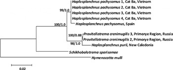
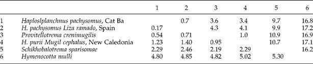
Phylogenetic relationships showed that H. pachysomus from Vietnam was closely related to H. pachysomus from Spain (fig. 2). Specimens of H. pachysomus from Vietnam (present study) and H. pachysomus from Spain (sequence IDs: FJ211225 for 18S and FJ211242 for 28S) differ by 0.17% and 0.68% by 18S rDNA and 28S rDNA, respectively (table 4). Combined data showed genetic differentiation of 0.35%. The divergence values were higher when Vietnamese and Spanish sequences of H. pachysomus were compared with sequences of H. purii: 1.23 and 1.4% by 18S rDNA and 3.38 and 4.05% by 28S rDNA.
Phylogenetic tree of the family Haplosplanchnidae based on the analysis of combined 18S rRNA (complete) and of 28S rRNA (partial) gene sequences; nodal numbers indicate bootstrap values/posterior probabilities for Maximum Likelihood/Bayesian Inference algorithms.

Molecular differentiation (%) of the species of the family Haplosplanchnidae by combined complete 18S rRNA gene (below the diagonal) and partial 28S rRNA gene (above the diagonal) sequences.

Adult specimens of Provitellotrema Pan, Reference Pan1984 found in L. haematocheila agreed well with the morphometric data for P. crenimugilis (table 3). The only difference between the present material and P. crenimugilis was the morphology of the eggs. Pan (Reference Pan1984) noted that the eggs of P. crenimugilis were unembryonated. The specimens found in this study contained eggs with miracidia in the distal loops of the uterus, whereas eggs were unembryonated in the proximal portions of the uterus. Zhukov (Reference Zhukov1971) described Haplosplanchnus bivitellosus Zhukov, Reference Zhukov1971 based on adult specimens from the intestine of Mugil cephalus from Posjet Bay, Sea of Japan, and of L. haematocheila (syn. Mugil so-iuy) from Liaohe River, China (Zhukov, Reference Zhukov1971). Pan (Reference Pan1984) synonymized H. bivitellosus with H. purii (Srivastava, Reference Srivastava1939). However, these species, in spite of many morphological similarities, differ significantly in size of the body, ventral sucker and caecum (table 3). The adult worms described by Pan (Reference Pan1984) as P. crenimugilis and the present material are similar to H. bivitellosus metrically (table 3) and in most morphological features. The only difference between P. crenimugilis and H. bivitellosus was in the morphology of the vitellarium. According to Zhukov (Reference Zhukov1971), the vitellarium of H. bivitellosus forms two tape-like structures, extending from the mid-level of the caecum to the level of the testis. The vitellarium in P. crenimugilis, according to Pan (Reference Pan1984) and our data, consists of minimal medium-sized follicles located anterior to the ventral sucker. Two wide vitelline ducts extend from the vitellarium up to the level of the ovary. In the present study, analysis of the slides showed that vitelline follicles of some specimens are poorly differentiated and only vitelline ducts are visible; these may be erroneously considered to represent the vitellarium. Therefore, it is possible that H. bivitellosus and P. crenimugilis are conspecific.
Comparative molecular analysis of P. crenimugilis and H. pachysomus from Vietnam and Spain showed 0.81% and 0.95% of differentiation by complete sequences of the 18S rRNA gene and 3.62% and 4.29% by partial sequences of the 28S rRNA gene. Sequence differentiation of P. crenimugilis and H. pachysomus from Vietnam and Spain by combined 18S rDNA and 28S rDNA sequence data was 1.88% and 2.23%, respectively. On the other hand, P. crenimugilis was closer to H. purii from New Caledonia (data from GenBank, table 1). Genetic differentiation values between these species were 0.81% and 1.01% by complete sequences of the 18S rRNA gene and partial sequences of the 28S rRNA gene, respectively. Genetic differentiation between these two species by combined sequence data was 1.02%. Phylogenetic analysis showed P. crenimugilis in the same cluster as H. purii, which was highly differentiated from the H. pachysomus cluster (fig. 2). These data allow us to suggest that P. crenimugilis and H. purii are different species of the same genus. Moreover, differentiation between these two trematodes may be caused by: (1) wide geographical distance (Primorye Region and New Caledonia); and (2) definitive host specificity – L. haematocheila for P. crenimugilis and M. cephalus for H. purii (Blasco-Costa et al., 2008, unpublished, data from GeneBank). Nevertheless, final taxonomical conclusions about these two species can only be made after more detailed morphological analysis and molecular studies using more representative samples.
Financial support
This study was partially supported by the President of the Russian Federation (project number 394.2014.4) and the Vietnamese National Foundation for Development of Science and Technologies (project number 106.15-2012.8).
Conflict of interest
None.