Introduction
Takotsubo cardiomyopathy is characterised by akinesis and ballooning of the left ventricular apex during contraction of the otherwise normal base of the heart. Often triggered by acute physical and/or emotional stress and a subsequent surge in circulating catecholamines, it is most common in women older than 55 years of age and has been rarely described in the paediatric population.Reference Deshmukh, Kumar and Pant 1 We describe the first case of a previously healthy 7-month-old infant who developed Takotsubo cardiomyopathy after presumed non-accidental trauma and review the recent paediatric case reports and literature regarding long-term prognosis and follow-up.
Case report
A 7-month-old previously healthy female presented to the emergency room after reportedly being found non-responsive by the caretaker. The morning of admission, she rolled off the changing table and hit her head on a hardwood floor. There was no loss of consciousness and she cried immediately. Four hours later, she was reportedly found unresponsive. She received cardiopulmonary resuscitation for 2 minutes before return of spontaneous circulation. CT scan of the head showed a right parafalcine acute subdural haematoma and loculated anterior right frontal subdural haematoma with local mass effect as well as concern for diffuse cerebral oedema/diffuse hypoxic ischaemic injury. Chest X-ray showed bilateral infiltrates concerning for pulmonary contusions as well as other concerning signs of trauma.
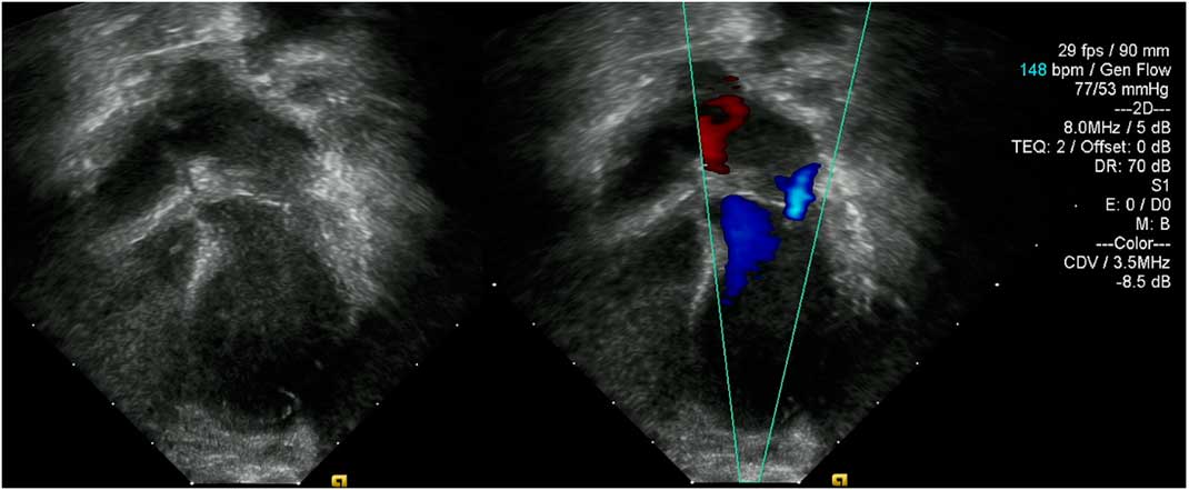
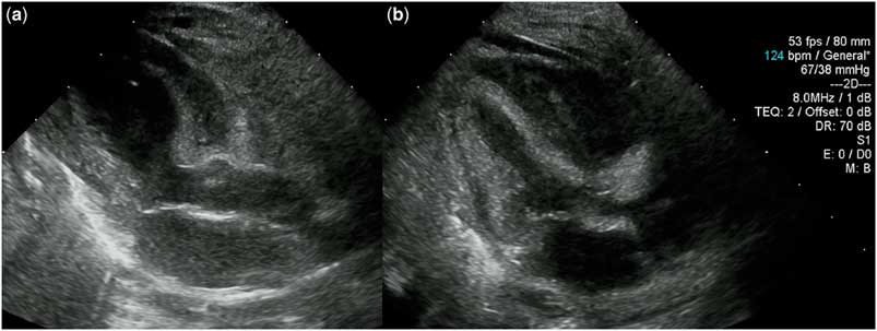
An echocardiogram was completed while the patient was on epinephrine at 0.05 mcg/kg per minute to assess cardiac function and to search for any anatomical lesion that could explain her unwitnessed cardiac arrest. The apical half of the left ventricle was globally akinetic with paradoxical motion and normal contractility at the base of the heart. There was also moderately restrictive diastolic function with trivial mitral regurgitation (Fig 1). The ejection fraction was 25% by Simpson method. Cardiac enzymes revealed a mildly elevated troponin at 0.94 ng/ml. A baseline electrocardiogram showed a normal sinus rhythm with an indeterminate QRS axis and right ventricular hypertrophy. The infant’s neurologic status deteriorated and repeat head CT demonstrated hypoxic ischaemic injury of the cerebral hemispheres and cerebellum with increased brain swelling. Urine drug screen was negative. A repeat echocardiogram while on epinephrine 0.05 mcg/kg per minute and milrinone 0.25 mcg/kg per minute was completed 40 hours after the initial assessment and showed an ejection fraction of 50–55% with marked improvement and normalisation of the apical-mid left ventricular akinesis. Diastolic function remained abnormal (Fig 2). Her evolving clinical picture lead to brain death protocol evaluation and she was subsequently pronounced brain dead and support was withdrawn. Autopsy did not show any evidence of pheochromocytoma or myocarditis. In light of the clinical history and examination findings, the child abuse specialist concluded that the event was highly suspicious for non-accidental/inflicted trauma.
Initial Apical 4 chamber view via transthoracic echocardiogram showed left ventricular apical ballooning but without significant mitral regurgitation.

Initial echocardiographic images from parasternal long axis view show dilation of the apical portion of the left ventricle (a), with normalization 40 hours later (b). Both images were taken during end-systole.

Discussion
Takotsubo cardiomyopathy is a transient, reversible cardiomyopathy that is usually triggered by an acute emotional or physical stress. A recent study estimated that it is diagnosed in about 0.02% of all hospitalisations in the United States,Reference Deshmukh, Kumar and Pant 1 and the prevalence in children is extremely rare. Reported triggers in the paediatric population range from stress during a natural disasterReference Bajolle, Basquin and Lucron 2 to infectionReference Fabi, Testa and Gesuete 3 to neurologic injury.Reference Hong, Glater-Welt and Siegel 4 Direct myocardial toxicity from elevated catecholamines,Reference Wittstein, Thiemann and Lima 5 abnormalities in endothelial vasodilation and constriction,Reference Martin, Prasad and Rihal 6 and multi-vessel epicardial coronary artery spasmReference Lacy, Contrada and Robbins 7 have all been implicated in the pathogenesis of the disease process.
In adults, Takotsubo often mimics an acute coronary syndrome with chest pain, shortness of breath, and syncope as the most common presenting symptoms. In children, however, atherosclerotic disease is rare and Takotsubo is more likely to mimic an acute myocarditis syndrome. An inciting event and improvement in morphology and function should aid the practitioner in distinguishing Takotsubo from congenital left ventricular aneurysm, which has a high risk for spontaneous rupture and should be surgically repaired. Other aetiologies to consider include illicit drug use and pheochromocytoma.
There is no universally accepted diagnostic definition for Takotsubo cardiomyopathy, but our patient did meet all criteria proposed by the Mayo clinic in 2010,Reference Madhavan and Prasad 8 including transient akinesia of the left ventricular apical segment with regional wall-motion abnormalities extending beyond a single epicardial vascular distribution, absence of obstructive coronary disease, electrocardiogram, and troponin abnormalities, and no evidence of myocarditis or pheochromocytoma. In this case, the inciting event was both physical and emotional stress that stemmed from presumed non-accidental trauma and subsequent acute subdural haematoma. Nevertheless, her cardiac function and left ventricular akinesia resolved within 36 hours. She was supported on low-dose epinephrine at the time of the initial and follow-up echocardiograms. While epinephrine and other catecholamine-derived inotropic agents are commonly used for support of blood pressure and ventricular function in the setting of cardiac arrest, administering exogenous catecholamine to a patient with Takotsubo could potentially exacerbate the disease process and delay recovery.
The diagnostic challenge for every patient who presents with an unwitnessed cardiac arrest and decreased ventricular function is determining whether the decreased function contributed to the cardiac event, for example primary cardiomyopathy and subsequent tachyarrhythmia, or whether it is the result of cardiac arrest and subsequent cardiopulmonary resuscitation triggered by a non-cardiac aetiology. To our knowledge, this is the first case of Takotsubo cardiomyopathy secondary to non-accidental trauma. It highlights the importance of maintaining a low threshold of suspicion for non-accidental trauma in any patient who presents with an unexplained cardiac event. It also emphasises the importance of frequent serial echocardiograms to assess for potential rapid recovery of the myocardium. While ventricular function is frequently decreased post-cardiac resuscitation and cardiac biomarkers may be elevated, a dyskinetic left ventricle and the setting of a neurological injury or acutely stressful event should raise suspicion for Takotsubo cardiomyopathy. A recent adult study suggests that despite normalisation of left ventricular ejection fraction and serum biomarkers, patients often have persistent heart failure symptoms, reduced peak oxygen consumption, and abnormal strain and deformation analysis.Reference Scally, Rudd and Mezincescu 9 In addition, there is a risk of recurrence reported both in adults and in paediatric populations.Reference Srivastava, Parent and Hurwitz 10 In light of this, we recommend that all children with Takotsubo cardiomyopathy be followed by a paediatric cardiologist for the development and/or persistence of long-term sequalae.
Financial support
This research received no specific grant from any funding agency, or from commercial or not-for-profit sectors.
Conflicts of interest
None.
Ethical standards
The authors assert that all procedures contributing to this work comply with the ethical standards of the relevant national guidelines on human experimentation and with the Helsinki Declaration of 1975, as revised in 2008, and has been approved by the institutional committee.