Introduction
Non-traumatic bone fractures in cancer patients are usually pathological fractures caused by bone metastases. In contrast, clavicle stress fractures may occur as a result of atrophy of the trapezius muscle after neck dissection in which the accessory nerve is structurally or functionally damaged.Reference Cummings and First 1 – Reference Hao and Chang 3 Atrophy of the trapezius muscle causes dropped shoulder syndrome, so that external force would be loaded onto the medial portion of the clavicle, which results in fracture.
In the literature, most cases of medial clavicle stress fractures were reported to have no significant displacement and were amenable to conservative treatment.Reference Throckmorton and Kuhn 4 , Reference van der Meijden, Gaskill and Millett 5 Herein, we report a case of clavicle stress fracture accompanied by osteomyelitis and a pectoralis major abscess, which occurred after modified radical neck dissection with accessory nerve preservation. Surgical intervention and prolonged hospital care were required in this case, suggesting that post-operative radiotherapy and/or local infection followed by tracheostomy may pose a risk of serious clavicle fracture after neck dissection.
Case report
A 71-year-old man was referred to our hospital for right-sided tongue cancer and a laryngeal tumour, with right vocal fold paralysis. Partial glossectomy and laryngeal tumour biopsy under direct laryngoscopy were performed, followed by temporary tracheostomy. Sulbactam/ampicillin was administered for post-operative cellulitis around the tracheostomy. Pathological examination revealed a T1N0M0 tongue squamous cell carcinoma and laryngeal chondroma.
Two months after the initial treatment, right-sided modified radical neck dissection was performed for submandibular lymph node metastases, in which the accessory nerve, sternocleidomastoid muscle and internal jugular vein were all preserved. Post-operative shoulder rehabilitation was carried out to prevent joint contracture. Pathological examination revealed two lymph node metastases with extracapsular spread, and thus post-operative radiotherapy (total dose of 66 Gy, including whole neck irradiation of 46 Gy and submandibular boost irradiation of 20 Gy) was performed.
Four weeks after the radiotherapy, the patient was urgently admitted to our hospital for a high fever and right-sided precordial swelling. On inspection, the sternoclavicular joint demonstrated redness, swelling and tenderness (Figure 1). The trapezius muscle had atrophied, the right shoulder had dropped (Figure 1) and elevation of the right upper limb was poor. Body temperature was 39.2 °C and blood pressure was 160/75 mmHg, with a pulse rate of 115 bpm. Laboratory data were as follows: white blood cell count = 15 700 /μl, neutrophil count = 13 738 /μl, haemoglobin = 13.2 g/dl and C-reactive protein = 10.63 mg/dl.
Regarding physical findings, the sternoclavicular joint shows redness, swelling and tenderness. The trapezius muscle is atrophied and the right shoulder is dropped.

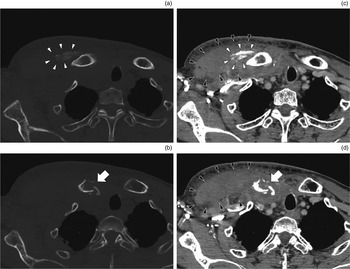
Computed tomography (CT) revealed a fracture of the medial clavicle (Figure 2b and d) and swelling of the pectoralis major muscle (Figure 2c and d), with ectopic calcification (Figure 2a and c).
(a & b) Axial bone window and (c & d) axial, contrast-enhanced soft tissue window computed tomography images show that the medial clavicle is fractured, with slight displacement (arrows). The pectoralis major muscle is swollen (black arrowheads) and ectopic calcification can be observed (white arrowheads).

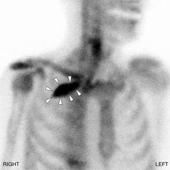
Technetium-99 m hydroxymethylene diphosphonate bone scintigraphy demonstrated extraosseous accumulation into the area corresponding to the calcified area observed on CT (Figure 3). This accumulation was considered to indicate myositis ossificans, which developed following the clavicle fracture. Sternoclavicular joint arthritis was absent, as indicated by the absence of abnormal accumulation (Figure 3).
Technetium-99 m hydroxymethylene diphosphonate bone scintigraphy shows extraosseous accumulation in the right precordium (arrowheads) in the area corresponding to the calcified area observed on computed tomography. No abnormal accumulation of the sternoclavicular joints is apparent.

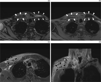
In the T1-weighted magnetic resonance images (Figure 4a), the bone marrow of the right clavicle showed a low intensity, in contrast to the left clavicle, whereas the opposite findings (moderate intensity in the right clavicle and low intensity in the left clavicle) were observed in T1-weighted, fat-suppressed images (Figure 4b). These decreases in fat intensity of the right clavicle suggested the presence of osteomyelitis. Gadolinium-enhanced, T1-weighted images (Figure 4c and d) showed an ill-defined, ring-enhancing lesion in the pectoralis major muscle, suggesting abscess formation.
(a) Axial, T1-weighted, (b) axial, T1-weighted, fat-suppressed, and (c) axial and (d) coronal, T1-weighted, fat-suppressed, gadolinium-enhanced magnetic resonance images show: (a) low intensity (white arrows) in the bone marrow of the right clavicle in contrast to the left clavicle (arrowheads), indicating the loss of fat; (b) moderate intensity (white arrows) in the right clavicle and low intensity (arrowheads) in the left clavicle in fat-suppressed images; and (c & d) an ill-defined, ring-enhancing lesion (black arrows) in gadolinium-enhanced images showing the pectoralis major muscle.

These imaging and laboratory data indicated a clavicle fracture, with osteomyelitis and a pectoralis major abscess, rather than a simple stress fracture or a pathological fracture due to metastasis. No locoregional recurrence of tongue cancer was noted.
Although meropenem (3.0 g/day) was partially effective for the symptoms, C-reactive protein did not reduce to within the normal range. The swollen precordium was punctured, but the content failed to drain. Accordingly, drainage surgery was performed on the 22nd hospital day. The abscess cavity was opened, the purulent content was discharged, the calcified part of the pectoralis major muscle was excised for pathological examination and a continuous-suction drain was placed near the sternoclavicular joint. Pathological examination revealed granulation tissue and ossification of the muscle, but no malignancy. Bacterial culture from the abscess was negative.
The drain was removed on the 25th hospital day, and shoulder rehabilitation recommenced on the 29th hospital day. The laboratory test findings, precordial swelling and fever were improved with meropenem administration for a total of 51 days. The antibiotic was changed to levofloxacin, and the patient was discharged on the 67th hospital day. At seven months after discharge, no recurrence of clavicular osteomyelitis or tongue cancer was observed.
Discussion
We report a rare case of a clavicle stress fracture with severe complications, which required surgical intervention and long-term hospitalisation, occurring after neck dissection with preservation of the accessory nerve. We assume that post-operative radiotherapy and/or local infection may have been causally related to this fracture.
Non-traumatic bone fracture occurring in cancer patients is usually a pathological fracture caused by bone metastases. Bone metastasis is the second most frequent form of metastasis after pulmonary metastasis, and accounts for 22 per cent of distant metastasis in head and neck cancer. Metastases to the hip, long bones and vertebrae are common.Reference Ferlito, Shaha, Silver, Rinaldo and Mondin 6 As a unique fracture of head and neck cancer, a clavicle stress fracture but not a pathological fracture may occur after neck dissection.
A clavicle stress fracture will mostly occur in cases where the accessory nerve has been cut,Reference Cummings and First 1 , Reference Ord and Langdon 2 but rarely in cases where the accessory nerve has been preserved.Reference Hao and Chang 3 When the trapezius muscle atrophies after accessory nerve damage, because of pulling or resection during neck dissection,Reference Sobol, Jensen, Sawyer, Costiloe and Thong 7 the clavicle is pulled forward and downward by the subclavius muscle, the pectoralis major muscle and the weight of the upper limb, resulting in dropped shoulder syndrome. External force is thereby loaded onto the medial portion of the clavicle because the clavicle is fixed to the sternum at the sternoclavicular joint. These changes are considered to be the mechanism underlying clavicular complications, including clavicle stress fractures.Reference Cummings and First 1 – Reference Hao and Chang 3 , Reference Gorman, Stone and Keats 8 , Reference Cantlon and Gluckman 9 Sobol et al. reported that the proportion of cases showing moderate or severe paralysis of the trapezius muscle was 100 per cent in radical neck dissection cases, 65 per cent in modified radical neck dissection cases and 22 per cent in supraomohyoid neck dissection cases.Reference Sobol, Jensen, Sawyer, Costiloe and Thong 7 Accordingly, clavicle fractures may occur if the accessory nerve is functionally damaged, or even when it is structurally preserved, as in the present case.
In most cases, a medial clavicle stress fracture can be conservatively treated.Reference Throckmorton and Kuhn 4 , Reference van der Meijden, Gaskill and Millett 5 However, in the case of an infected clavicle fracture, long-term antibiotic administration and/or surgery may be required, as in the present case.
Regarding sternoclavicular joint osteomyelitis in relation to head and neck disease, Vu et al. have described 10 cases occurring after tracheostomy, 6 cases occurring after radiotherapy and 1 case occurring after claviculectomy, suggesting that these are risk factors for sternoclavicular joint osteomyelitis.Reference Vu, Yammine, Al-Hakami, Hier and Black 10 Therefore, we consider that, in the present case, whole neck irradiation and/or cellulitis after tracheostomy might have played a role in the onset of clavicular osteomyelitis. Osteomyelitis itself might not be the cause of the clavicle stress fracture; however, we assume that the occurrence of a stress fracture in the infected clavicle caused the serious complication of abscess formation, which necessitated surgical intervention and long-term antibiotic administration.
In conclusion, clavicle stress fractures may occur as a result of atrophy of the trapezius muscle after neck dissection, irrespective of whether the accessory nerve is functionally damaged or structurally preserved. Clavicle fractures may be complicated by osteomyelitis and abscess formation, necessitating surgery and long-term antibiotic administration, in cases exposed to risk factors such as radiotherapy, tracheostomy and contiguous infection.
-
• Clavicle stress fracture may occur as a result of trapezius muscle atrophy after neck dissection
-
• This may happen irrespective of whether the accessory nerve is functionally damaged or structurally preserved
-
• Clavicle fracture may be complicated by osteomyelitis and abscess formation
-
• This requires surgery and long-term antibiotic administration in cases exposed to risk factors
-
• These risk factors include radiotherapy, tracheostomy and contiguous infection
Acknowledgements
The authors thank Drs R Wakasugi and K Hirokawa for treating the patient.