Introduction
Posterior fossa anomalies, Haemangiomas of the head and neck, Arterial, Cardiovascular, and Eye anomalies and ventral developmental defect is a rare syndrome, which was coined in 19961. It is commonly associated with long-segment aortic coarctation at an unusual location of the aortic arch with an adjacent aortic aneurysm. It can also involve brachiocephalic, cervical, and cerebral vessel anomalies Reference Metry, Heyer and Hess2,Reference Bayer, Frommelt and Blei3 . However, an associated ductus proximal to a mid-transverse aortic arch coarctation is extremely rare. We report the case of a 4-day-old girl with Posterior fossa anomalies, Haemangiomas of the head and neck, Arterial, Cardiovascular, and Eye anomalies and ventral developmental defect syndrome associated with aortic coarctation at the mid-transverse aortic arch, torturous large aneurysm, and a large ductus arteriosus proximal to the coarctation. We performed the aortic reconstruction without cardiopulmonary bypass through the midline sternal approach, and she is doing well.
Case report
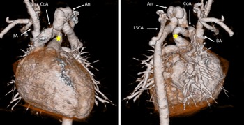
A Japanese girl born at 39 weeks 6 days, gestational maturity weighing 3.14 kg, was referred to our hospital at birth for a continuous cardiac murmur, Levine III, heard at the upper sternum. A haemangioma was also noted on the left side of the face and neck. Her left brachial and bilateral femoral pulses were weak, and blood pressure measured 82/40 mmHg in the right arm and 43/28 mmHg in the right leg. Chest X-ray showed cardiomegaly with a cardiothoracic ratio of 59%. Echocardiography revealed mid-transverse aortic coarctation with peak velocity of 3.0 m/s and a large ductus arteriosus with left-to-right shunt proximal to the coarctation. Chest CT showed a left aortic arch, coarctation at mid-portion of the transverse aortic arch, an adjacent tortuous aortic aneurysm located distally, and a large ductus arteriosus located proximally to the coarctation (Fig 1). The left common carotid artery was not identified, and the left subclavian artery originated distally from the descending aorta. No intracardiac defects were identified, except a patent fossa ovalis.
Preoperative enhanced 3-dimensional CT scan (Left: AP view, Right: PA view).
An = Aneurysm of the aorta; BA = Brachiocephalic artery; CoA = Coarctation of the aorta; LSCA = Left subclavian artery; ★ = Ductus arteriosus

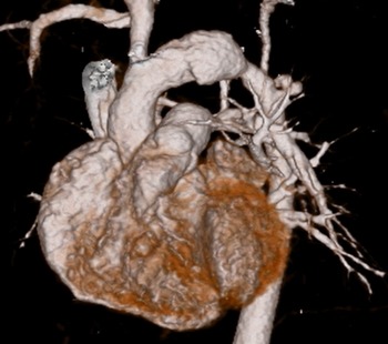
Gradually, congestive heart failure with tachypnoea progressed, and at 4 days of age, she was operated on through the midline sternal approach. The ascending aorta, aortic arch, descending aorta, and ductus arteriosus were carefully exposed without cardiopulmonary bypass. A narrow left common carotid artery was identified at the aortic aneurysm. The ductus arteriosus was ligated and divided, and aortic clamps were placed distal to the origin of the brachiocephalic artery and just proximal to the origin of the left subclavian artery. The narrow left common carotid artery was also clamped. The coarctation and its distal aortic aneurysm were completely excised, and the left common carotid artery was ligated since its lumen was obstructed without blood return. The distal aortic wall was thickened. The aortic reconstruction was performed by direct anastomosis of the posterior wall, and the anterior wall was augmented by 0.625% glutaraldehyde tanned pericardium. No pressure gradient between the right and left arms and lower legs and no neurological deficits were observed. Post-operative CT showed no residual obstruction of the aortic arch (Fig 2). MRI brain demonstrated a defect in the left vertebral artery and hypoplasia of the posterior cranial fossa. She is 4 years 8 months old, short-statured (-2.3 SD), but doing well without re-stenosis post-operatively.
Postoperative enhanced 3-dimensional CT scan.

Discussion
The Posterior fossa anomalies, Haemangiomas of the head and neck, Arterial, Cardiovascular, and Eye anomalies and ventral developmental defect acronym was coined by Frieden et al in 1996 to describe the association among malformations of the Posterior fossa malformations, Haemangiomas, Arterial anomalies, Coarctation of the aorta and cardiac defects, Eye anomalies, and sternal malformations or malformations of the abdominal midline Reference Frieden, Reese and syndrome1 . Its embryonic development has not been well clarified yet. Cervical and cerebral arterial, and cardiovascular anomalies are the most common and important associations Reference Metry, Heyer and Hess2 . Aortic coarctation is the most common cardiac anomaly (14.5%–19% of Posterior fossa anomalies, Haemangiomas of the head and neck, Arterial, Cardiovascular, and Eye anomalies and ventral developmental defect, 45% of cardiac anomalies) Reference Metry, Heyer and Hess2,Reference Bayer, Frommelt and Blei3 . Usually, coarctation of the aorta locates at the aortic isthmus. In the Posterior fossa anomalies, Haemangiomas of the head and neck, Arterial, Cardiovascular, and Eye anomalies and ventral developmental defect syndrome, however, it can exist at various regions of the aorta, more so in the transverse aortic arch. It involves long segments and is mostly associated with dilatation, aneurysms, and a torturous aorta at distal to the coarctation Reference Metry, Heyer and Hess2–Reference Sharma, Reddy and Ganigara5 .
Additionally, ductus arteriosus usually connects to the descending aorta distal to the coarctation and supplies the lower body: ductus dependent circulation. In our patient, ductus arteriosus was connected proximally to the coarctation; therefore, congestive heart failure progressed due to high pulmonary blood flow soon after birth. To the best of our knowledge, these associations have not been reported hitherto. Surgical interventions depend on regional morphology. The patient’s aortic arch was reconstructed by direct anastomosis of the posterior wall, along with anterior wall augmentation by auto-pericardium. In many patients with Posterior fossa anomalies, Haemangiomas of the head and neck, Arterial, Cardiovascular, and Eye anomalies and ventral developmental defect syndrome, graft interposition and patch augmentation are necessary in case of extensive aortic narrowing and aneurysms Reference Caragher, Scott and Siegel4,Reference Mery6,Reference Al-Khaldi, Alhabshan and Tamimi7 ; therefore, further observation of the reconstructed aortic arch is mandatory.
Acknowledgements
None.
Financial Support
This research received no specific grant from any funding agency, commercial or not-for-profit sectors.
Conflicts of Interest
None.
Ethical Standards
This article does not contain any studies with human participants.
Informed consent
The participant provided informed consent.