INTRODUCTION
A water-borne tropical disease, schistosomiasis, is caused by trematode worms infecting humans and some other mammals (Gryseels et al. Reference Gryseels, Polman, Clerinx and Kestens2006). These worms can be of several species collectively known as schistosomes. Schistosomes are remarkable: they live for years, sometimes decades, in what is putatively a very hostile environment – the blood of vertebrates – yet they solicit little if any reaction from either of their hosts' two defence systems: the immune response and mechanisms of haemostasis. To what factors is the prolonged survival, in spite of such potential adversity, attributable? Sophisticated mechanisms of immune evasion? Effective mechanisms for repairing damaged parasites? Are these the reasons it has been so difficult to develop an effective vaccine? Or, have we been targeting antigens that normally cannot be accessed by the immune effectors because they are presented like some bacterial antigens (McCallum et al. Reference McCallum, Schoenhals, Laakso, Clarke and Whitfield1989), some distance from exposed susceptible membranes? And, is this why it has proved to be extremely difficult to generate immune responses that alone can damage adult worms?
None of the above is an original question, but perhaps it will be useful to take a fresh look at the nature of the adaptation of the parasite to the host that may, in turn, allow the questions to be posed differently. The concept that the parasite shares a highly complex and intimate relationship with host tissues has already been well articulated in recent reviews written from an immunological perspective (Mountford and Trottein, Reference Mountford and Trottein2004; Capron et al. Reference Capron, Riveau, Capron and Trottein2005). For example, in the article by Capron et al. (Reference Capron, Riveau, Capron and Trottein2005), we read “where there is permanent cross-talk involving the parasite and multiple host factors, a single regulatory mechanism can hardly account for the complex picture occurring in these infections”. Our ideas of host and parasite interactions need to take account of this complexity. Other recent reviews (Verjovski-Almeida et al. Reference Verjovski-Almeida, DeMarco, Martins, Guimarães, Ojopi, Paquola, Piazza, Nishiyama, Kitajima, Adamson, Ashton, Bonaldo, Coulson, Dillon, Farias, Gregorio, Ho, Leite, Malaquias, Marques, Miyasato, Nascimento, Ohlweiler, Reis, Ribeiro, Sá, Stukart, Soares, Gargioni, Kawano, Rodrigues, Madeira, Wilson, Menck, Setubal, Leite and Dias-Neto2003; Hu et al. Reference Hu, Brindley, McManus, Feng and Han2004; Skelly and Wilson, Reference Skelly and Wilson2006; Van Hellemond et al. Reference Van Hellemond, Retra, Brouwers, van Balco, Yazdanbacksh, Shoemaker and Tielens2006; Liu et al. Reference Liu, Lu, Hu, Wang, Cui, Chi, Yan, Wang, Song, Xu, Wang, Zhang, Zhang, Wang, Xue, Brindley, McManus, Yang, Feng, Chen and Han2006) have emphasized the value of current efforts to sequence the schistosome genome (hereafter referred to as ‘the genome project’), and sequence data of individual molecules will help determine not only the identity, but also the location of proteins in the membrane or cytosol. The absence of homologues of many schistosome proteins in other genomes notwithstanding (Van Hellemond et al. Reference Van Hellemond, Retra, Brouwers, van Balco, Yazdanbacksh, Shoemaker and Tielens2006), these reviews have emphasized the potential value of studies of the proteome, lipidome and glycome in elucidating membrane structure and identifying vaccine candidates and potential drug targets.
With a wealth of new information becoming available from the application of new technologies, it is appropriate to reconsider some perspectives of host-schistosome interactions. We wish to portray the parasite as not so much engaged in an evolutionary arms race with the host but more as one of ‘peace with its host’, in which both contribute to well being. (The commonly expressed concept of the evolutionary arms-race is, of course, appropriate in some contexts, for example, with regard to mechanisms determining drug-resistance.) One extreme of this viewpoint is to consider the parasite so perfectly adapted that the host environment is not at all hostile. Remarkably, more than 2 decades ago, Davies et al. (Reference Davies, Hall, Targett and Murray1980) went so far as to suggest that the host immune system may in fact encourage the growth and development of parasites, a notion that is consistent with more recent observations indicating that helminth infections may be ‘good for us’ (reviewed by Doenhoff, Reference Doenhoff, Lachmann and Oldstone2006). In this review, the ideas expressed by Davies et al. (Reference Davies, Hall, Targett and Murray1980) will serve as a basis for a reconsideration of some of the features of schistosomes that adapt them to life in the mammalian host. We will examine the way in which these parasites interact with the mammalian environment and the extent to which this environment, particularly the immune defence system, serves as a ‘welcome mat’, as envisioned by Davies et al. (Reference Davies, Hall, Targett and Murray1980). In doing so, we may need to re-evaluate the function of molecules that we have assumed to be involved in immune evasion of the parasite. For example, the generally accepted implication of the ‘host antigen’ hypothesis is that adsorbed host molecules render the parasite largely undetectable by the immune system of the host (Smithers et al. Reference Smithers, Terry and Hockley1969; Clegg, Reference Clegg, Porter and Knight1974). First formulated to suggest that schistosomes survive in the face of an active inmmune response because of the presence of blood group antigens on their surfaces, this hypothesis has been expanded to include the presence of virtually every other host molecule subsequently found to be in or on the parasite, it being assumed that these all contribute to immune evasion. But each molecule may have a quite distinct function which, if understood, could help us understand some other features of adaptation.
Many of the topics covered in this review have been studied for more than 30 years, using biochemical, molecular biological, immunological, biophysical, cell biological and physiological approaches. However, it has been appreciated that some early studies, for example, those of the membrane structure and turnover, need to be reappraised using modern techniques, taking into account new understanding drawn from genomics, proteomics, genetics, biochemistry and biophysics, employing more precise microscopical techniques (Skelly and Wilson, Reference Skelly and Wilson2006). A multidisciplinary approach continues to be necessary if we wish to fully understand the complexity of host-parasite interactions. With this multidisciplinary attitude in mind, we will explore aspects of the surface membrane and tegument, and the excretory system of the Schistosoma cercaria, schistosomulum, immature and adult parasite and the egg. We will be guided by recent reviews and will draw also on knowledge of analogous mechanisms in cells of other organisms to suggest further directions in research on schistosomiasis.
THE ADULT WORM
Paired schistosome male and female worms can live for a long time, Schistosoma mansoni and S. japonicum mainly in the portal vein and S. haematobium in veins of the urogenital system. Although the literature is unclear as to the exact length of time worms can survive (half-lives of several years have been estimated and some individual cases of several decades recorded (Gryseels et al. Reference Gryseels, Polman, Clerinx and Kestens2006), the relatively prolonged survival of worms in the bloodstream of the host suggests a very successful adaptation to the environment. Because the control of schistosomiasis by immunological means has been a clear objective for a long time, most research has focused on the interactions of the parasite with the immune system. Consequently, the influence of, and interactions between the parasite and other host factors, such as hormones, blood pressure, blood iron, ion and gas concentrations, and the multifarious factors involved in blood clotting, have received much less attention. However, it is known that the parasite requires cholesterol, iron, essential fatty acids, purines, essential amino acids and vitamins, all of which it acquires from the host bloodstream (Rumjanek, Reference Rumjanek, Rollinson and Simpson1987). That the parasite also responds to changes in the metabolism of the host is indicated by results from the genome project, showing that the parasite has genes for receptors for host hormones (Verjovski-Almeida et al. Reference Verjovski-Almeida, DeMarco, Martins, Guimarães, Ojopi, Paquola, Piazza, Nishiyama, Kitajima, Adamson, Ashton, Bonaldo, Coulson, Dillon, Farias, Gregorio, Ho, Leite, Malaquias, Marques, Miyasato, Nascimento, Ohlweiler, Reis, Ribeiro, Sá, Stukart, Soares, Gargioni, Kawano, Rodrigues, Madeira, Wilson, Menck, Setubal, Leite and Dias-Neto2003). To date, the respective locations of the receptors that facilitate uptake of different host factors by the parasite are unknown, but have been assumed to be in or on the surface membrane (Davies and Pearce, Reference Davies and Pearce1995; LoVerde et al. Reference LoVerde, Niles, Osman, Wu, Secor and Colley2005).
The morphology of the adult parasite is highly complex, and the great morphological differences between male and female worms (i.e. dimorphism) are reflected and accentuated by similarly great differences in gene expression (Grevelding et al. Reference Grevelding, Sommer and Kunz1997; LoVerde et al. Reference LoVerde, Niles, Osman, Wu, Secor and Colley2005; Fitzpatrick and Hoffmann, Reference Fitzpatrick and Hoffmann2006) and metabolism (Cornford, Reference Cornford1985; Rumjanek, Reference Rumjanek, Rollinson and Simpson1987). The organ systems (gut, nervous system and excretory system) of male and female worms have different distributions in the whole organism and may have very different mechanisms regulating metabolic pathways. For example, the excretory system is developed into a highly complex organelle in both sexes, but its regulation and response to drugs is different between males and females (Sato et al. Reference Sato, Kusel and Thornhill2002; Oliveira et al. Reference Oliveira, Kusel, Ribeiro and Coelho2006). However, descriptions of this and other features of female worms are relatively rare, as work has often only been done on the male.
The schistosome surface
The surface membrane of infective larvae (cercariae) is a single bilayer covered by a thick glycocalyx. The glycocalyx is lost and the membrane transforms into a double bilayer during the penetration of mammalian skin, and except for the occurrence of some multiple bilayer regions this structure remains a double bilayer during the development of the parasite into the adult stage in the mammalian host (Hockley and McLaren, Reference Hockley and McLaren1973). The structure and other details of the double bilayer membrane were described by. McLaren (Reference McLaren1980) in a classical book-length review of the schistosome surface. There are excellent, more recent reviews by Skelly (Reference Skelly, Secor and Colley2005), Van Hellemond et al. (Reference Van Hellemond, Retra, Brouwers, van Balco, Yazdanbacksh, Shoemaker and Tielens2006), and Skelly and Wilson (Reference Skelly and Wilson2006) on the proteins, enzymes, carbohydrates and host molecules exposed by the parasite to the host environment, and by Jones et al. (Reference Jones, Gobert, Zhang, Sunderland and McManus2004) on the cytoskeletal structures in the surface.
The structure of the schistosome surface membrane
Box 1 outlines some features of the plasma membranes of mammals and other organisms that may serve as a useful comparison for what is known about the surface of schistosomes. Do the individual membranes of the double membrane have the same structure as conventional plasma membranes? As in the classical work of Singer and Nicolson (Reference Singer and Nicolson1972) on the fluid mosaic structure of biological membranes, evidence about the schistosome membrane must be drawn from a number of disciplines.
BOX 1 Brief review of mammalian cell membrane structure
Singer and Nicolson proposed in 1972 the fluid mosaic model for the structure of biological membranes, based on a large amount of data derived from biophysical and biochemical techniques. The bilayer of lipids was considered to be largely fluid, with most lipids moving with high rates of lateral diffusion, and proteins within the bilayer either diffusing more slowly than the lipids, or being immobile due to their being tethered by cytoskeletal proteins. This major advance was supported by overwhelming evidence from many plasma and internal cytoplasmic membranes. However, this model has now been modified since some lipids have been shown to aggregate together in regions called lipid domains or lipid rafts. The predominant lipids in the rafts are cholesterol and sphingomyelin. Such aggregates associate with proteins with a glycolipid anchor (GPI-linked proteins (Magee and Parmryd, Reference Magee and Parmryd2003)). Signalling molecules and cytoskeletal proteins are also found in these rafts. The evidence for the existence of the rafts, which can vary in size is drawn from numerous disciplines, ranging from cell fractionation to confocal microscopy. There is much experimental evidence that membrane rafts are regions of (1) entry and release of some viruses, (2) exocytosis from mast cells, (3) T and B cell signalling and (4) assembly of complex membrane bound enzymes, such as nitric oxide synthase.
Part of the cytoskeleton of the eukaryotic cell acts as a support to the plasma membrane. This is most clearly seen in the mammalian erythrocyte. The cytoskeleton is involved in many other activities, such as signalling processes (Funatsu et al. Reference Funatsu, Kumanogoh, Sokawa and Maekawa2000).
Signalling across membranes is well described for most mammalian membranes, but where the membrane has a complex structure, such as the double membrane of the gram negative bacterium, the nucleus, the mitochondrion, or the chloroplast signalling processes occur but are less well understood (Livigni et al. Reference Livigni, Scorziello, Agnese, Adornetto, Carlucci, Garbi, Castaldi, Annuziato, Avvedimento and Feliciello2006).
Membrane proteins exposed to the external environment are those involved in transport of ions and metabolites, hormone and growth factor receptors, histocompatibility molecules and a variety of enzymes. Many of these proteins are glycosylated and undergo conformation changes on binding to ligand.
Diffusion of hydrophilic molecules through or within membranes can be a process of simple diffusion, facilitated diffusion, or active transport, while hydrophobic compounds readily dissolve in the lipid bilayer and transfer from there into the cytoplasm of the cell.
There is evidence for the existence of aqueous pores in some membranes. Hepatocyte plasma membranes have pores containing aquaporin 9, which are increased in number after exposure to glucagons (Gradilone et al. Reference Gradilone, Garcia, Huebert, Tietz, Larocca, Kierbel, Carreras, Larusso and Marinelli2003). Gram-negative bacterial membranes have porins that makes them permeable to small molecules. The mitochondrial outer membrane also contains porins, which assemble to form pores that allow access of molecules up to 10 kDa to the inner membrane (Freitag et al. Reference Freitag, Neupert and Benz1982). The inner membrane can develop permeability transition pores (Kroemer et al. Reference Kroemer, Zamzami and Susin1997).
The nuclear membrane of many cells has pores visible by electron microscopy. The chloroplast outer membrane contains a protein OEP 16 which allows access to a variety of small molecules, such as triosephosphate and dicarboxylic acid.
Many cell surface membranes are covered with a layer of carbohydrate rich molecules forming a glycocalyx. This may have a protective function as in the enterocytes, and the surface pH properties of MDCK cells (Kovbasnjuk and Spring, Reference Kovbasnjuk and Spring2000) may be profoundly modified by its presence. The glycocalyx can act as a transducer of fluid shear stress across the membrane (Thi et al. Reference Thi, Tarbell, Weinbaum and Spray2004). The adenosine receptor can modify the glycocalyx properties on ligand binding.
Turnover of the plasma membrane of mammalian cells can occur by ‘ectodomain shedding’ of integral membrane proteins, release or shedding of receptors, shedding of intact membrane vesicles, and endocytosis of membrane components, with degradation in lysosomes or proteosomes. The incorporation and removal of membrane proteins can be measured and some authors suggest synchronous insertion of membrane proteins while others suggest that low molecular weight proteins behave differently from those of high molecular weight. The carbohydrate moieties of membrane glycoproteins can have a half-life much less than the rest of the molecule. Glycoproteins and glycolipids in membrane lipid rafts have a different half-life than non-raft macromolecules.
Lipids themselves have very different half-lives according to their function, and their ability to interact with serum proteins or other cells.
Repair of plasma membranes can involve the exocytosis of lysosomes and is regulated by calcium ions (Reddy et al. Reference Reddy, Caler and Andrews2001) and endosomes can be involved in fusion also (Keefe et al. Reference Keefe, Shi, Feske, Massol, Navarro, Kirchhausen and Lieberman2005) during perforin-induced repair. The involvement of vesicle-associated proteins such as SNAP-25, synaptobrevin and syntaxin have been implicated in plasma membrane resealing in sea urchin eggs and 3T3 fibroblast cells. Exocytosis is a process involving budding, targeting and docking, which are highly regulated at each step. Families of proteins, called SNAREs, give rise to this specificity. Each vesicle SNARE (v-SNARE) selectively interacts with its counterpart, target-membrane SNAREs (t-SNAREs) in vesicle release and fusion with membranes (plasma membrane or other compartments).
The functions of surface membrane enzymes can range from phosphate transport (alkaline phosphatase), sodium and potassium transport (Na+ K+ ATPase), nucleotide transport (5′ nucleotidase), signalling (adenyl cyclase, sphingomyelinase) to cytoplasmic organelles, cytoskeleton and enzyme pathways.
Electron microscopy
Hockley and McLaren (Reference Hockley and McLaren1973) showed that the surface of the adult is a double bilayer, the outer bilayer being prominent in electron micrographs because it bound uranium salts, which was, in turn, possibly due to its high phospholipid content. Using an electron microscope, the juxtapositioning of the two bilayers gave the appearance of a pentalaminate structure. However, very few cytoskeletal structures could be seen beneath the inner bilayer, and the properties of the narrow area between the two lipid bilayers were not defined.
Using freeze fracture methods, Hockley et al. (Reference Hockley, McLaren, Ward and Nermut1975) carried out a painstaking study of the distribution of intramembranous particles in all of the monolayers in the double bilayer of the adult worm, studied either as fresh tissue or after fixation in glutaraldehyde. These authors showed that the innermost and outermost monolayers of adult worms contained the majority of intramembranous particles, and they speculated that the outermost monolayer had the properties of an inner monolayer of a mammalian cell (Hockley et al. Reference Hockley, McLaren, Ward and Nermut1975). However, this proposal is not considered likely, since, although some of the particles in the outermost monolayer may have been derived from the host (i.e. were host antigens), the study by Harnett et al. (Reference Harnett, Kusel and Barrowman1985) showed the presence of parasite-specific antigens in the outer surface of the glutaraldehyde-fixed adult male worm. Subsequently, it has been shown that the outermost monolayer of schistosomes is rich in lectin-binding sites and has a glycocalyx (Schmidt, Reference Schmidt1995), thus being distinct from a cytoplasmic monolayer.
The conclusions from electron microscopy and freeze-fracture studies are thus that the double bilayer consists of 2 apposed bilayers, with a glycocalyx on the outermost monolayer, but a region of very uncertain composition between the bilayers.
Identification of surface components
Early studies (Snary et al. Reference Snary, Smith and Clegg1980; Roberts et al. Reference Roberts, MacGregor, Vojvodic, Wells, Crabtree and Wilson1983) were carried out with the enzyme lactoperoxidase, which, in the presence of hydrogen peroxide and radioactive iodine, catalyses the labelling of surface protein and lipid. The advantage of using radioactive iodine and lactoperoxidase is that only proteins that are exposed in the outer monolayer become labelled, since lactoperoxidase has a molecular weight of 90 kDa and will not penetrate membrane pores. Therefore, the iodine should not label constituents of inner bilayers. Using this method, Roberts et al. (Reference Roberts, MacGregor, Vojvodic, Wells, Crabtree and Wilson1983) showed that the outer bilayer of freshly perfused adult worms carried 13 proteins that could be labelled. After renewing the outer bilayer in vitro, as a result of turnover induced by cationised ferritin, they showed that 3 additional proteins were accessible to the labelling. These proteins were obscured by the presence of molecules, proposed to be derived from the host.
Through the application of genomic and proteomic technologies, Braschi et al. (Reference Braschi, Curwen, Ashton, Verjovski-Almeida and Wilson2006) were able to identify proteins in various membrane fractions. They fractionated purified tegumental membranes by 3 consecutive chemical extractions to yield a final pellet (designated 4) that contained most of the integral membrane proteins. Three other fractions contained (1) cytosolic proteins, (2) cytoskeletal proteins and (3) membrane-associated proteins. The identity of many proteins in each of these fractions was established by proteomic analysis. The presence of a calcium pump (fraction 4) and several calcium-binding proteins (fractions 1 and 3) suggested a role for calcium in the regulation of tegumental function, evidence for which has also come from biochemical data (Cunha et al. Reference Cunha, Meyer-Fernandes and Noel1992). No cytokine receptors, such as the TGFβ receptor (Davies and Pearce, Reference Davies and Pearce1995; LoVerde et al. Reference LoVerde, Niles, Osman, Wu, Secor and Colley2005) were found, and the glucose transporter SGPT1 was found, but not SGPT4. Braschi and Wilson (Reference Braschi and Wilson2006) went on to evaluate the location of some surface-exposed proteins by labelling with biotin reagents which, although membrane impermeant, may diffuse through membrane pores. Two biotin reagents, a short and a long form, were used and labelled proteins were isolated in sequential chemical steps to yield 5 fractions. The proteins were isolated from each extract after binding to a streptavidin column. Possible locations for some of the labelled proteins could be suggested. Thus, 3 enzymes, alkaline phosphatase, phosphodiesterase and ATP-diphosphohydrolase were located in the inner bilayer and it was proposed that phosphorylated substrates gained access to these enzymes, as did the biotin reagents, through aqueous pores. The presence of pores, measuring 13·4 angstroms in diameter, had previously been suggested by Davies and Pearce (Reference Davies and Pearce1995). There is no direct evidence for these pores. Indeed, the work of El Ridi and Tallima (Reference Tallima2006) suggests that alterations in the sphingomyelin metabolism are sufficient to alter the exposure of enzyme proteins, antigens and receptors. Thus, pores may not exist! Box 2 suggests some experiments to determine whether the pores have reality.
BOX 2 We suggest here some experiments that might be carried out to determine the existence and size of the pores
(1) The size of the pores: we assume they will not be larger than 30 kDa (Braschi and Wilson, Reference Braschi, Curwen, Ashton, Verjovski-Almeida and Wilson2006).
(a) We assume that alkaline phosphatase, acetylcholinesterase and ATP-diphosphohydrolase are on the inner membrane. The use of phosphorylated or acylcholine substrates of increasing molecular size, or the normal substrate attached to dextrans or peptides of increasing size should show a cut off point in hydrolysis when the pore size is exceeded.
(b) We assume that both the outer and inner membrane carry net negative charges, so the penetration of the pores by positively charged tetramethyl rhodamine poly-L-lysines, or other peptides of different sizes should be detectable by fluorescence or confocal microscopy, provided that the negative charges on the outer surface were neutralized, for example by poly-L-lysines or cationized ferritin of size larger than 30 kDa. This could only be done on fixed worms, since positively charged peptides can stimulate turnover or damage the surface.
(c) Sheets of isolated membrane, or whole vesicles might be subjected to negative staining. This may reveal the pores (Dourmashkin et al. Reference Dourmashkin, Deteix, Simone and Henkart1980; Hayashi et al. Reference Hayashi, Hironaka, Shikata, Ogawa, Ota, Wada, Kamata, Ota and Makino2001).
(d) Using monoclonal antibodies against epitopes on the enzymes in the inner bilayer, it should be possible to detect binding of Fv fragments (ca 12 kDa), and not of whole antibody, if the Fv fragment penetrates the pore.
(e) The pore proteins may be associated with caveolae and caveolin (Bàthori et al. Reference Bàthori, Parolini, Tombola, Szabò, Messina, Oliva, De Pinto, Lisanti, Sargiacomo and Zoratti1999).
(2) Presence of proteins capable of forming pores.
From the schistosome proteome, an aquaporin 9 has been found. Porins and claudins might be expected and models for the assembly of mitochondrial transition pores, bacterial and chloroplast pores might be considered. Electrophysiological techniques have been used on vesicles from endoplasmic reticulum (Simon et al. Reference Simon, Blobel and Zimmerberg1989).
The pores might be expected to be very dynamic, since Cornford (Reference Cornford1985) showed pairing of male and female parasites stopped any transport of acidic amino acids. This could have been due to a rapid closure of pores. Antibodies against such pore proteins may inhibit transport of small molecular weight compounds.
It is possible that some mechanism coupled to the release of ATP, causes ATP binding cassette proteins, such as CFTR, to form a pore (Gadsby and Nairn, Reference Gadsby and Nairn1999). A synthetic peptide forms voltage-gated porin-like ion channels in lipid bilayer membranes (Thundimadathi et al. Reference Thundimadathil, Roeske and Guo2005).
Tetraspanins and annexins were located between the bilayers, tethering of the two bilayers being their suggested function (Braschi and Wilson, Reference Braschi and Wilson2006). Sm29, a secreted protein, was also placed between the bilayers. Host proteins, such as IgM, IgG3, IgG1 and complement component C3, were located in the outer bilayer. Altogether, 24 proteins were grouped according to possible functions, such as membrane structural proteins, membrane enzymes, transporters, cytoskeletal proteins and cytosolic proteins (Braschi and Wilson, Reference Braschi and Wilson2006).
It is of interest to compare these very recent results with the earlier evidence obtained using the lactoperoxidase method. Roberts et al. (Reference Roberts, MacGregor, Vojvodic, Wells, Crabtree and Wilson1983) reported that 13 proteins could be labelled with lactoperoxidase on freshly perfused worms, and 3 additional proteins were detected after stimulation of membrane turnover. As lactoperoxidase cannot penetrate into the inner membrane, these proteins are very likely to be located in the outer bilayer, though there have been reports of internal proteins being labelled by this method (for example, in Aeromonas (Dooley and Trust, Reference Doolley and Trust1988)), but usually lactoperoxidase and biotin labelling give similar results (Apps et al. Reference Apps, Percy and Perez-Castineir1989). All of the proteins detected employing the lactoperoxidase method have molecular weights corresponding to those found in the study by Braschi et al. (Reference Braschi and Wilson2006). However, in this study, only 4 proteins (IgM, IgG3, IgG1 and complement C3 (C3c/C3dg fragments) were located in the outer bilayer. This discrepancy could perhaps be resolved by using precise methods (cf. Braschi et al. Reference Braschi and Wilson2006) to identify which proteins are present in the outer bilayer, following their isolation by the methods described in Roberts et al. (Reference Roberts, MacGregor, Vojvodic, Wells, Crabtree and Wilson1983), in which cationised ferritin is allowed to stimulate the turnover of this layer.
Alkaline phosphatase is a glycoprotein that occurs in the outer monolayer of mammalian plasma membranes (Hirano et al. Reference Hirano, Iiizumi, Mori, Toyoshi, Sugiura and Iino1985). Braschi and Wilson (Reference Braschi and Wilson2006) detected this enzyme in the inner bilayer of the schistosome. Thermodynamic considerations, which allow very little mixing of hydrophobic and hydrophilic moieties (Singer and Nicolson, Reference Singer and Nicolson1972), indicate that its carbohydrate chain is unlikely to cross into the outer bilayer. Thus, the bulk of the molecule may lie in the space between the bilayers. This argument holds for any other glycoprotein suggested to be located in the inner bilayer, such as ATP-phosphohydrolase, acetylcholinesterase and the heavy subunit of the amino acid transporter (Skelly, Reference Skelly, Secor and Colley2005).
Some proteins that were expected to be labelled were not detected by Braschi and Wilson (Reference Braschi and Wilson2006), for example, the glucose transporter SGPT4, the amino-acid transporters, and caveolin. This could have been due to the low abundance of these molecules in the membrane preparation, the insensitivity of the technique, or the protected state or burial of the lysines and other amino-groups with the potential to react with the biotin probes.
The inner bilayer of the double bilayered surface of schistosomes appears to have conventional lipid membrane properties. The outer bilayer has been shown to be secreted on to the inner bilayer and has been termed the membranocalyx (Wilson and Barnes, Reference Wilson and Barnes1977; Roberts et al. Reference Roberts, MacGregor, Vojvodic, Wells, Crabtree and Wilson1983). The freeze fracture studies of Hockley et al. (Reference Hockley, McLaren, Ward and Nermut1975) suggest that the majority of intramembranous particles are found in the outermost monolayer of the outer bilayer (membranocalyx) and innermost monolayer of the inner bilayer. Further work on the isolation of the outer bilayer (membranocalyx) is needed to explain why few proteins are found in the outer monolayer using the biotin labelling technique. The function of some molecules in the space between the two bilayers may be to anchor the outer to the inner bilayer; the properties of the tetraspanins and annexins, found there by Braschi and Wilson (Reference Braschi and Wilson2006), would be ideal for this since both tetraspanins and annexins interact with membranes and could serve as molecules connecting the bilayers (Braschi et al. Reference Braschi and Wilson2006). Since many membrane enzymes and receptors are glycoproteins (Skelly and Wilson, Reference Skelly and Wilson2006), the hydrophilic glycanic residues are likely to be located in the aqueous environment within this inter-membrane space.
Lipid composition
Evidence of the presence and quantity of specific lipids is of crucial relevance for understanding the structure and physiology of the membrane (Allan et al. Reference Allan, Payares and Evans1987; Brouwers et al. Reference Brouwers, Skelly, Van Golde and Tielens1999). There is a large amount of sphingomyelin in the isolated membrane (Allan et al. Reference Allan, Payares and Evans1987), and it may have a stabilizing role with cholesterol, as was shown for vesicles and monolayers, and for mammalian membranes (Albi et al. Reference Albi, Peloso and Magni1999). Sphingomyelin can act as a signalling molecule (Testi, Reference Testi1996) as well as a molecule that can stabilize membrane rafts. Liberation of ceramide from sphingomyelin can stimulate vasodilator pathways in mammalian smooth muscle, as well as activate vesicle membrane fusion pathways and pathways for calcium release. Fluorescence quenching studies showed that sphingomyelin was likely to be present in the outermost monolayer of the adult membrane (Redman and Kusel, Reference Redman and Kusel1996), so that any signalling event would have to cross both bilayers. The proportion of the phosphatidylcholine fraction of the surface membrane is not very different from other plasma membranes (Allan et al. Reference Allan, Payares and Evans1987). Brouwers et al. (Reference Brouwers, Skelly, Van Golde and Tielens1999) showed that this fraction is rapidly labelled with radioactive fatty acids, and these fatty acids can be transferred after deacylation to form triglycerides. Little radioactive lipid was found in the growth medium, and it was concluded that no rapid release of surface membrane was occurring by sloughing. This finding is different from that of Kusel et al. (Reference Kusel, Mackenzie and McLaren1975), who postulated that sloughing occurred in the form of small vesicles. Further work is required to resolve this discrepancy.
The lipid 5-octadecenoic fatty acid is rare in nature. Its presence in the worm is almost entirely restricted to the single phosphatidylcholine species, 1-palmitoyl-2-(5-octadecenoyl) phosphatidylcholine, which is abundant in, and confined to the outer tegumental membrane. Although schistosomes do not synthesize fatty acids de novo, the parasite probably manufactures 5-octadecenoic acid in the cells beneath the tegument (Brouwers et al. Reference Brouwers, Versluis, Van Golde and Tielens1998) as it is absent from the host's blood. The octadecenoic acid may have a signalling function in the parasite (Brouwers et al. Reference Brouwers, Versluis, Van Golde and Tielens1998) or have an effect on host red cell or endothelial cell membranes, or even in signalling between parasites. In mammalian cells, phosphatidylcholine has both a structural and a signalling function (Billah, Reference Billah1993). This lipid is likely to be a major component of both bilayers, so any signalling pathway involving phosphatidylcholine could act through a single or a double bilayer.
The uptake and distribution of fatty acids in the tissues of schistosomula and adult worms may involve Sm14, a fatty acid-binding protein that has been a potential vaccine candidate and whose crystal structure has been determined (Angelucci et al. Reference Angelucci, Johnson, Baiocco, Miele, Brunori, Valle, Vigorosi, Troiani, Liberti, Cioli, Klinkert and Bellelli2004). These authors found an interesting increase in affinity of fatty acids for this protein at a low pH, and suggested that this property could allow it to transfer fatty acids from host plasma to worm tissues (Angelucci et al. Reference Angelucci, Johnson, Baiocco, Miele, Brunori, Valle, Vigorosi, Troiani, Liberti, Cioli, Klinkert and Bellelli2004). A low pH is expected in the microenvironment of the inner membrane surface, but it may be influenced by the membrane-bound carbonic anhydrase that is present in the membrane fraction (Braschi and Wilson, Reference Braschi and Wilson2006). Moffat and Kusel (Reference Moffat and Kusel1992) showed an uptake and transfer of fluorescent fatty acids from the membrane to the gut. Lipid bodies, small membrane-bound lipid droplets, are thought to be crucial in intracellular signalling and regulation of metabolism in mammalian cells, but their function in schistosomula has not been established. In leucocytes, these bodies are sites of eicosanoid metabolism (Weller and Dvorak, Reference Weller and Dvorak1994) and thus they may be very relevant factors facilitating early schistosome migration, since eicosanoids have been shown to inhibit the exit of Langerhans cells from the skin (Angeli et al. Reference Angeli, Faveeuw, Roye, Fontaine, Tessier, Capron, Wolowczuk, Capron and Trottein2001).
Cholesterol is also found in the isolated tegument (Allan et al. Reference Allan, Payares and Evans1987). It seems that cholesterol can be taken up from medium containing bovine serum albumin as a carrier (Haseeb et al. Reference Haseeb, Eveland and Fried1985). Cholesterol can stabilize membranes by interacting with sphingomyelin, and is often found in lipid rafts in mammalian plasma membranes associated with sphingomyelin, glycolipid and proteins anchored by the GPI moiety. Cholesterol can be shown to be in the outer monolayer of the adult membrane by staining with fluorescent filipin (El Ridi et al. Reference El Ridi, Tallima, Mohamed and Montash2004). The manner by which cholesterol is inserted in schistosome membranes is different from that in erythrocytes, because streptolysin O, a bacterial toxin specific for cholesterol and that lyses erythocytes, does not damage the schistosomulum or adult surface (Kusel et al. Reference Kusel, Stones and Harnett1981). Amphotericin B is a polyene antibiotic from Streptomyces nodosus and binds to sterols in membranes. It can damage the schistosomulum surface, but not the adult surface (Kusel et al. Reference Kusel, Stones and Harnett1981). Therefore, it can be concluded that the lipid-protein organization of the adult membrane is very different from that of the schistosomulum and both are distinct from the erythrocyte membrane.
The organization of proteins and lipids in the bilayers
Probes have been employed to dissect the organization of membranes. Such probes have included (i) fluorescent molecules whose structures allow access to different regions and different depths of the bilayers, (ii) enzymes with specificities towards various lipids, a variety of toxins which bind to particular lipids, and (iii) some proteins and fluorescent lectins which bind to specific sugars on glycoproteins and glycolipids (Kusel and Gordon, Reference Kusel and Gordon1989).
In investigations of the lipid phase of the double bilayer of living parasites, membrane-active compounds (Dingle and Lucy, Reference Dingle and Lucy1965), and a variety of detergents have been used as disruptive agents to worm surfaces labelled with radioactive chromium and iodine-labelled lectin (e.g. wheat germ agglutinin), to assess the extent and nature of the damage so induced (Kusel et al. Reference Kusel, Stones and Harnett1982). From this work, it was suggested that the bilayers were composed of domains, which might be considered to be equivalent to the lipid rafts and appear to be universally present in membranes (Simons and Ikonen, Reference Simons and Ikonen1997). Rafts are enriched in proteins with glycosyl phophatidylinositol (GPI)-anchors. In the model proposed by Braschi and Wilson (Reference Braschi and Wilson2006), all GPI moieties are located in the inner bilayer and, by extrapolation from other organisms and mammalian cells (Magee and Parmryd, Reference Magee and Parmryd2003), might be expected to be associated with rafts. Racoosin et al. (Reference Racoosin, Davies and Pearce1999) treated surface membranes with detergents and demonstrated the existence of a fraction that is detergent-resistant, a feature of lipid rafts. Furthermore, they identified caveolin, a raft-associated protein in mammalian membranes, as a molecule that concentrates in this fraction. The importance of the heterogeneity of rafts in cells has been emphasized by Pike (Reference Pike2004), who suggested that the observed great variability in raft composition was dependent upon the structure of the proteins present in the membrane at a particular time. Some rafts are cholesterol-free, and the composition of all rafts can undergo alteration depending on changes in environmental conditions. The existence of rafts in schistosome surface membranes is of great interest, since their presence may explain not only the great variability in lipid composition between individual parasites (Tallima et al. Reference Tallima and El Ridi2005), but also heterogeneity on the surface of an individual schistosome. Different rafts are thought to have different functions in the membrane, and a non-genetic variability in the surface of schistosomes may thus occur due to the dynamic nature of the rafts. The process of transmembrane signalling in lipid rafts is described in Box 3. The heterogeneity in surface properties among individual parasites is illustrated clearly by the patterns of binding of antibody by ‘clones’ of schistosomula (Vieira and Kusel, Reference Vieira and Kusel1991), and in the range of susceptibilities of individual lung stages in a population to treatment with methyl-beta-cyclodextrin that removes cholesterol from the surface membrane (El Ridi et al. Reference El Ridi, Tallima, Mohamed and Montash2004). Many other examples of this heterogeneity are described in the article by Jones and Kusel (Reference Jones and Kusel1989).
BOX 3 Receptor signalling in membrane lipid rafts
The hypothesis that the fluid bilayered membrane (Singer and Nicolson, Reference Singer and Nicolson1972) contained lipid rafts is described in Box 1. The biological processes mediated by rafts are numerous and also listed there. Since many of the receptor proteins and ion channels found in the schistosome surface may be associated with rafts, we summarize here some literature from mammalian cells concerning rafts and receptor signalling in rafts.
The demonstration that receptors are located in rafts has often depended on (a) their presence in detergent-resistant membrane fractions (b) the change in receptor activity when cholesterol is removed from the cell most commonly by methyl-beta-cyclodextrin (MβCD) and (c) coprecipitation of receptor and effector signalling molecules (such as Syk, Fyn, phospholipase C).
Some authors are sceptical about the value of (a) and (b) and concentrate on observations from confocal and electron microscopy, often comparing evidence from both (Brown, Reference Brown and Grenfell2001; Gaus et al. Reference Gaus, Gratton, Kable, Jones, Gelissen, Kritharides and Jessup2003).
In the mast cell, it was shown that the detergent-resistant fraction contained IgE receptor, Thy1 and all effector signalling molecules. Electron microscopy showed that cross-linking of IgE receptor induced aggregation into domains, which did not contain Thy1 molecules. The IgE receptor containing domains contained some but not all effector signalling molecules, and also proteins involved in endocytosis (Wilson et al. Reference Wilson, Pfeiffer and Oliver2002, Reference Wilson, Steinberg, Liederman, Pfeiffer, Surviladze, Zhang, Samelson, Yang, Kotula and Oliver2004). The raft organization is seen as regulating the signalling process.
Epidermal growth factor receptor is less active in rafts than when it is released from rafts by MβCD, and so here the rafts are inhibiting activity of the receptor (Pike and Casey, Reference Pike and Casey2002; Roepstorff et al. Reference Roepstorff, Thomsen, Sandvig and van Deurs2002). The protective effects of rafts can be seen when, after ligand cross-linking by the IgE receptor, the src kinase Lyn is concentrated in rafts, which maintain its activity, as the raft environment prevents it from being dephosphorylated.
The cytoskeleton is intimately involved in the assembly and organization of the rafts (Holowka et al. Reference Holowka, Sheets and Baird2000, Reference Holowka, Gosse, Hammond, Han, Sengupta, Smith, Wagenknecht-Wiesner, Wu, Young and Baird2005), which is pictured as a ‘platform’. This is powerfully seen when the binding of ligand to the T-cell receptor is studied, and the term ‘immunological synapse’ describes the complexity of the interactions (Villalba et al. Reference Villalba, Bi, Rodriguez, Tanaka, Schoenberger and Altman2001; Valensin et al. Reference Valensin, Paccani, Ulivieri, Mercati, Pacini, Patrussi, Hirst, Lupetti and Baldari2002).
All tissue cells appear to have rafts involved in transmembrane signalling, and there are mechanisms to produce very large rafts or ‘super-rafts’ due to the presence of galectin, a lectin that binds galactose in intestinal brush border cells (Braccia et al. Reference Braccia, Villani, Immerdal, Niels-Christiansen, Nystrom, Hansen and Danielsen2003).
Ion channels often function in rafts (Janas et al. Reference Janas, Priest and Malhotra2005) and the carbohydrate hyaluronan (containing hyaluronic acid) binds to its receptor CD44 and the resulting signalling requires rafts (Singleton and Bourguinon, Reference Singleton and Bourguignon2004).
Signalling molecules in schistosomes
We can see from the above that rafts are likely to be involved in the organization of signalling in the tegument and other tissues. What receptors and signalling molecules have been reported in schistosomes?
Growth factor receptors (receptor tyrosine kinases) are likely to be important in male–female signalling, and epidermal growth factor receptor has been cloned (Ramachandran, Reference Ramachandran, Skelly and Shoemaker1996). The insulin receptor has been detected. A similar molecule with a venus fly trap module is known (Saule et al. Reference Saule, Vicogne, Delacre, Macia, Tailleux, Dissous, Auriault and Wolowczuk2005; Vicogne et al. Reference Vicogne, Pin, Lardans, Capron, Noel and Dissous2003).
The TGF beta receptor (transforming growth factor beta, a receptor serine threonine kinase) has been shown to be functional in a heterologous system (Davies et al. Reference Davies, Shoemaker and Pearce1998) and can be activated by protein 14-3-3. Protein 14-3-3 is described in the main text.
Molecules down stream from these receptors have been cloned – the small G protein Ras, GAP (GTPase activator protein), and mitogen activated protein (MAP) and MAP kinases have been shown to be developmentally regulated in the male-female interaction (Schussler et al. Reference Schussler, Grevelding and Kunz1997).
Non-receptor tyrosine kinases are important in many growth processes and TK5 is homologous to Fyn from the Src family (Kapp et al. Reference Kapp, Schussler, Kunz and Grevelding2001), and TK4 corresponds to a Syk protein (Knobloch et al. Reference Knobloch, Kunz and Grevelding2006) and TK3 is an active Src-like kinase (Kapp et al. Reference Kapp, Knobloch, Schussler, Sroka, Lammers, Kunz and Grevelding2004). Sm Fes, a fes/fps tyrosine kinase has been found (Oliveira et al. Reference Oliveira, Rodrigues, Romanha and Bahia2004) localized in the tegument in intact larval parasites (Bahia et al. Reference Bahia, Mortara, Kusel, Andrade, Ludolf, Kuser, Avelar, Trolet, Dissous, Pierce and Oliveira2007).
Receptors (e.g. TNF alpha receptors) that can lead to sphingomyelin or phosphatidyl choline breakdown have not been found as yet, but many of the enzymes relevant to the metabolism of these lipids have been shown to be active (Redman et al. Reference Redman, Kennington, Spathopoulou and Kusel1997; Brouwers et al. Reference Brouwers, Skelly, Van Golde and Tielens1999; Tallima et al. Reference Tallima and El Ridi2005).
If we consider the surface of the adult male schistosome, for instance, the membrane occupies a large surface area, covering a large number of various sensory organs, spines and tubercles and is greatly pitted and folded (McLaren, Reference McLaren1980). The same applies to earlier life-cycle stages, such as the schistosomula and lung stage, but not the eggshell. To cover so many structures and present such a pitted surface with many curves and folds, membrane composition is likely to vary throughout the worm surface, leading to differences in membrane permeability in different areas (Kusel and Gordon, Reference Kusel and Gordon1989) Rafts in the membrane are likely to allow for such versatility in membrane structure and these ideas have been reviewed for mammalian cells by Morris et al. (Reference Morris, Cox, Mombelli and Quinn2004). We hypothesize that rafts of various compositions and structures account for the variability in expression of an antigen or a receptor among individual schistosomes. There were clear differences in membrane fluidity parameters between tail, dorsal and head regions of the surface of the individual adult male. This may be due to variations in protein/lipid composition or the distribution under the bilayered membrane of the cytoskeleton. These variations may explain why certain areas are more readily damaged by poly-L-lysine than others (Jones et al. Reference Jones, Helm and Kusel1988). Differences between the dorsal surface and the gynaecophoric canal are exemplified by the morphology and the different antigens expressed (Osman et al. Reference Osman, Niles, Verjovski-Almeida and LoVerde2006). This heterogeneity may also explain why it was not possible to select for parasites highly resistant to protective immunity (Lewis et al. Reference Lewis, Heiny and Sher1985) and to praziquantel (Cioli et al. Reference Cioli, Botros, Wheatcroft-Francklow, Mbaye, Southgate, Tchuente, Troini, El-Din, Sabra, Alton, Engels and Doenhoff2004), since the expression of an antigen or a calcium channel might depend not only on the genetic make-up of the parasite, but also on heterogeneity of its distribution on the surface.
The effects of praziquantel on the surface
Treatment of adult S. mansoni worms with praziquantel (PZQ) leads to the exposure of surface antigens that are buried in the membrane and not normally exposed to the bloodstream (Harnett and Kusel, Reference Harnett and Kusel1986; Doenhoff et al. Reference Doenhoff, Sabah, Fletcher, Webbe and Bain1987; Modha et al. Reference Modha, Lambertucci, Doenhoff and McLaren1990). Most of the proteins whose epitopes are exposed following PZQ-treatment are likely to be located in the inner bilayer (Braschi and Wilson, Reference Braschi and Wilson2006). Some of these proteins are GPI-linked proteins and are thus most likely to be associated with lipid rafts (Braschi and Wilson, Reference Braschi and Wilson2006). PZQ may thus be acting directly with constituents of lipid rafts (Redman et al. Reference Redman, Kennington, Spathopoulou and Kusel1997). Alternatively, by interacting with calcium channels in the surface (Greenberg, Reference Greenberg2005) and by inducing calcium influx, PZQ induces a high intracellular calcium concentration, causing the rafts to aggregate through the action of calcium ions. However, preliminary studies using fluorescently labelled PZQ has not revealed the drug in membrane rafts or other membrane structures (Kusel et al. Reference Kusel, Oliviera, Coelho, Mattos, Reis, Lima and Ribeiro2006).
A new impetus to examine the molecular interactions with calcium comes from the work of Pica-Mattoccia et al. (Reference Pica-Mattoccia, Valle, Basso, Troiani, Vigorosi, Liberti, Festucci and Cioli2007) (see section on the cytoskeleton). These authors were able to show that the disruption of the actin cytoskeleton by cytochalasin D ablated the effects of praziquantel. Some of the implications of this observation may indicate that calcium channels are regulated by the organization of actin in the membrane. To date, the role of rafts in these interactions is unknown.
Fluorescent probes and the use of fluorescence recovery after photobleaching to measure membrane fluidity
Experimental evidence about the structure of the double bilayer from use of fluorescent lipid probes, enzymes or bacterial toxins is difficult to interpret because of the possible leakiness of the outer bilayer due to its aqueous pores. Based on the study by Braschi and Wilson (Reference Braschi and Wilson2006), it is assumed that the pores cannot allow the access of molecules larger than 30 kDa; it is also assumed that hydrophobic molecules will not readily diffuse through the aqueous pores.
A widely used lipophilic fluorescent probe is octadecanoylaminofluorescein (AF18), which is known to become inserted in the outer monolayer of mammalian cells. It is relatively stable in this location, as it does not translocate across the bilayer into the inner monolayer for several hours. In schistosomes, AF18 remains in the outer monolayer as trypan blue quenches the fluorescence on AF18-labelled parasites (trypan blue quenches any fluorescence emission from molecules with which it collides). However, only fluorescence located in the outer monolayer will be quenched, whereas any in inner monolayers will not be close enough to come into contact with trypan blue (Foley et al. Reference Foley, MacGregor, Kusel, Garland, Downie and Moore1986). Further evidence of the surface monolayer location of AF18 is the observation that most of it is removed from the labelled parasite by bovine serum albumin (BSA) (the probe is taken up by the hydrophobic pockets of the BSA) (Redman and Kusel, Reference Redman and Kusel1996). Another fluorescent dye applied to mammalian cells is dioctadecylindocarbocyanine (DiIC18). This dye was shown to locate to an inner monolayer of the schistosome, since it could not be quenched by trypan blue (Foley et al. Reference Foley, MacGregor, Kusel, Garland, Downie and Moore1986).
The fluidity of a biological membrane relates to two parameters, the rate of lateral diffusion of a particular molecule and the percentage of those molecules moving in the plane of the membrane (Singer and Nicolson, Reference Singer and Nicolson1972; Foley et al. Reference Foley, MacGregor, Kusel, Garland, Downie and Moore1986). Fluorescence recovery after photobleaching (FRAP) has been applied to schistosomes to measure these parameters. The details of this technique are summarized in the article by Kusel and Gordon (Reference Kusel and Gordon1989). It was found that the schistosome surface had a partially fluid outer monolayer, but that one of the inner monolayers was non-fluid (Foley et al. Reference Foley, MacGregor, Kusel, Garland, Downie and Moore1986; Kusel and Gordon, Reference Kusel and Gordon1989). This has implications for the nature of the signalling processes in the membrane, because receptors in the schistosome may have to function in regions where the lipid infrastructure is relatively immobile (Box 3). The immobility of membrane lipids supports the notion that some areas of membrane are organized into ‘lipid rafts’ and that these may be present in considerable numbers (Racoosin et al. Reference Racoosin, Davies and Pearce1999). Assuming that the outer bilayer of the schistosome contains aqueous pores, the receptors in the inner bilayer, which bind to ligands diffusing through the pores, may be in lipid rafts and exert their signalling function in this environment. The involvement of rafts in membrane signalling is described in Box 3.
Fluorescent dyes, such as BoDipy sphingomyelin, localized largely in the outer monolayer (Redman and Kusel, Reference Redman and Kusel1996), whereas a precursor of this probe, BoDipy ceramide, rapidly diffused through the double bilayer and was converted to the BoDipy sphingomyelin, which could be found in lysosome-like vesicles. Fluorescent cholesterol and fluorescent fatty acids also diffused rapidly through the double bilayer, to be found in the gut and other tissues (Moffat and Kusel, Reference Moffat and Kusel1992). El Ridi et al. (Reference El Ridi, Tallima, Mohamed and Montash2004) used the fluorescent surface-active drug filipin to measure cholesterol in the surface of lung schistosomula; amphotericin B and other membrane-active compounds have been used to examine the surface of other life-cycle stages (Kusel et al. Reference Kusel, Stones and Harnett1981, Reference Kusel, Stones and Harnett1982).
Recent studies (Tallima et al. Reference Tallima and El Ridi2005; El Ridi and Tallima, Reference El Ridi and Tallima2006) suggest that raft dynamics are involved in the exposure of surface antigens via a sphingomyelinase-sphingomyelin synthesis cycle. Unsaturated fatty acids stimulate the neutral sphingomyelinase that leads to exposure of surface antigens of the lung stages of S. mansoni and S. haematobium. Thus, the exposure of surface receptors and other functional proteins may be intimately associated with raft structures. The significance of heterogeneity in raft structure has been discussed previously (Pike, Reference Pike2004).
Surface carbohydrates and glycoconjugates
In our discussion of membrane structure thus far, it has been implicit that the surface membrane separates the parasite from the host environment. However, when the organization of carbohydrates and glycoconjugates in a glycocalyx on the surface is considered, it becomes evident that the glycocalyx may have the function of ‘connecting’ the parasite with its host, since it will contain numerous host proteins and may act as transducers of mechanical signals into the internal tissues of the schistosome (provided it is similar to the glycocalyx of some mammalian cells). Indeed, the glycocalyx of mammalian epithelial cells has been shown to transduce signals (Kovbasnjuk and Spring, Reference Kovbasnjuk and Spring2000; Platts and Duling, Reference Platts and Duling2003).
The work of Schmidt (Reference Schmidt1995), showing clear evidence of a meshwork of carbohydrate on the schistosome surface, has stimulated us to consider the possibility of an extensive glycocalyx comprised of carbohydrates interacting with host glycolipids and the glycanic residues that might extend from surface membrane glycoproteins, such as Sm200 (MacGregor et al. Reference MacGregor, Stott and Kusel1985; MacGregor and Kusel, Reference MacGregor and Kusel1989; Sauma et al. Reference Sauma, Tanaka and Strand1991; see Box 4). There is little experimental evidence to support any of the proposed functions of the glycocalyx in schistosomes, except for the clear ability of some of the parasites' glycoconjugates to stimulate antibody production (MacGregor and Kusel, Reference MacGregor and Kusel1989), with demonstrable binding of these antibodies to the surface of the live schistosome in vitro. There is some preliminary evidence for the presence of a microenvironment in the glycocalyx (Kusel and Gordon, Reference Kusel and Gordon1989). Thus, fluorescence recovery after photobleaching showed that a proportion of fluorescent dextran molecules, which diffuse freely in solution, were immobile on the adult worm surface. Clearly, more work needs to be done to demonstrate the physiological effects of the glycocalyx.
BOX 4 The glycocalyx: possible functions
Taking examples from mammalian cells, we can summarize the possible functions of the glycocalyx on the adult schistosome surface as follows.
(a) The prevention of adhesion of erythrocytes and other cells.
(b) To act as a sensor for the blood flow.
(c) To transmit adhesion forces through the membrane when the sucker adheres to the endothelial cell of the portal vein.
(d) To modulate the immune responses.
(e) To mediate contact signals between male and female and perhaps between individuals, leading to quorum sensing.
(f) To experience antibody binding, with or without C fixation.
(g) Prevention of platelet aggregation.
(h) Creation of a surface microenvironment.
The puzzle of Lewis-X (LeX)
LeX is a carbohydrate grouping, Gal (beta1-4)[Fuc(a1-3)]GlcNAc (Kannagi et al. Reference Kannagi, Nudelman, Levery and Hakamori1982) which is found in the tissues, surface membranes, gut, and proteins (circulating cationic antigen, CCA, and circulating anionic antigen, CAA) secreted from the gut of S. mansoni (see Remoortere et al. Reference Remoortere, Hokke, van Dam, van Die, Deelder and van den Eijinden2000; Robijn et al. Reference Robijn, Wuhrer, Kornelis, Deelder, Geyer and Hokke2005). LeX also occurs in the mammalian host on erythrocytes (Kannagi et al. Reference Kannagi, Nudelman, Levery and Hakamori1982) and on the surfaces of a variety of cells (Stocks et al. Reference Stocks, Albrechtsen and Kerr1990; Forsyth et al. Reference Forsyth, Simpson and Levinsky1989). It is also found on the lipopolysaccharide of Helicobacter pylori (Aspinall et al. Reference Aspinall, Monteiro, Shaver, Kurjanczyk and Penner1997). The precise location of LeX in the surface membrane of the schistosome adult is unclear, since the epitope has been revealed only on tissue sections. It might be confined to the inner bilayer of the surface membrane or buried in the glycocalyx. In the schistosomulum, both LeX and a selectin have been detected on the surface (Nutten et al. Reference Nutten, Trottein, Gounni, Papin, Capron and Capron1997).
There is also evidence that LeX (a) might stimulate a protective immune response (Nyame et al. Reference Nyame, Lewis, Doughty, Correa-Oliveira and Cummings2003), (b) generates antibodies (Nyame et al. Reference Nyame, Debose-Boyd, Long, Tsang and Cummings1998) that damage immune effector cells (Van Dam et al. Reference Van Dam, Claas, Yazdanbakhsh, Kruize, van Keulen, Ferreira, Rotmans and Deelder1996b), (c) binds to dendritic cells to produce a Th2 immune response (Meyer et al. Reference Meyer, van Liempt, Imberty, van Kooyk, Geyer, Geyer and van Die2005), (d) may be important in migration of schistosomula and sensing the environment (Skelly and Wilson, Reference Skelly and Wilson2006), and (e) that it mediates binding to endothelial cells (Robijn et al. Reference Robijn, Wuhrer, Kornelis, Deelder, Geyer and Hokke2005; Remoortere et al. Reference Remoortere, Hokke, van Dam, van Die, Deelder and van den Eijinden2000; Lejoly-Boisseau et al. Reference Lejoly-Boisseau, Appriou, Seigneur, Pruvost, Tribouley-Duret and Tribouley1999). Weak binding forces have been measured during interactions between chains containing LeX. This information suggests that the presence of multiple LeX chains could maintain a definite shape (Eggens et al. Reference Eggens, Fenderson, Toyokuni, Dean, Stroud and Hakomori1989; de la Fuente et al. Reference de la Fuente, Eaton, Barrientos, Menendez and Penades2005). Therefore, these residues may play a role in maintaining the structure of the glycocalyx of the adult female (Wuhrer et al. Reference Wuhrer, Koeleman, Fitzpatrick, Hoffmann, Deelder and Hokke2006b) and in other tissues. Another possible role for LeX is in signal transduction, after binding to a receptor. This function has been shown during mammalian sperm acrosomal reaction (Hanna et al. Reference Hanna, Kerr, Shaper and Wright2004) and in monocyte activation (Rudnicka et al. Reference Rudnicka, Czkwianianc, Planeta-Malecka, Jurkiewicz, Wisniewska, Cieslikowski, Rozalska, Wadstrom and Chmiela2001; Lo et al. Reference Lo, Golenbock, Sass, Maskati, Xu and Silverstein1997). There is an exciting possibility that the surface glycans may be involved in the schistosome male–female interaction (as proposed by Wuhrer et al. 2006 b).
The schistosome egg produces several macromolecules that contain LeX, including the glycoprotein IPSE (Wuhrer et al. Reference Wuhrer, Koeleman, Fitzpatrick, Hoffmann, Deelder and Hokke2006b) that is a very potent inducer of IL-4 production by basophils (Schramm et al. Reference Schramm, Falcone, Gronow, Haisch, Mamat, Doenhoff, Oliveira, Galle, Dahinden and Haas2003). The basophil activation process depends on IPSE binding to IgE in the cell membrane. However, the LeX moiety itself does not bind to the IgE, but may have a role in maintaining the structure of IPSE. Another egg product consists of LeX itself (lacto-N-fucopentaose III), which is a potent inducer of IL-10 from B cells (Okano et al. Reference Okano, Satoskar, Nishizaki and Harn2001).
Studies of H. pylori (Vandenbroucke-Grauls and Appelmelk, Reference Vandenbroucke-Grauls and Appelmelk1998; Chmiela et al. Reference Chmiela, Wadstrom, Folkesson, Planeta, Czkwianianc, Rechcinski and Rudnicka1998; Taylor et al. Reference Taylor, Fedorak and Sherburne1999) suggest that, although a mild autoimmune response to LeX is stimulated, it does not correlate with the severity of the lesions caused by this bacterium (Rudnicka et al. Reference Rudnicka, Czkwianianc, Planeta-Malecka, Jurkiewicz, Wisniewska, Cieslikowski, Rozalska, Wadstrom and Chmiela2001), and some uninfected children had circulating antibodies against LeX (Chmiela et al. Reference Chmiela, Wadstrom, Folkesson, Planeta, Czkwianianc, Rechcinski and Rudnicka1998). Some authors have suggested that the LeX residues in the lipopolysaccharide are important for adhesion of the bacterium to the gastric cells (Appelmelk et al. Reference Appelmelk, Monteiro, Martin, Moran and Vandenbroucke-Grauls2000).
The lactosamine glycans Gal (beta1-4) GlcNAc (LacNAc) and GalNAc (beta1-4) GlcNAc (LacdiNAc) are similar to the Lewis residues in that they occur on cells of the mammalian host (Schmidt, Reference Schmidt1995) and in many tissues of S. mansoni (see Schmidt, Reference Schmidt1995; Remoortere et al. Reference Remoortere, Hokke, van Dam, van Die, Deelder and van den Eijinden2000; Robijn et al. Reference Robijn, Wuhrer, Kornelis, Deelder, Geyer and Hokke2005). What are the functions of these glycan residues, which are identical to those of the host? Do they constitute an immunoevasion strategy on the part of the pathogen to avoid recognition? Is there a value in stimulating, to some extent, an autoimmune response to induce inflammation to enhance parasitism, or is there a strategy of immunomodulation?
The role of the glycans produced by pathogens is a puzzle, not least because their synthesis requires an enormous expenditure of energy – one ATP molecule for the addition of each glycanic residue! How do they manage to stimulate antibody responses at all, since there are identical epitopes in the host? The presence of antibodies against these epitopes may have an intriguing biological function, which has yet to be determined.
Antibody and complement binding to the surface of the adult parasite
From the time the parasite enters the bloodstream of its host (2–3 days after skin penetration), its surface will be in constant contact with all the components of blood, including potentially damaging complement, fatty acids and the effector arm of the immune response (Clegg, Reference Clegg, Porter and Knight1974). No blood clots are formed around the migrating parasites and, thus, it is concluded that the parasite does not activate the blood coagulation (haemostasis) cascades in the host. It has been shown in vitro that, after 2 days, the parasite produces a surface that does not bind host immunoglobulin (although it does bind some other host molecules (Clegg, Reference Clegg, Porter and Knight1974). The cercaria of S. mansoni is rapidly killed by complement (Tavares et al. Reference Tavares, Gazzinelli, Mota-Santos and Dias de Silva1978) due to the presence of the glycocalyx, but the loss of this coat during penetration prevents massive complement fixation on the surface of the schistosomulum. Tavares et al. (Reference Tavares, Gazzinelli, Mota-Santos and Dias de Silva1978) suggested that there was substantial heterogeneity in vitro in the damage to some schistosomula by antibody-mediated complement attack and to others by the alternative pathway.
The activation of complement by different stages of development depends on the source of the serum complement (Ruppel et al. Reference Ruppel, Rother, Vongerichten and Diesfeld1983). Binding of complement to the surface occurs in vitro, but it is much less evident in vivo (Ruppel et al. Reference Ruppel, Rother, Vongerichten and Diesfeld1983). The role of regulatory proteins on the ability of complement to damage the surface of S. mansoni has been reviewed (Skelly, Reference Skelly2004), and it was concluded that their role in vivo is not yet understood. The role of complement during S. japonicum infection is very different from its effects in S. mansoni (Wang et al. Reference Wang, Kirschfink and Ruppel2006). Complement can damage the surface of the schistosomulum of S. mansoni, whereas in S. japonicum its binding facilitates the transformation from cercaria to schistosomulum.
Immediately after removal from the host live, intact lung stages and adults of S. mansoni are found to have very little specific anti-schistosome antibody on their surfaces that can be detected using immunofluorescence or radioimmuno-assays (Goldring et al. Reference Goldring, Sher, Smithers and McLaren1977b; Harnett et al. Reference Harnett, Kusel and Barrowman1985; El Ridi et al. Reference El Ridi, Tallima, Mohamed and Montash2004 but see McIntosh et al. Reference McIntosh, Jones, Dunne, McKerrow and Pleass2006). However, using the biotin labelling technique (Braschi and Wilson, Reference Braschi and Wilson2006), surface-bound IgM, IgG1 and IgG3 were detected, as was a complement C3 degradation product, indicating a fixation of complement and a subsequent inactivation of C3; some of this binding may be non-specific (Braschi and Wilson, Reference Braschi and Wilson2006). It is not clear why so little antibody binds to the surface of the immature and adult stages compared with the early schistosomulum stage (Clegg, Reference Clegg, Porter and Knight1974). A recent study (El Ridi and Tallima, Reference Tallima2006) suggests that the equilibrium between the synthesis and breakdown of sphingomyelin in the membrane regulates the expression of surface antigens and allows small nutrient molecules access to receptors buried in the membrane.
A long-standing and influential hypothesis is that host antigens in the membrane, in some manner, prevent the access of cells, antibody and complement to parasite antigens and membranes (Smithers et al. Reference Smithers, Terry and Hockley1969; Clegg and Smithers, Reference Clegg and Smithers1972; Clegg, Reference Clegg, Porter and Knight1974; McLaren and Terry, Reference McLaren and Terry1982). There are a number of candidate host molecules involved in this adaptation, but the major components are thought to be glycolipids (Clegg, Reference Clegg, Porter and Knight1974; Goldring et al. Reference Goldring, Kusel and Smithers1977a). To test the hypothesis, it is necessary to remove the host molecules without damaging the surface membrane and to measure the exposure of parasite antigens. To accomplish this effect, Roberts et al. (Reference Roberts, MacGregor, Vojvodic, Wells, Crabtree and Wilson1983) added cationized ferritin, which stimulates the shedding of the outer bilayer (membranocalyx). This procedure allowed the surface proteins to be readily labelled using the lactoperoxidase technique, and revealed 2 proteins that could not be labelled without the shedding of the outer bilayer. The authors suggested that these 2 proteins could be obscured by host molecules. Harnett et al. (Reference Harnett, Kusel and Barrowman1985) used glutaraldehyde to fix the adult male worms and found that this process decreased the detectability of host antigen epitopes, while concurrently increasing detectability of parasite epitopes. These authors concluded that host erythrocyte-specific antigens did prevent access of host immunoglobulin to parasite antigens. Schmidt (Reference Schmidt1995) used a gentle fixation procedure, employing osmium and ferrocyanide instead of the widely used uranium salts. Glutaraldehyde treatment time was much shorter (1 h) than in the protocol of Hockley and McLaren (Reference Hockley and McLaren1973). The electron microscopy of gold-labelled lectins showed a covering of carbohydrate, which is rich in N-acetyl-lactosamine epitopes. This finding is of great significance because the host also has surface lactosamine; thus, the covering is likely to be non-immunogenic, a process termed glycomimesis (Schmidt, Reference Schmidt1995). This layer is synthesized by the schistosome, since lectin staining with identical specificities as the tegument was observed in the tegumental cytoplasm and within the cells nourishing the tegument (cytons) (Schmidt, Reference Schmidt1995). Furthermore, the binding pattern of the lectins did not resemble that on the surface of host erythrocytes, and no sialic acid was detected. Removal of this coat can occur readily, in an energy independent process, and it is possible that, in the studies of Harnett et al. (Reference Harnett, Kusel and Barrowman1985), the glutaraldehyde treatment removed the glycan layer and also displaced the host antigens. Thus, both the molecular arrangement of the glycocalyx loosely bound to the surface and the membrane-bound host antigens may be obscuring parasite antigens.
The question arises as to whether the host antigens and the glycan coat containing N-acetyl lactosamine epitopes have the sole, traditionally accepted function of concealing parasite antigenicity, or whether they have some other functional role in the membrane. It is possible they have an involvement in membrane signalling or in maintaining the surface glycan coat, which may be protective or allow the development of a microenvironment; other functions are possible (see Box 4). The surface membrane cannot be considered in isolation and the study by Schmidt (Reference Schmidt1995) showed that an extensive glycan layer on the outer surface might form a glycocalyx, which itself might form a microenvironment, in which the properties of the ‘unstirred’ layer might differ considerably from the ‘bulk phase’. The fluidity properties of this layer have been investigated (Kusel and Gordon, Reference Kusel and Gordon1989); the freedom of dextran molecules to diffuse is limited at the parasite surface. It has been suggested that the glycocalyx may act as a vital link between the environment and the parasite, and that any perturbation of its structure might adversely affect the parasite. Very little work has been carried out to investigate the role of the glycocalyx, but preservation of its intactness is likely to be crucial for the well being of the parasite, as has been shown for the endothelial cell (Ryan and Ryan, Reference Ryan and Ryan1984).
The exposure of antigens on the surface of the adult worm may be affected by their orientation in the membrane. This orientation can depend on the lipid content of the membrane, and the research findings from several groups have suggested that the host antigens may not play a role in reducing parasite antigen expression. Dean (Reference Dean1977) showed that schistosomula cultured in the absence of host molecules had a decreased expression of antigens at the surface, suggesting that changes in the orientation of the epitopes in the membrane, rather than host antigen acquisition, diminished their antigenicity. Other workers (Tallima et al. Reference Tallima and El Ridi2005; El Ridi and Tallima, Reference Tallima2006) showed that the activation of sphingomyelinase by unsaturated fatty acids exposed parasite antigens without the involvement of host antigens. Thus, changes in lipid composition alone may be sufficient to alter antigenicity. Tarrab-Hazdai et al. (Reference Tarrab-Hazdai, Camacho, Mendelovic and Schechtman1997) adopted a novel approach by showing that a decrease in tegumental membrane potential increased the ability of antibody and complement to damage the schistosome surface. Thus, the maintenance of the tegumental membrane potential by a Na+/K+ ATPase pump is important for the concealment of parasite antigen and, incidentally, also for maintaining the membrane potential of muscle systems (Bricker et al. Reference Bricker, Pax and Bennett1982).
It thus appears that the manner in which proteins are deployed at the schistosome surface can depend on a variety of factors, including local lipid structure, membrane potential and the presence of host molecules. All of these factors will, in turn, be dependent on rates of metabolism in the schistosome. The worm surface is indeed a dynamic structure!
Surface membrane potential
Maintenance of the surface charge (a manifestation of the surface membrane potential) by the schistosome has considerable biological significance – it may determine the binding of cells and serum proteins; and it is a reflection of the integrity of the membrane and the rate of tegumental metabolism. Factors relevant to surface membrane potential include the surface pH, which will have an influence on the uptake of essential amino acids, and the inner membrane pH, which will regulate fatty acid transport (Angelucci et al. Reference Angelucci, Johnson, Baiocco, Miele, Brunori, Valle, Vigorosi, Troiani, Liberti, Cioli, Klinkert and Bellelli2004). Disruption of membrane potential has been shown to result in the exposure of surface proteins and thus allow immune damage (Pearce et al. Reference Pearce, Zilberstein, James and Sher1986; Tarrab-Hazdai et al. Reference Tarrab-Hazdai, Camacho, Mendelovic and Schechtman1997). Breternitz et al. (Reference Breternitz, Kusel, Lutz, Buehrle and Ruppel1992) showed that the re-acquisition of membrane potential reflected repair of membrane that had been damaged by bacterial cytotoxin. It may also play a role in the interaction between the male and the female surfaces and between the parasite and host cells. The membrane potential of the schistosome surface was studied by Fetterer et al. (Reference Fetterer, Pax, Bennet and Bennett1981), who measured the effects of oubain, an inhibitor of the Na+/K+ ATPase. These authors showed that the membrane potential is generated by this enzyme, and is an addition to the dipole potential due to the phospholipids. Thus, the excretion of sodium ions and the import of potassium ions, using ATP as the energy source, generate the membrane potential.
Depolarization usually leads to the entry of calcium, which changes energy production (Mendonca-Silva et al. Reference Mendonca-Silva, Novozhilova, Cobbett, Silva, Totten, Maule and Day2006). Measurements of the potentials of various compartments in the male schistosome have been made (Bricker et al. Reference Bricker, Pax and Bennett1982); these authors demonstrated that the tegumental membrane potential was correlated with the muscle potential. The correlation between tegument and muscle potential changes might be explained by junctional complexes between tegumental cell bodies and muscle cell bodies.
High concentrations of K+ ions introduced into the culture medium depolarize the surface membrane and lead to entry of calcium. An example of depolarization due to high K+ ions is described in the study by Mendonca-Silva et al. (Reference Mendonca-Silva, Novozhilova, Cobbett, Silva, Totten, Maule and Day2006), who used isolated muscle from the adult male worm and showed that >12 mm K+ depolarized the sarcolemma membrane. This resulted in an influx of calcium ions through voltage-gated calcium channels, and could be inhibited by nicardipine. The repolarization of the membrane occurred during the removal of intracellular K+ by K+ pumps in the membrane.
The surface membrane potential of adult male schistosomes was shown to be dependent upon the presence of glutamine and phosphate. In a culture medium lacking in amino acids, the membrane was depolarized and the membrane potential could be restored by the addition of glutamine (Lane et al. Reference Lane, Pax and Bennett1987). Phosphate ions were also essential for the maintenance of the potential. When glutamine was absent, the membrane potential was determined by the external pH. In its presence, the potential was pH-independent (Lane et al. Reference Lane, Pax and Bennett1987).
The intra-tegumental pH has been studied by Pax and Bennett (Reference Pax and Bennett1990). These authors showed a dependence on aerobic metabolism and the possible presence of a Na+/H+ transporter. There was little evidence of a regulatory role for a CO2/bicarbonate buffer system, thus making the function of the carbonic anhydrase found in the surface membrane (Braschi and Wilson, Reference Braschi and Wilson2006) a puzzle.
A recent study has demonstrated the presence of calcium and other ion channels in the surface membrane (Greenberg, Reference Greenberg2005). Calcium channels are a potential target for the drug praziquantel, and their integrity is maintained by a cytoskeleton sensitive to cytochalasin D (Pica-Mattoccia et al. Reference Pica-Mattoccia, Valle, Basso, Troiani, Vigorosi, Liberti, Festucci and Cioli2007). In mammalian cells, calcium channels have been shown to be linked to the cytoskeleton (Cioffi et al. Reference Cioffi, Wu, Alexeyev, Goodman, Zhu and Stevens2005). Direct measurements of the number and activity of ion channels in the surface have been attempted by Robertson et al. (Reference Robertson, Kusel and Martin1997). The channels examined included a non-selective cation channel (360 pS), a K+ channel (with a conductance of 115 pS in high-K conditions), and a Cl-selective channel (20 pS). The currents of these ion-channels may cross the double bilayer of the outer tegumental membrane. Day et al. (Reference Day, Bennett and Pax1992) measured ion channels in isolated patches from the adult female schistosome and found that the 295 pS conductance was by far the most commonly occurring conductance, but it appears to result from cooperativity among several channels, the unitary conductance for the channel averaging 95 pS. Alterations in the Na+ or K+ concentration ratios changed the reversal potential for this conductance, whereas alterations in the Cl− concentration did not. From this study, it was concluded that this channel is selective for Na+ or K+ over Cl−, and it appears to be a non-selective cation channel. Further work is required to establish whether the ion channels open across both bilayers, are cooperative between the two bilayers or are restricted to one bilayer. The link between membrane potential and muscle contraction has been demonstrated (Bricker et al. Reference Bricker, Pax and Bennett1982; Mendonca-Silva et al. Reference Mendonca-Silva, Novozhilova, Cobbett, Silva, Totten, Maule and Day2006), and the requirement for phosphate in the medium for expression of full membrane potential has also been shown (Lane et al. Reference Lane, Pax and Bennett1987). The need for phosphate by the excretory system (Kusel et al. Reference Kusel, Oliviera, Coelho, Mattos, Reis, Lima and Ribeiro2006) may suggest that the function of this system is also linked to surface membrane potential. In addition, the schistosome can induce an increase in the number of voltage-dependent (L-type) calcium channels in the endothelial cells of the portal vein of infected mice (Silva et al. Reference Silva, Lenzi, Silva, Paulo and Noel2004), which may, in turn, increase the calcium concentration in the environment of adult parasites living in the portal vein.
Surface membrane turnover
The phrase ‘membrane turnover’ refers to a very complex series of key cell physiological events. To begin with membrane proteins, a variety of mechanisms for their turnover have been found in different cell types. A summary of some work on membrane turnover in mammalian and other cells is given in Box 5.
BOX 5 Membrane turnover
Kaplan et al. (Reference Kaplan, Unkeless and Cohn1979) showed that the turnover rates of membrane proteins could be measured accurately and varied according to molecular weight. The average time spent in the membrane was about 80 h for proteins larger than 50 kDa. Smaller proteins stayed in the membrane for a much shorter period. The mechanism of turnover was suggested to be one or several of those listed here (shedding, endocytosis non-lysosomal methods (via ubiquitin) exocytosis). Vesicles carrying proteins are incorporated rapidly into the cholangiocyte plasma membrane, a process sensitive to cAMP and requiring actin (Doctor et al. Reference Doctor, Dahl, Fouassier, Kilic and Fitz2002). This mechanism contributes to turnover. In hepatoma cells, but not liver cells, the glycan moiety of the membrane glycoproteins had a much shorter half-life than the protein component. A process of deglycosylation of surface glycoproteins was suggested (Tauber et al. Reference Tauber, Park, Becker, Geyer and Reutter1989).
GPI-specific phospholipase C release of proteins from membrane (Muller et al. Reference Muller, Dearey, Korndorfer and Bandlow1994) is a mechanism stimulated by insulin and glimeperide in adipocytes and is dependent on the glucose concentration. Turnover of yeast MDR is very rapid and requires vacuolar proteolysis. No proteasome activity is required (Egner et al. Reference Egner, Mahe, Pandjaitan and Kuchler1995). Turnover of alpha-factor in yeast requires ubiquitinylation, and degradation by the proteosome (Roth and Davis, Reference Roth and Davis1996).
Thus, in different cell types it can be seen that all different kinds of mechanism for membrane protein turnover can occur.
A particular motivation for the study of membrane turnover in schistosomes has been that, since the adult parasite is long-lived, any membrane proteins lipids and carbohydrates released into the bloodstream from the surface membrane may be immunogenic and could be central to inducing protective immunity. A very good account of research on schistosome membrane turnover is given in Skelly and Wilson (Reference Skelly and Wilson2006). Much of the in vitro work may have been studying membrane damage (Kusel and MacKenzie, Reference Kusel, Mackenzie and McLaren1975; Kusel et al. Reference Kusel, Mackenzie and McLaren1975; Wilson and Barnes, Reference Wilson and Barnes1977; Roberts et al. Reference Roberts, MacGregor, Vojvodic, Wells, Crabtree and Wilson1983). The membrane-derived molecules released from the parasite were analysed in a very imaginative way by Cutts and Wilson (Reference Cutts and Wilson1997). In this study, adult S. mansoni pairs or single male and females were surgically transferred into naïve mice, and the identity of antigens released was predicted/inferred based on the specificity of the antibodies stimulated. Dominant antigens released from the worms ranged from 14 to 208 kDa. The origin of most of these antigens was shown by immunofluorescence to be the gut (i.e. 32, 38, 53 and 67 kDa antigens) and/or the tegument (i.e. 14, 18, 19, 22 and 24 kDa antigens). Monoclonal antibodies made against the tegumental antigens showed some cross-reactivity with other tissues, such as the protonephridial (excretory) system (Riengrojpitak et al. Reference Riengrojpitak, Vojvodic, Boot and Wilson1989), indicating excretory antigens may be released from both the surface membrane and the latter system. This possibility must be examined further by molecular techniques, for example, by whole mount in situ hybridization. Other in vivo work measuring the loss of host erythrocyte antigens (Saunders et al. Reference Saunders, Wilson and Coulson1987) revealed a turnover time of 131 h. This result is similar to estimates based on the rates of exchange of host antigens measured by a very sensitive radioimmunoassay in experiments in which mouse-derived worms were transferred to the portal veins of monkeys (Smithers et al. Reference Smithers, Terry and Hockley1969).
Kusel and Mackenzie (Reference Kusel and Mackenzie1975) detected ‘soluble’ membrane-derived antigens in culture medium in vitro. These may represent the ‘ectodomain shedding’ reported for mammalian cells (Blobel, Reference Blobel2002) and may have been mediated by surface proteases. Perez and Terry (Reference Perez and Terry1973) showed massive tegumental changes during damage of the surface with anti-host antigen antibody; the damage caused by antibody and complement induced the production of membraneous vesicles in what is likely to be a repair process.
Membrane synthesis and turnover has also been studied using a detergent fractionation technique to separate the ‘membranocalyx’ fraction from the inner membrane fraction (Zhou and Podesta, Reference Zhou and Podesta1989). Examination of the fractions by electron microscopy and by biochemical analysis demonstrated effects of complement C3 and serotonin on the synthesis of each fraction. Synthesis of the membranocalyx was stimulated by C3, and the synthesis of the inner bilayer by serotonin (Young and Podesta, Reference Young and Podesta1986). Sands and Kusel (Reference Sands and Kusel1992) showed that serotonin, however, had an effect on the fluidity of the outer monolayer (the ‘membranocalyx’), indicating that the response of the membrane can be complex.
The identification of the protease calpain in the membrane (Siddiqui et al. Reference Siddiqui, Zhou, Podesta, Karcz, Tognon, Strejan, Dekaban and Clarke1993) suggested that this molecule is involved in membrane assembly. Braschi and Wilson (Reference Braschi and Wilson2006) also suggested that calpain was located in the surface membrane, but to the inner bilayer and suggested that it was involved in cytoskeletal remodelling. Siddiqui et al. (Reference Siddiqui, Phillips, Charest, Podesta, Quinlin, Pinkson, Lloyd, Pompa, Villalovos and Paz2003b) have developed a DNA immunization procedure using the large subunit of calpain (Sm-p80). This work was based on the imaginative idea that an inhibition of calpain may interfere with membrane synthesis. This immunization protocol gave about 40% protection (Siddiqui et al. Reference Siddiqui, Phillips, Charest, Podesta, Quinlin, Pinkson, Lloyd, Pompa, Villalovos and Paz2003a).
Cytoskeleton
At the proximal region of the surface membrane adjacent to the cytoplasm (innermost monolayer), interactions with the cytoskeleton are to be expected, which has been the subject of a fine review by Jones et al. (Reference Jones, Gobert, Zhang, Sunderland and McManus2004). Although the function of many cytoskeletal proteins is not clear, microtubules, actin, severin and fimbrin have been found in preparations of the worm surface (Braschi et al. Reference Braschi, Curwen, Ashton, Verjovski-Almeida and Wilson2006), such that important membrane-cytoskeleton interactions are to be expected, particularly during organelle fusion or endocytosis. An interesting suggestion by Jones et al. (Reference Jones, Gobert, Zhang, Sunderland and McManus2004) is that adult worms suffer a collapse of the cytoskeleton when removed from the host. This proposal is consistent with the extreme fragility of the worm surface in vitro, compared with its apparent robustness in vivo, alluded to by Skelly and Wilson (Reference Skelly and Wilson2006). Studies of the cytoskeleton of worms fixed in vivo are awaited with interest. In their excellent review of the cytoskeleton, Jones et al. (Reference Jones, Gobert, Zhang, Sunderland and McManus2004) stated: “Schistosomes express many of the classic cytoskeletal and motor proteins found in eukaryotes, but it is most plausible that we do not observe them in the distal tegument simply because we have not looked for them, because we do not know how to look for them, or because we cannot preserve the tegument adequately”. A recent paper by Pica-Mattoccia et al. (Reference Pica-Mattoccia, Valle, Basso, Troiani, Vigorosi, Liberti, Festucci and Cioli2007) has now opened up a new phase in the understanding of cytoskeletal function in schistosomes.
The aim of the work of Pica-Mattoccia et al. (Reference Pica-Mattoccia, Valle, Basso, Troiani, Vigorosi, Liberti, Festucci and Cioli2007) was to provide evidence from the living schistosome that praziquantel acts on calcium channels. This hypothesis was supported by results from a study of the interaction of praziquantel with L-type calcium channels in schistosomes expressed in a heterologous system (Xenopus) (Greenberg, Reference Greenberg2005). Pica-Mattoccia et al. (Reference Pica-Mattoccia, Valle, Basso, Troiani, Vigorosi, Liberti, Festucci and Cioli2007) initially tested this hypothesis by incubating living male worms for 1 h in growth medium containing calcium channel blockers, and subsequently incubating the culture with praziquantel overnight, washing and then observing for a further 7 days. This protocol showed that certain blockers (nifedipine and nicardipine) functioned to partially, but significantly inhibit the effect of praziquantel, suggesting that an ‘L’ type calcium channel was involved in the activity of praziquantel. Interestingly, nifedipine and nicardipine incompletely inhibited the effect of praziquantel, suggesting that, although considerable homology exists between mammalian and schistosome channels, these do not behave in the same way, an important lesson learnt from studying homologous channels in different organisms. Pica-Mattoccia et al. (Reference Pica-Mattoccia, Valle, Basso, Troiani, Vigorosi, Liberti, Festucci and Cioli2007) argued that, since mammalian L-type calcium can be regulated by the actin cytoskeleton, cytochalasin D and other inhibitors of actin function might inhibit the action of praziquantel. These authors found that cytochalasin D completely obviated the effects of praziquantel; thus, the calcium channel appeared to be regulated by the actin cytoskeleton. Although many details of this regulation are unknown, this discovery links the cytoskeleton with a vital function in the schistosome membrane. Therefore, investigations into the mechanism by which actin regulates the calcium channel will reveal a great deal about all aspects of membrane structure and function. Lipids also may be involved here, since lipid rafts have been implicated in calcium channel activity (Davies et al. Reference Davies, Douglas, Hendrich, Wratten, Tran, Foucault, Koch, Pratt, Saibil and Dolphin2006). Lipid–protein interactions will also need to be considered.
Endocytosis
In vitro studies of schistosomula or adult worms have yielded little evidence for the occurrence of endocytosis in the surface membrane. Cushley and Kusel (Reference Cushley and Kusel1987) could not demonstrate any entry of ricin into either stage of the parasite, although binding of fluorescent ricin to the surface of living worms was shown. Ribeiro et al. (Reference Ribeiro, Coelho, Vieira, Powell and Kusel1998) showed the uptake of a fluorescent marker for endocytosis, called FM 1-43, into vesicles, but no uptake of protein could be shown. Tan et al. (Reference Tan, Thornhill, Al-Adhami, Akhkha and Kusel2003) could find endocytosis in the excretory system of the schistosomulum, while damage to the surface leads to uptake of external protein by the excretory epithelium of adult worms (Oliveira et al. Reference Oliveira, Kusel, Ribeiro and Coelho2006). However (Thors et al. Reference Thors, Jokiranta, Meri, Kairemo, Meri and Linder2006), using an assay for the detection of host immunoglobulins in freshly perfused adult worms, demonstrated their presence within the worm close to the tegument. Uptake by endocytosis, or ingestion followed by endocytosis by gut cells, may have occurred.
The lysosome/autophagosome system seems to have similar properties to that of the mammalian cell. However, there may be significant differences between schistosomes and mammals (Al-Adhami et al. Reference Al-Adhami, Noble, Sharaf, Thornhill, Doenhoff and Kusel2005). Soluble N-ethyl maleimide sensitive factor attachment receptor proteins (SNARE), which mediate vesicle fusion in mammalian cells, were not found in the labelling studies conducted by Braschi and Wilson (Reference Braschi and Wilson2006).
In dealing with the host antigen hypothesis, some possible alternative functions for the host molecules (e.g. signalling, or forming a glycocalyx which might be additional to or instead of immune concealment/disguise) have been considered. Host-derived protease inhibitors, such as serpins (Modha and Doenhoff, Reference Modha and Doenhoff1994) and alpha-2 macroglobulin (Damian et al. Reference Damian, Greene and Hubbard1973) may also have distinct roles, such as the inhibition of damage from host proteases or to regulate membrane-bound parasite proteases involved in such processes as membrane turnover. Whether host-derived proteases such as contrapsin (Modha and Doenhoff, Reference Modha and Doenhoff1994) have functions distinct from those expressed by inherent schistosome genes (Blanton et al. Reference Blanton, Licate and Aman1994) remains to be determined. Similarly, complement binding is not necessarily related to immune evasion, as Young and Podesta (Reference Young and Podesta1986) showed that C3 can increase membrane turnover and may be activating signalling cascades.
The excretory system
The excretory system is extraordinarily well developed in adult male and female schistosomes. It has extensive ramifications very close to the tegument, and carries epithelial cells close to the surface membrane (Figs 1 and 2D and E). Its structure will be discussed briefly, followed by its functions, which may be very diverse in relation to the surface, the tegument and the worm as a whole.
Top panel shows, by fluorescence microscopy, the labelling of collecting ducts and branches of the excretory system of male adult Schistosoma mansoni with the fluorescent dye resorufin. Side branches issuing from the collecting ducts end very close to the surface (S). Bottom panel shows a male adult Schistosoma mansoni by bright field microscopy, with a clear view of the collecting ducts of the excretory system.
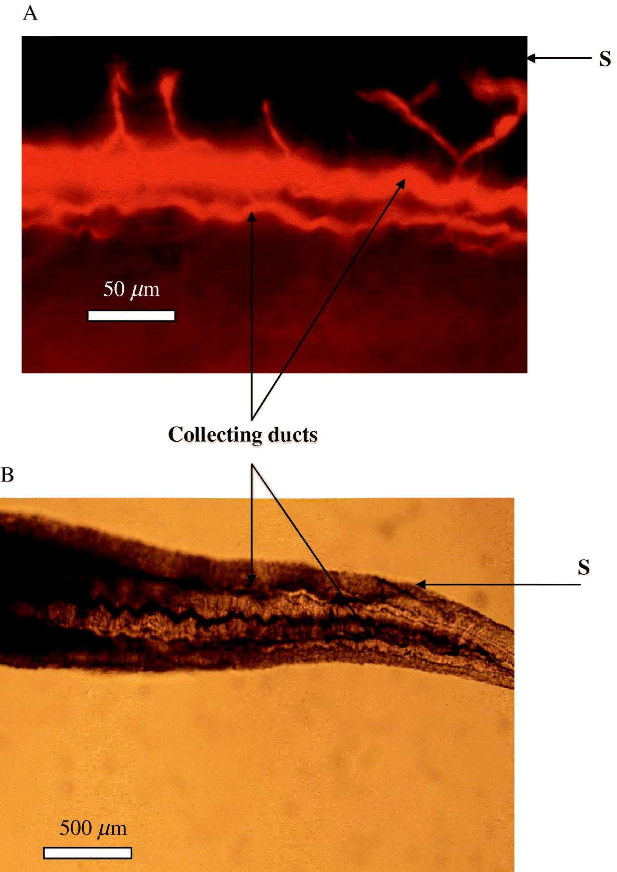
(A) Schistosomulum produced by syringe passage (Colley and Wikel, Reference Colley and Wikel1974) stained with LysoTracker to show the presence of large lysosomes (acid-rich vesicles) (LY) (Al-Adhami et al. Reference Al-Adhami, Noble, Sharaf, Thornhill, Doenhoff and Kusel2005). (B) Schistosomulum produced by syringe passage (Colley and Wikel, Reference Colley and Wikel1974) without fluorescence, stained with methylene blue. (C) Schistosomulum produced by syringe passage (Colley and Wikel, Reference Colley and Wikel1974). This has been slightly damaged by poly-L-lysine in the presence of Texas red-labelled bovine serum albumin (BSA) (Tan et al. Reference Tan, Thornhill, Al-Adhami, Akhkha and Kusel2003), and shows uptake of the fluorescent protein through the nephridiopore (N) into the excretory tubules (Tub) and adjacent tissue. (D) Adult worms incubated with poly-L-lysine, in the presence of Texas red-labelled bovine serum albumin (BSA). This shows slight areas of damage indicated by the blue fluoresecence of Hoechst 33258, and uptake of the fluorescent protein into the excretory tubules (Tub) in the damaged areas. (E) Higher magnification of the labelled tubules is also shown. (G) Indicates uptake of the fluorescent protein by the activities of the gut.

The excretory system of adult S. mansoni (both female and male) has been well described morphologically (Wilson and Webster, Reference Wilson and Webster1974; Bogers et al. Reference Bogers, Nibbeling, van Marck and Deelder1994). In brief, the functional unit is the flame cell, which leads into a fine tubule lined with epithelial cells. These tubules connect to larger tubules, similarly lined, until the tubules combine to form a large ciliated collecting duct. The collecting duct empties its contents into the external environment through a nephridiopore distal to a muscular contractile vesicle. The function of the system is assumed to be excretory, and the beating of the flame cells has been shown to vary with the osmotic pressure of the medium surrounding the parasite (Wilson and Webster, Reference Wilson and Webster1974). Some studies (Finken et al. Reference Finken, Sobek, Symmons and Kunz1994; Schechtman et al. Reference Schechtman, Winnen, Tarrab-Hazdai, Ram, Schinder, Grevelding, Kunz and Arnon2001; Skelly and Shoemaker, 2001 b; Wippersteg et al. Reference Wippersteg, Kapp, Kunz, Jackstadt, Zahner and Grevelding2002) have indicated the presence of signalling molecules in the excretory system, which may be of importance both in the response of the parasite to the host, and to the interactions between adult male and female schistosomes. Mecozzi et al. (Reference Mecozzi, Rossi, Lazzaretti, Kady, Kaiser, Valle, Cioli and Klinkert2000) have demonstrated that calcineurin is localized on the flame cells of the schistosomula and adult worms, and Schechtman et al. (Reference Schechtman, Winnen, Tarrab-Hazdai, Ram, Schinder, Grevelding, Kunz and Arnon2001) found protein 14-3-3, a molecule that regulates many signalling pathways, in the protonephridial system of the female schistosome.
P glycoproteins (P-gp) have also been detected in schistosomes (Bosch et al. Reference Bosch, Wang, Tao and Shoemaker1994; Greenberg, personal communication). For instance, Sato et al. (Reference Sato, Kusel and Thornhill2002, Reference Sato, Kusel and Thornhill2004) have shown that the excretory system of S. mansoni functions with the aid of P-gp and multi-resistance proteins (MRP) to pump out particular fluorescent substrates. This group of proteins is often over-expressed in drug-resistant tumour cells, and their function is to reduce the concentration of cytotoxic drugs in the cytoplasm of these cells (Box 6). The role of phosphorylation in the regulation of these activities is unclear, but the finding that 14-3-3 is present in the female excretory system (Schechtman et al. Reference Schechtman, Winnen, Tarrab-Hazdai, Ram, Schinder, Grevelding, Kunz and Arnon2001) suggests an interaction leading to the regulation of the P-gp activity. Furthermore, as P-gps can transport steroids (Borst et al. Reference Borst, Zelcer and van Helvoort2000), they may be involved in male–female interactions. The presence of active P-gps and multi-resistance proteins in the excretory system invites the speculation that their activity might respond to changes in the environment. Thus, these molecules could act as the sensors that monitor environmental change. In this context, Greenberg (personal communication) has shown by Western blotting that praziquantel treatment appears to induce the expression of P-gp molecules.
BOX 6 P-glycoproteins and multiresistance proteins
These proteins belong to a large family of ABC transporters that have numerous functions in normal eukaryotic and prokaryotic cells. They can transport inorganic and organic ions, xenobiotics, a variety of hydrophobic compounds, peptides, lipids, and proteins (Stavrovskaya, Reference Stavrovskaya2000; Dahl et al. Reference Dahl, Sylte and Ravna2004).
The P glycoproteins in eukaryotes are 170 kDa membrane proteins, having 12 transmembrane domains and a cytoplasmic nucleotide-binding domain.
P glycoproteins can recognize and transport an enormous number of chemical compounds, and attempts to classify these have been based on the number of binding sites on the molecule (Dahl et al. Reference Dahl, Sylte and Ravna2004). P glycoproteins can be regulated in their amount and activity by a variety of environmental agents but the response depends on the cell type or tissue. For example, reactive oxygen species can up or down regulate P glycoprotein activity depending on the tissue and context (Sukai and Piquettte-Miller, Reference Sukai and Piquette-Miller2000). Some drug-resistant mammalian cells with high levels of P-glycoproteins can upregulate these molecules in other cells in their vicinity by soluble factors (Eliseenkova et al. Reference Eliseenkova, Ktitorova, Kakpakova, Sidorova and Stavrovskaya1998) a kind of quorum sensing.
The multi-resistance proteins have a molecular weight of 190 kDa, and can pump organic anions, and cations and compounds complexed with or co-transported with glutathione, glucuronic acid and other adducts.
Excretory system in adult male and female worms
When adult worms in culture medium are labelled with the fluorescent dye resorufin, the excretory system of the male is clearly and rapidly revealed (Fig. 1). Unpaired females also show clear excretory system activity. However, it was observed that, although some dye had penetrated the surface of paired females, they were rarely labelled in their collecting ducts and tubules (Oliveira et al. Reference Oliveira, Kusel, Ribeiro and Coelho2006). These findings suggest that the male is inhibiting the excretory activity of the female worm. When the male was damaged by a drug (e.g. praziquantel) or suffered a metabolic inhibition by cooling, the activity of the female excretory system increased substantially from its previous state, even though it remained paired with the male. Therefore, we hypothesize that, when paired, the male profoundly decreases the diffusion of resorufin into the female excretory system. Cornford (Reference Cornford1985) reported that acidic amino acids were not absorbed by paired adults, although single unpaired males and females showed full transport activity. Some mechanism involving changes in membrane protein conformation may occur when pairing takes place, with signals being transmitted to transport and excretory systems.
The importance of external calcium and phosphate ions
The activity of the adult excretory system is clearly seen in RPMI medium when the parasites are labelled with resorufin. In a medium without calcium ions (e.g. Dulbecco's Modified Eagle's Medium, DMEM), the excretory tubules are inactive. However, with the addition of calcium ions, the tubules regain their activity (Kusel et al. Reference Kusel, Oliviera, Coelho, Mattos, Reis, Lima and Ribeiro2006). Similarly, phosphate ions are also essential (Kusel et al. Reference Kusel, Oliviera, Coelho, Mattos, Reis, Lima and Ribeiro2006). This evidence suggests that the external surface can respond and transport external calcium and phosphate ions (Kusel et al. Reference Kusel, Oliviera, Coelho, Mattos, Reis, Lima and Ribeiro2006). A membrane-bound calcium and phosphate sensor may be present (Garrett et al. Reference Garrett, Capuano, Hammerland, Hung, Bron, Herbert, Nemeth and Fuller1995). There are some candidates that might be involved in the calcium/phosphate effect on the excretory system. Reports of surface enzymes sensitive to calcium have been in the literature for some time (Cunha et al. Reference Cunha, Meyer-Fernandes and Noel1992) and recent proteomic data for the surface membrane (Braschi and Wilson, Reference Braschi and Wilson2006) suggest the presence of a calcium ATPase (Accession no. C604128.1). Alkaline phosphatase has been implicated in the transport of phosphate through the membrane (Peerce et al. Reference Peerce, Fleming and Clarke2003). However, there may be a different explanation for the apparent linkage between the surface membrane and the excretory system. It has been shown that the tegumental membrane potential depends crucially on the presence of glutamine and an optimum phosphate concentration (Lane et al. Reference Lane, Pax and Bennett1987). Thus, the membrane potential of the excretory tubules may be linked to the surface in the same way as are the muscle membranes. Depolarization of the surface membrane may lead to depolarization and lack of function of P-gps.
Effect of removal of cholesterol from the surface
It has been shown that treatment of the adult male worm with methyl-beta-cyclodextrin (MCD), which disrupts lipid rafts in cell membranes (El Ridi et al. Reference El Ridi, Tallima, Mohamed and Montash2004), caused the excretory system to become active in its excretion of resorufin (Oliveira et al. Reference Oliveira, Kusel, Ribeiro and Coelho2006). This finding could be explained by suggesting that the disruption of rafts in the bilayer(s) of the surface alters calcium transport into the tegumental cytoplasm, altering the activity of the excretory system. It has been shown for blood cells (Unruh et al. Reference Unruh, Li, Mutch, Sharia, Grigoriou, Sanval, Brown and Deans2005) that the function of CD20 (which is putatively a store-operated calcium channel) is affected by the integrity of lipid rafts. However, the observation may mean that the P-gps are directly involved in cholesterol metabolism. This is a possibility, since P-gps are known to pump steroids (Debry et al. Reference Debry, Nash, Neklason and Metherall1997). P-gps are present in rafts in multidrug-resistant mammalian cells, and alteration of cholesterol levels can cause a change in activities and in raft localization (Troost et al. Reference Troost, Lindenmaier, Haefeli and Weiss2004).
Damage to the surface by poly-L-lysine
Incubation with damaging concentrations of poly-L-lysine has been shown to rapidly induce activity of the excretory system (Kusel et al. Reference Kusel, Oliviera, Coelho, Mattos, Reis, Lima and Ribeiro2006), which may be due to an influx of calcium and phosphate ions into the tegumental cytoplasm. It could not be replicated by the calcium ionophore ionomycin, although the latter did give a stimulus that caused sustained activity of the excretory system. Repair of the membrane may involve removal of Ca++ to low levels by intracellular pumps, as indicated by the repair of mammalian membranes, which needs a low calcium ion concentration (Reddy et al. Reference Reddy, Caler and Andrews2001).
The effects of praziquantel
Praziquantel completely inhibits the function of the excretory system of the adult male schistosome, as demonstrated by resorufin staining (Oliveira et al. Reference Oliveira, Kusel, Ribeiro and Coelho2006). The mechanism is likely to be due to the profound inhibition of the male excretory system by the excess of calcium entering the worm tissues or released from intracellular stores. The male parasite can recover rapidly from this inhibition, perhaps by pumping calcium ions from tissues, either out of the worm or back into intracellular stores. The enzyme systems responsible for the pumping of calcium ions are unknown, but there are numerous candidate membrane-bound enzymes represented in the transcriptome (Verjovski-Almeida et al. Reference Verjovski-Almeida, DeMarco, Martins, Guimarães, Ojopi, Paquola, Piazza, Nishiyama, Kitajima, Adamson, Ashton, Bonaldo, Coulson, Dillon, Farias, Gregorio, Ho, Leite, Malaquias, Marques, Miyasato, Nascimento, Ohlweiler, Reis, Ribeiro, Sá, Stukart, Soares, Gargioni, Kawano, Rodrigues, Madeira, Wilson, Menck, Setubal, Leite and Dias-Neto2003). These enzymes are vital for the function of the excretory system. Further work will elucidate their identity. In human submandibular gland cell membranes, store-operated calcium (SOC) channels are located in rafts associated with caveolin (Lockwich et al. Reference Lockwich, Liu, Singh, Jadlowiec, Weiland and Ambudkar2000). It would be interesting to establish whether caveolin in the schistosome membrane (Racoosin et al. Reference Racoosin, Davies and Pearce1999) is associated with calcium channels of this type. Recent work has suggested that the actin cytoskeleton may control the activity of SOC channels (Cioffi et al. Reference Cioffi, Wu, Alexeyev, Goodman, Zhu and Stevens2005) and the adenyl cyclase associated with caveolin (Head et al. Reference Head, Patel, Roth, Murray, Swaney, Niesman, Farquhar and Insel2006).
Pica-Mattoccia et al. (Reference Pica-Mattoccia, Valle, Basso, Troiani, Vigorosi, Liberti, Festucci and Cioli2007) have shown that the treatment of adult male schistosomes with cytocalasin D (which binds to the end of F-actin chains) prevents the action of praziquantel and, by implication, inactivated calcium channels, which are the putative sites of action of PZQ (Greenberg, Reference Greenberg2005). This exciting finding suggests that the cytoskeleton of the adult schistosome has a function in the control of the activity of calcium channels, so that, if the cytoplasmic concentration increases, the dissociation of the actin cytoskeleton prevents further ingress of calcium in a feedback loop with the calcium channels. This finding might explain why immature stages are less susceptible to praziquantel, if the feedback mechanism has not developed in these forms. In contrast to male worms after treatment with PZQ, adult females, that were paired with males, showed increased activity of their excretory systems (Oliveira et al. Reference Oliveira, Kusel, Ribeiro and Coelho2006). The female has a system that is much less sensitive to the calcium ion concentration. This finding may explain why unpaired female worms are less susceptible to PZQ than unpaired male worms (Gonnert and Andrews, Reference Gonnert and Andrews1977). This seems also to be the case for cercariae and parasites between 7 days and 4 weeks old (Kusel et al. Reference Kusel, Oliviera, Coelho, Mattos, Reis, Lima and Ribeiro2006).
Membrane damage and repair and the activity of the excretory system
The damage and repair of the adult schistosome membrane has been studied by electron microscopy (Perez and Terry, Reference Perez and Terry1973; Wilson and Barnes, Reference Wilson and Barnes1977; Roberts et al. Reference Roberts, MacGregor, Vojvodic, Wells, Crabtree and Wilson1983; Popiel et al. Reference Popiel, Irving and Basch1985) and, recently, by using the uptake of fluorescent proteins as an indication of the production of lesions by poly-L-lysine (Oliveira et al. Reference Oliveira, Kusel, Ribeiro and Coelho2006). When such lesions were induced by poly-L-lysine in schistosomula (Tan et al. Reference Tan, Thornhill, Al-Adhami, Akhkha and Kusel2003), it was concluded that the excretory system absorbed environmental protein into its tubules through the nephridiopore, and this protein was endocytosed and transferred into the body of the parasite. As a consequence of damage to adult worms fluorescent proteins were endocytosed from the tegumental cytoplasm into the epithelial cells of the excretory system and passed into the nephridiopore (Wippersteg et al. Reference Wippersteg, Ribeiro, Liedke, Kusel and Grevelding2003). It is possible that the excretory system is acting as an organ system active in the repair of the lesion. Certainly, the activity in pumping resorufin is increased by surface membrane damage (Oliveira et al. Reference Oliveira, Kusel, Ribeiro and Coelho2006) and thus might be expected to remove ions and metabolic end products, for example, lactic acid. However, it is possible that the excretory epithelium removes denatured proteins, lipids and carbohydrates that are released due to worm damage, and has an anti-apoptotic effect by reducing the concentration of effector molecules, such as cytochrome c. Further work, to establish the fate of such molecules within the parasite, would be of great importance and may reveal new vesicle cycling mechanisms.
The repair of the surface membrane is likely to involve vesicle fusion, lipid synthesis and turnover (Redman et al. Reference Redman, Kennington, Spathopoulou and Kusel1997; Brouwers et al. Reference Brouwers, Skelly, Van Golde and Tielens1999) as well as protein and carbohydrate chain synthesis. Few studies have examined repair as such, but the rapid turnover rates reported for the surface membrane may have been a reflection of inadvertently inflicted damage. Electron microscopy of tissues subsequent to lesions (cuts) made in the tegument (Popiel et al. Reference Popiel, Irving and Basch1985) has shown the involvement of the membranous bodies in the repair process. Evidence from mammalian cells suggests that lysosomes can be involved in membrane repair in calcium-linked exocytosis (Reddy et al. Reference Reddy, Caler and Andrews2001). Many cytoplasmic vesicle structures may be involved (Keefe et al. Reference Keefe, Shi, Feske, Massol, Navarro, Kirchhausen and Lieberman2005). In schistosomes, it is possible that vesicles cycled from the excretory system, lysosomes and other vesicles may be involved in membrane repair. In mammalian cells damaged by perforin many organelles contribute to the repair process (Keefe et al. Reference Keefe, Shi, Feske, Massol, Navarro, Kirchhausen and Lieberman2005), so that the cell undergoes apoptosis rather than necrosis. Clearly, much more work is required to address this topic in schistosomes. Very few biochemical studies have been carried out on membrane repair. The results of an in vitro study of sphingomyelin synthesis (Redman et al. Reference Redman, Kennington, Spathopoulou and Kusel1997) have suggested a rapid rate of membrane repair but another study (Brouwers et al. Reference Brouwers, Skelly, Van Golde and Tielens1999) did not report any such evidence. This is an important field for further investigations.
CERCARIAE AND SCHISTOSOMULA
The recognition and penetration of the mammalian skin (Stirewalt, Reference Stirewalt1974) is the first step in a complex process of transformation from a free–living cercaria to a schistosomulum, which migrates from the skin through a variety of tissues and finally locates in the bloodstream of the host. Cercariae form a population with variable metabolic properties, and they penetrate and migrate at different rates. Cercariae of S. japonicum migrate much more rapidly than those of S. mansoni (see Gui et al. Reference Gui, Kusel, Shi and Ruppel1995).
The penetration of human skin by schistosome cercariae has been studied by Whitfield et al. (Reference Whitfield, Bartlett, Khammo, Brain, Brown, Marriott and Clothier2003), who showed that the cercarial tail often penetrates the epidermis and is not cast off, as is usually found for other mammalian skins and other trematode species. Whitfield et al. (Reference Whitfield, Bartlett, Khammo, Brain, Brown, Marriott and Clothier2003) suggested that this behaviour may have relevance to the phenomenon of concomitant immunity, since the antigens from the tail musculature or its very active excretory system (Stirewalt, Reference Stirewalt1974) may induce a specific immune response in the host. A stimulating discussion on cercarial penetration of human and other skins was published by McKerrow (Reference McKerrow2003) and Curwen and Wilson (Reference Curwen and Wilson2003).
Enzymes are required from the parasite to complete the migration of the schistosomulum, and present evidence shows that those of S. japonicum could be very different from those found in S. mansoni and S. haematobium (see Chlichlia et al. Reference Chlichlia, Schauwienold, Kirsten, Doenhoff, Fishelson and Ruppel2005). Intriguingly, it seems that, as a result of a natural infection, no antibodies are produced in the host against the cercarial elastase (Bahgat et al. Reference Bahgat, Franklow, Doenhoff, Li, Ramzy, Kirsten and Ruppel2001). This enzyme hydrolyses specific anti-parasite immunoprecipitating antibodies, putatively IgG (Darani et al. Reference Darani, Curtis, McNeice, Price, Sayers and Doenhoff1997) and also IgE (Pleass et al. Reference Pleass, Kusel and Woof2000). When antibody responses were raised in mice against elastase, some protection against challenge infection was achieved (Darani et al. Reference Darani, Curtis, McNeice, Price, Sayers and Doenhoff1997).
Signals from the host must be received for normal development. Careful recent studies (Knudsen et al. Reference Knudsen, Medzihradszky, Lim, Hansell and McKerrow2005; Curwen et al. Reference Curwen, Ashton, Sundaralingam and Wilson2006) have built on the much earlier work of Stirewalt (Reference Stirewalt1974) on the process by which the cercariae enter the host skin and on the involvement of the acetabular glands in this process. Curwen et al. (Reference Curwen, Ashton, Sundaralingam and Wilson2006) have identified new peptidases and proteases likely to be involved in skin penetration. Furthermore, venom allergens similar in homology to chemokines, and potassium channel blockers may affect the T cell response to the cercariae (Curwen et al. Reference Curwen, Ashton, Sundaralingam and Wilson2006). In view of the high calcium ion concentration in the secretions, it was interesting that Knudsen et al. (Reference Knudsen, Medzihradszky, Lim, Hansell and McKerrow2005) identified a calcium-binding protein, calcium ATPase, and calreticulin. Paramyosin was also detected, and Deng et al. (Reference Deng, Gold, LoVerde and Fishelson2003) demonstrated an important role for parasite surface-bound paramyosin in inhibiting complement activation. Schistosomula have also a surface bound elastase (Ghendler et al. Reference Ghendler, Parizade, Arnon, McKerrow and Fishelson1996) which may be related to the above-mentioned secreted elastase, but the function of which has not been fully investigated.
During penetration the cercaria releases from membrane, glands or the excretory system phospholipids which were identified as lysophosphatidylserine and which can interact with Toll-like receptor 2 on dendritic cells (Van der Kleij et al. Reference Van der Kleij, Latz, Browers, Kruize, Schmitz, Kurt-Jones, Espevik, de Jong, Kapsenberg, Golenbock, Tielens and Yazdanbakhsh2002). This interaction allows presentation of parasite antigens in a mode that favours the induction of regulatory T cells and the production of IL-10. This very interesting work shows us how important it is to keep an open mind about the function of parasite molecules. Lysophosphatidylserine can act as a detergent and one possible function is that it lyses host cells, and one might therefore have speculated that if it reacts with membranes of effector cells and destroys them, it functions as a mechanism of immune evasion. For example, lysophosphatidylcholine produced by adult worms has been shown to lyse erythrocytes (Golan et al. Reference Golan, Brown, Cianci, Furlong and Caulfield1986; Brouwers et al. Reference Brouwers, Skelly, Van Golde and Tielens1999). However, it seems from the work of Van der Kleij et al. (Reference Van der Kleij, Latz, Browers, Kruize, Schmitz, Kurt-Jones, Espevik, de Jong, Kapsenberg, Golenbock, Tielens and Yazdanbakhsh2002) that lysophosphatidylserine has a signalling function. Thus the earliest stage of the parasite is adapted to modulate the host's immune response in an extremely sophisticated manner. This is further seen as eicosanoids are also released and these can interact with dendritic cells to prevent their migration to lymph nodes and also to favour a Th2 response (Angeli et al. Reference Angeli, Faveeuw, Roye, Fontaine, Tessier, Capron, Wolowczuk, Capron and Trottein2001). These molecules are synthesized from arachidonic acid that may be derived from the skin of the host or from membrane phospholipids in the cercariae, the arachidonic acid being originally derived from the snail or from linoleic acid (Fusco et al. Reference Fusco, Salafsky and Delbrook1986). Thus, the host, especially through skin-derived arachidonic acid, is contributing to the survival of the parasite in its modulation of the immune response! This is a good example of the ‘welcome mat’ concept that was envisaged by Davies et al. (Reference Davies, Hall, Targett and Murray1980). The transfer of macromolecules in vitro to the membrane of the schistosomulum from red blood cells and from radioactively-labelled epithelial cells has been observed by Goldring et al. (Reference Goldring, Kusel and Smithers1977a). The glycocalyx fixes complement very vigorously and because it is shed rapidly, there is little surface membrane damage. Where complement binds to membranes, and this is relevant to the metabolism of the adult as well as the schistosomulum, sublytic membrane attack complexes (MAC) can signal across the membrane and have a growth effect (Bohana-Kashtan et al. Reference Bohana-Kashtan, Ziporen, Donin, Kraus and Fishelson2004).
It is not clear how active the excretory system is during its development in the schistosomulum. Tail loss from the cercaria exposes the nephridiopore and damaged schistosomula take up protein through this orifice (Tan et al. Reference Tan, Thornhill, Al-Adhami, Akhkha and Kusel2003) and this uptake may occur during skin penetration. No excretion of resorufin has been found in schistosomula at this stage (Kusel et al. Reference Kusel, Oliviera, Coelho, Mattos, Reis, Lima and Ribeiro2006). A range of molecules having profound effects on the host are released from the schistosomulum during migration and from the adult when in the portal vein and its vascular ramifications in the gut and liver. What is the origin of these molecules in the structures of the parasite?
The immunomodulatory lysophosphatidylserine is produced by the adult worms and eggs and perhaps schistosomula (Van der Kleij et al. Reference Van der Kleij, Latz, Browers, Kruize, Schmitz, Kurt-Jones, Espevik, de Jong, Kapsenberg, Golenbock, Tielens and Yazdanbakhsh2002), but the mechanism of release of the molecule has not been studied. Similarly, the lung stage produces ‘excretory products’ which can decrease the permeability of lung endothelial cells and have an anti-inflammatory effect, but their origin is not known (Trottein et al. Reference Trottein, Descamps, Nutten, Dehouck, Angeli, Capron, Cecchelli and Capron1999a, Reference Trottein, Nutten, Angeli, Delerive, Tessier, Capron, Staels and Capronb). The authors suggested these molecules from the parasites may be eicosanoids and prostaglandins. Angeli et al. (Reference Angeli, Faveeuw, Roye, Fontaine, Tessier, Capron, Wolowczuk, Capron and Trottein2001) described the formation of prostaglandin D2 (PGD2) by schistosomula and the inhibition that this excretory product induces on the Langerhans cells in the skin. These authors also mentioned that PGD2 synthase is secreted into the skin during penetration, and this could increase PGD2, a molecule derived from the host by the enzyme secreted by the parasite. It transpired that PGD2 synthase was in fact a glutathione S-transferase isoform (Herve et al. Reference Herve, Angeli, Pinzar, Wintjens, Faveeuw, Narumiya, Capron, Urade, Capron, Riveau and Trottein2003).
Neuropeptides, morphine-like compounds and renin-angiotensin peptides are secreted by all stages of the parasite and these compounds may have an immunomodulatory function (Salzet et al. Reference Salzet, Capron and Stefano2000; Duvaux-Miret et al. Reference Duvaux-Miret, Stefano, Smith, Mallozzi and Capron1992). An apoptosis-inducing molecule of about 23 kDa molecular weight is secreted and induces apoptosis in the effector cells which gather around the parasite (Chen et al. Reference Chen, Rao, He and Ramaswamy2002). It is a distinct possibility that the excretory system and its epithelia are actively involved in the release of many of these molecules, and further research is needed to determine whether this is the case. If so, regions of the excretory system may be targets of protective immunity. Currently nothing is known unequivocally about those compounds excreted through the nephridiopore.
Other molecules can be taken up from the skin cells during penetration, and there is a massive release of glycocalyx during the penetration process. Skin lipids, proteins (Smith and Kusel, Reference Smith and Kusel1979) and blood group substances can be incorporated into the membrane. This means that the skin schistosomulum is a very different organism from the mechanically produced form. No proteomic comparisons have been made between skin-transformed and mechanically transformed schistosomula, but DNA arrays of cultured and lung-derived schistosomula have been compared (Chai et al. Reference Chai, McManus, McInnes, Moertel, Tran, Loukas, Jones and Gobert2006). Certain genes (e.g. heat shock protein 70, the anti-inflammatory Sm16 and paramyosin) are expressed at a higher level in organisms taken from the living host when compared with the in vitro-transformed and cultured schistosomula. The acquisition of, or interaction with, molecules from the host may be a cause of transmembrane signalling and thereby cause changes in transcription. Shaker et al. (Reference Shaker, Wu, El-Shobaki, Ashour, Khattab, Draz, Kamel and Wu1998) have shown increases in the rates of cell division in parasites cultured in portal blood compared with those maintained in peripheral blood and they showed that portal blood contains a factor of 38 kDa which stimulates cell division in cultured 20-day-old worms. Rocha and Coelho (Reference Rocha and Coelho1980) inoculated schistosomula directly into the portal system of mice and these showed an enhanced rate of development compared to those schistosomula derived from the normal transcutaneous infection process. During migration through skin schistosomula can stimulate dermal endothelial cells to produce IL-7, which in turn can stimulate the development of the parasite and increase its fecundity (Wolowczuk et al. Reference Wolowczuk, Nutten, Roye, Delacre, Capron, Murray, Trottein and Auriault1999). It is not currently understood how the secreted molecules are involved in the development of the parasite during and after penetration. Some could be vaccine candidates but their suitability in this regard depends on their accessibility to the immune effectors. The cercarial secretions, if injected into the skin, do not protect against an infection (Harn et al. Reference Harn, Cianci and Caulfield1989; Fallon et al. Reference Fallon, Teixeira, Neice, Williams, Hellewell and Doenhoff1996). The penetration of the skin and subsequent migration can be enhanced if cercarial extracts or the medium in which mechanically-transformed cercariae are incubated are injected into the penetration site prior to exposure of the skin to cercariae (Fallon et al. Reference Fallon, Teixeira, Neice, Williams, Hellewell and Doenhoff1996), possibly because constituents in the injected fluid, particularly proteases, induce an inflammation which involves influx of neutrophils and eosinophils (Teixeira et al. Reference Teixeira, Doenhoff, McNeice, Williams and Hellewell1993). Also released into the skin are factors that decrease inflammation such as the anti-inflammatory protein Sm16 (Rao and Ramaswamy, Reference Rao and Ramaswamy2000) and eicosanoids (Angeli et al. Reference Angeli, Faveeuw, Roye, Fontaine, Tessier, Capron, Wolowczuk, Capron and Trottein2001). Migration of the parasite through host tissues appears to be a delicate balance between pro- and anti-inflammatory effects of molecules released by the parasite (Mountford and Trottein, Reference Mountford and Trottein2004). Harrop and Wilson (Reference Harrop and Wilson1993) cultured S35 methionine-labelled schistosomula and showed that in the first 24 h few proteins were synthesized. Between 1 and 7 days numerous proteins (from <20 kDa to >100 kDa) were synthesized and released. These were immunogenic, but many were unidentified and their function unknown.
During early transformation, remarkable changes occur in the structure of the cercarial membrane, as first described by Hockley and McLaren (Reference Hockley and McLaren1973). Some of the molecular events involved in this process have been elegantly described by Skelly and Shoemaker (Reference Skelly and Shoemaker2001a). For example, relocation of the glucose transporter SGTP4 from intracellular vesicles into the surface membrane by membrane fusion. Other changes in the membrane may result in the transient expression of particular proteins that are not usually found in mature membranes (Skelly and Wilson, Reference Skelly and Wilson2006). These molecules could be ‘cryptic’ antigens and possible vaccine targets in view of their brief expression on the surface. Tetraspanin in the membrane may be exposed at this stage and thus achieve some degree of protective immunity (Tran et al. Reference Tran, Pearson, Bethony, Smyth, Jones, Duke, Don, McManus, Correa-Oliveira and Loukas2006). Also, a molecule named Sm Fes, which is an fes/fps tyrosine kinase has been discovered in the tegument of both miracidium and cercaria and is suggested to have a signalling function during host penetration and parasite transformation (Bahia et al. Reference Bahia, Mortara, Kusel, Andrade, Ludolf, Kuser, Avelar, Trolet, Dissous, Pierce and Oliveira2007).
The profound environmental changes experienced by the parasite as it passes from fresh water to the skin and tissues of the mammalian host are accompanied by secretions from the acetabular glands and the re-organization of parasite tissues following these secretory processes. There are also surface membrane and cytoskeletal changes (Skelly and Shoemaker, Reference Skelly and Shoemaker2001a) as well as re-oganization of tissue near the nephridiopore after the loss of the tail of the cercaria, immediately following skin penetration. The tissue remodelling and perhaps the repair processes are likely to be carried out by the lysosomal/autophagosome system. This remodelling was first described for helminths by Threadgold and Arme (Reference Threadgold and Arme1974) and Bogitsh (Reference Bogitsch1975) in gastric epithelial cells. In mammalian cells and yeast, the lysosome and autophagosome pathway is induced by signals acting on PI3 kinase. This pathway is also present in the schistosomulum, as was shown by inhibitors of the PI3 kinase-stimulated vesicular pathway, such as wortmannin, 3-methyl adenine, and N-ethylmaleimide (Al-Adhami et al. Reference Al-Adhami, Noble, Sharaf, Thornhill, Doenhoff and Kusel2005). In this work, the inhibitors had to be present in the transforming fluid for them to be active, and thus may have entered the transforming parasite via the nephridiopore (Tan et al. Reference Tan, Thornhill, Al-Adhami, Akhkha and Kusel2003, Fig. 2C).
Lysosomes and autophagosomes have not been found in cercariae or miracidia, but they form rapidly during transformation of the cercariae into schistosomula (Al-Adhami et al. Reference Al-Adhami, Noble, Sharaf, Thornhill, Doenhoff and Kusel2005, Fig. 2A) or the miracidia into mother sporocysts (Mattos et al. Reference Mattos, Kusel, Pimenta and Coelho2006). The signals triggering the formation of these organelles are unknown, but are likely to be related to the effects of environmental change. The cultivation of schistosomula in Eagle's medium containing 5% foetal calf serum increases the number of these organelles, whereas culturing them in a starvation medium lacking amino acids leads to the formation of very large lysosomes. These observations were made using quantitative fluorescence with lysosome- and autophagosome-specific fluorescent probes, but Western blot analysis of parasite extracts lent support to the conclusion that there was an induction of lysosome specific (Lamp-1 and MAP-LC3) protein synthesis (Al-Adhami et al. Reference Al-Adhami, Noble, Sharaf, Thornhill, Doenhoff and Kusel2005).
The migration of schistosomula through tissues and the bloodstream brings the surface membrane into contact with a variety of host cell membrane-bound and plasma macromolecules. Some of these may act as signals for metabolic pathways or stimulate the parasite nervous system, hence signalling environmental change. This signalling can have a profound effect on gene expression, as was shown when in vitro-grown schistosomula were compared with those derived from the infected host using a transcriptomic approach (Chai et al. Reference Chai, McManus, McInnes, Moertel, Tran, Loukas, Jones and Gobert2006). Among the genes upregulated from in vivo parasites are those responding to stress (e.g. hsp70) and those associated with immune evasion, such as Sm16, suggested to be an anti-inflammatory protein. During development in the skin, the schistosomulum requires the presence of IL-7, in order to develop to maturity (Wolowczuk et al. Reference Wolowczuk, Nutten, Roye, Delacre, Capron, Murray, Trottein and Auriault1999). Later during development in the liver, the parasite also requires the presence of factors from a subset of CD4+ cells (Hernandez et al. Reference Hernandez, Kim, McKerrow and Davies2004). This is another example of a close interplay between the host and the parasite. Thus, it is necessary to be aware of these signals and explore the nature and location of their receptors in the future.
Lung schistosomula
The passage of schistosomula through the lung capillaries involves very close juxtaposition of the parasites' surface with the endothelial cell and this slows the passage (Wilson, Reference Wilson, Rollinson and Simpson1987). As discussed, excretions/secretions from the parasite can induce changes through signalling pathways in the endothelial cell (Trottein et al. Reference Trottein, Descamps, Nutten, Dehouck, Angeli, Capron, Cecchelli and Capron1999a). It is not known whether endothelial membrane components become transferred to the lung-stage parasite, but lipophilic compounds released from the migrating parasite can profoundly decrease the permeability of the endothelial plasma membranes and also reduce the amount of selectins and VCAM expressed by these cells (Trottein et al. Reference Trottein, Nutten, Angeli, Delerive, Tessier, Capron, Staels and Capron1999b). These anti-inflammatory signals balance an inflammatory response during migration in an immunized host. In the immunologically naive mouse and, to a greater extent in the infected mouse host, migrating schistosomula burst through the blood-air barrier and enter the alveoli, where most of them perish (Crabtree and Wilson, Reference Crabtree and Wilson1986, Reference Crabtree and Wilson1987).
An excellent example of how the results from transcriptomic studies can give insights into the biology of the migrating parasite is evident from the work of Dillon et al. (Reference Dillon, Feltwell, Skelton, Ashton, Coulson, Quail, Nikolaidou-Katsaridou, Wilson and Ivens2006). These authors used expressed sequence tag (EST) data for the S. mansoni lung stage to construct a microarray. They examined the up and down regulation of genes in a variety of life-cycle stages, and could relate their findings to the biology of the lung, liver and adult stages. There was an upregulation of proteases for haemoglobin digestion, saposin and pyrophosphatase in the lung compared with the adult stage. The upregulation of the cytoskeletal components in the lung stage was explained in the context of the body and tegumental flexibility of the migrating parasite. They also detected increased transcription for a gynaecophoral canal protein in the lung stage, which suggests that a triggering of sexual development occurs very early, before cell division has been initiated.
Many proteins are secreted by the lung schistosomulum (Harrop et al. Reference Harrop, Coulson and Wilson1999; Mountford et al. Reference Mountford, Harrop and Wilson1995). These molecules act as potent immunogens and are involved in the inflammatory response in an immunized host (Mountford et al. Reference Mountford, Harrop and Wilson1995). However, the lung-stage parasite does not bind antibody and is resistant to direct damage by the immune system. Tallima et al. (Reference Tallima and El Ridi2005) stressed the importance of the presence of lipid rafts in the surface. Removal of cholesterol with methyl-β-cyclodextrin can disrupt lipid rafts and increases antigen exposure from the lung-stage parasite in S. mansoni. Although methyl-β-cyclodextrin removed cholesterol from the lung-stage larvae in S. haematobium, no exposure of antigens was observed. However, corn oil did have the effect of exposing antigens in S. haematobium, and it was deduced that the unsaturated fatty acids in this oil were responsible for this effect (Tallima et al. Reference Tallima and El Ridi2005). It was observed that the unsaturated fatty acids caused sphingomyelin breakdown, leading to the antigen exposure. The hydrolysis of sphingomyelin stimulated by unsaturated fatty acids is of great importance in this species as this will lead to disruption of the integrity of lipid rafts. Tallima et al. (Reference Tallima and El Ridi2005) further showed that the incorporation of small amounts of unsaturated fatty acids in the diet of mice infected with S. haematobium cercariae caused a significant decrease in the size of the final worm burden. A high serum fatty acid concentration was predicted to decrease the worm burden in S. mansoni, since it was able to cause sphingomyelin degradation, antigen exposure and subsequent recognition by the immune system of the host, and this prediction was verified (see Tallima et al. Reference Tallima and El Ridi2005). A recent paper (El Ridi and Tallima, Reference Tallima2006) concluded that it is the equilibrium between the synthesis and breakdown of sphingomyelin that is crucial for the maintenance of surface membrane expression of receptors and the diffusion of nutrients, so that surface antigenicity is minimal.
While migrating through the lungs and other host tissues during the first few days after having penetrated through the skin, schistosomula have to pass through tiny capillaries. In fact, on arrival in the lungs early after skin penetration, the schistosomula are likely to have a diameter that is greater than that of the lung capillaries (Wilson, Reference Wilson, Rollinson and Simpson1987). It is not known whether host proteins can aid the process of migration through the lung capillaries, but a kininogenase (a protease of the kallikrein family) which hydolyses kallikrein to bradykinin has been found in association with the tegument of adult worms (Carvalho et al. Reference Carvalho, Lopes, Juliano, Coelho, Cunha-Melo, Beraldo and Pesquero1998). If lung-stage schistosomula also express this molecule, it might enhance parasite survival by dilating the blood capillaries and vessels through which the larvae need to pass.
The apparent failure of blood clotting to occur around the migrating schistosome larvae or the adult parasites is an intriguing feature of the schistosome-host relationship. The failure of schistosome worms to be recognized by platelets contrasts with the intensely thrombogenic properties of eggs (Ngaiza and Doenhoff, Reference Ngaiza and Doenhoff1990) and the innate ability of platelets to protect the host against larval infection (Stanley et al. Reference Stanley, Ngaiza, Wambayi, Lewis and Doenhoff2003b).
Factors that may contribute to this lack of blood clotting around the parasite are the thrombocytopaenia occuring during infection (Ngaiza and Doenhoff, Reference Ngaiza and Doenhoff1987; Reference Ngaiza and Doenhoff1990; Stanley et al. Reference Stanley, Ngaiza, Atieno, Jell, Francklow, Jackson, Parry and Doenhoff2003a), inhibition of blood Factor XIIa (Tsang et al. Reference Tsang, Hubbard and Damian1977; Foster et al. Reference Foster, Flanigan, DeStigter, Blanton, Dumenco, Gallagher and Ratnoff1992) and infection-induced increases in blood clotting times (Carvalho et al. Reference Carvalho, Mello, Soares, Bicalho, Lima e and Coelho2005). The worm surface nucleotide dihydrolase described by Vasconcelos et al. (Reference Vasconcelos, Nascimento, Meirelles, Verjovski-Almeida and Ferreira1993) may inhibit platelet activation by hydrolysing ADP and an anticoagulant peptide has been identified (Lin and He, Reference Lin and He2006). Surface proteoglycans may also prevent blood clots from forming (Robertson and Cain, Reference Robertson and Cain1985; Hamed et al. Reference Hamed, Maharem and El Guindy1997). The cholesterol content in surface membrane lipid rafts may also play an important role, as it does in some mammalian cells (Papanikolaou et al. Reference Papanikolaou, Papafotika, Murphy, Papamarcaki, Tsolas, Drab, Kurzchalia, Kaspar and Christophoridis2005).
Liver stage
When the parasite reaches the liver, it receives signals that enable it to grow. Female worms develop less rapidly than the male and complete development of the male depends on an active immune system (Harrison and Doenhoff, Reference Harrison and Doenhoff1983; Davies et al. Reference Davies, Grogan, Blank, Lim, Locksley and McKerrow2001). The female only develops to maturity in the presence of the male, but, although Harrison and Doenhoff (Reference Harrison and Doenhoff1983) showed that fecundity was delayed and/or inhibited by immunosuppression, in immunosuppressed mice the development of females is not any more retarded than it is in the absence of male worms (Hernandez et al. Reference Hernandez, Kim, McKerrow and Davies2004). While in the liver, the developing parasite stimulates Kupffer cells to induce Th2 responses and inspection of the Kupffer cells shows that considerable amounts of haemin have been taken up (Grimaud et al. Reference Grimaud, Borojevic and Araujo Dos1976). It would be interesting to know the effects of the haemin on antigen presentation, as antigen presentation has been shown to be a function of Kupffer cells (Hayashi et al. Reference Hayashi, Matsui, Tsutsui, Osada, Mohamed, Nakano, Kashiwamura, Hyodo, Takeda, Akira, Hada, Higashino, Kojima and Nakanishi1999), and schistosome-generated haemin may modify this presentation (Sherman and Lara, Reference Sherman and Lara1989; Taramelli et al. Reference Taramelli, Recalcati, Basilico, Olliaro and Cairo2000).
Once the parasite is in the portal vein it grows rapidly. However, presently, little is known about developments in the surface membrane. Early liver-stage parasites are resistant to the lethal effects of praziquantel (Cioli and Pica-Mattoccia, Reference Cioli and Pica-Mattoccia2003). Nevertheless, the drug does cause worms of this age to contract in vitro, but the recovery from the drug is rapid and there is little effect on the excretory system of the parasite, compared with that observed in the adult male schistosome (Kusel et al. Reference Kusel, Oliviera, Coelho, Mattos, Reis, Lima and Ribeiro2006). Some changes in membrane properties can be detected as the parasite enters the portal system. For example, Breternitz et al. (Reference Breternitz, Kusel, Lutz, Buehrle and Ruppel1992) showed that the 27-day-old form can repair the lesions caused by the bacterium Pseudomonas aeruginosa cytotoxin, as shown by the restoration of the resting membrane potential, whereas older forms succumb, being unable to restore the resting membrane potential. Changes in the function of the excretory system can also be measured (Kusel et al. Reference Kusel, Oliviera, Coelho, Mattos, Reis, Lima and Ribeiro2006). Male worms in the portal vein develop a significantly enlarged pair of collecting ducts that are highly sensitive to praziquantel. Amiri et al. (Reference Amiri, Locksley, Parslow, Sadick, Rector, Ritter and McKerrow1992) and Davies et al. (Reference Davies, Grogan, Blank, Lim, Locksley and McKerrow2001) described the importance of TNF-alpha in worm development in the portal system, but the receptor for this molecule on the worm is presently uncharacterized. It is possible that the P-glycoproteins of the excretory system are responsive to TNF-alpha, as are such molecules in mammalian cells (Sukai and Piquette-Miller, Reference Sukai and Piquette-Miller2000). Therefore, there are clear examples of the dependency of parasites in this location on the host immune system and cytokines, but many aspects remain to be discovered.
The tegument surface area is substantially increased by the pits and folds that it contains. Cornford (Reference Cornford and Oldendorf1979, Reference Cornford1985) showed that glucose and amino acids are transported through this surface, and the transporters have been identified (Skelly et al. Reference Skelly, Pfeiffer, Verrey and Shoemaker1999; Skelly and Shoemaker, Reference Skelly and Shoemaker1996).Very interesting work by Smyth et al. (Reference Smyth, Glanfield, McManus, Hacker, Blair, Anderson and Jones2006) has been carried out on molecular transporters for iron. What then is the importance of the gut in providing amino acids for the organism? The gut in both the male and female schistosome is very extensive; thus, it is to be expected that its ability to absorb nutrients is very considerable. The value of the gut for red blood cell degradation and for the nutrition of female worms has been demonstrated (Gupta and Basch, Reference Gupta and Basch1987), and, hence, the gut has considerable importance perhaps for a variety of nutrients (e.g. vitamins).
Adult schistosomes release material into the bloodstream (i.e. circulating antigens) in sufficient quantities for methods for their detection to have become useful in diagnosis of human infection (reviewed by Doenhoff et al. Reference Doenhoff, Chiodini and Hamilton2004). Two antigens, circulating anodic antigen and circulating cathodic antigen (respectively CAA and CCA) are excreted by larvae and worms (Van Dam et al. Reference Van Dam, Bogitsh, Vanzeyl, Rotmans and Deelder1996a) and there has been progress in elucidation of their respective structures (Bergwerff et al. Reference Bergwerff, VanDam, Rotmans, Deelder, Kamerling and Vliegenthart1994; Van Dam et al. Reference Van Dam, Bergwerff, Thomasoates, Rotmans, Kamerling, Vliegenthart and Deelder1994). It is not clear what benefit the parasite gains from the considerable energy expenditure required for synthesis of these proteoglycans, although Capron et al. (Reference Capron, Carlier, Nzeyimana, Minoprio, Santoro, Sellin and Capron1980) found an inverse relationship between circulating antigens and complement-dependent cytotoxic antibodies, and CAA has been shown to be a receptor for complement C1q (Van Dam et al. Reference Van Dam, Seino, Rotmans, Daha and Deelder1993), and may thus protect the parasite gut (where both CAA and CCA are synthesized) against complement-mediated attack. CAA shares Lewis-X epitopes with human granulocyte surface antigen and anti-Lewis-X antibodies thus evoked may help protect the parasite by mediating complement-dependent lysis of granulocytes (Van Dam et al. Reference Van Dam, Claas, Yazdanbakhsh, Kruize, van Keulen, Ferreira, Rotmans and Deelder1996b). Circulating immune complexes down-regulated the in vitro activity of PBMC, this activity being associated with a decrease in TNF-α production and increase in IL-10 production (Rezende et al. Reference Rezende, Silvateixeira, Drummond and Goes1997).
Whether they live in the portal system (S. mansoni and S. japonicum) or the vesicle plexus (S. haematobium), schistosome worms may constitute a community communicating with each other by their excretory or secretory products, a process that may affect the dynamics of the group as a whole (Brown and Grenfell, Reference Brown and Grenfell2001). This phenomenon has been termed ‘quorum sensing’ in bacteria (Sun et al. Reference Sun, Daniel, Wagner-Dobler and Zeng2004). Analogies may be drawn with pathogenic bacteria (Kalkum et al. Reference Kalkum, Lyon and Chait2003) and also malaria parasites which can influence their own development (Dyer and Day, Reference Dyer and Day2003). Thus, although there is very little published evidence, it is worth considering the possibility that schistosomes might also be able to respond to signals from other worms in their vicinity. Damaged individuals might produce wound hormones or danger signals to others of the group. Neuropeptides are known to stimulate cellular proliferation of planarian worms (Salo and Barguna, Reference Salo and Barguna1986).
There is substantial evidence that immunological tolerance to specific antigens is induced if the liver is the first major organ reached by those antigens (Knolle and Gerken, Reference Knolle and Gerken2000; Crispe, Reference Crispe2003). Tolerance-induction may thus contribute as an immune evasion strategy by schistosomes (i.e. S. mansoni and S. japonicum), particularly by those species resident in the portal bloodstream. In this context, it may be relevant that the intensity of human S. haematobium infection, as measured by urine egg excretion, appeared to decline more rapidly than those of S. mansoni and S. japonicum (literature cited by Agnew et al. Reference Agnew, Murare and Doenhoff1993).
Also of possible relevance was the observation that, unlike other inbred mouse strains studied, the WEHI 129/J strain became relatively resistant to reinfection with S. japonicum. This work was reviewed by Mitchell et al. (Reference Mitchell, Tiu and Garcia1991). Further analysis of these mice indicated that they suffered from spontaneous formation of porta-systemic anastomoses (Coulson and Wilson, Reference Coulson and Wilson1989); one explanation offered for the development of resistance in these mice is that incoming schistosome larvae (e.g. from both the infection and the reinfection) can by-pass the liver via the anastamoses and accumulate and begin growing inappropriately in the lungs, where the schistosomula die early (Wilson, Reference Wilson1990). The resistance-expressing WEHI 129/J mice were also found to have produced antibody responses particularly reactive with a parasite 26 kDa glutathione S-transferase (Tiu et al. Reference Tiu, Ehl, Walker, Garcia, Smith and Mitchell1986), and the recombinant form of this enzyme from S. haematobium is now being progressed through clinical trials as a vaccine candidate for humans (Capron et al. Reference Capron, Riveau, Capron and Trottein2005). The possibility that the anastamoses in WEHI 129/J mice provides an opportunity for schistosome antigens to by-pass tolerance-inducing mechanisms of the liver and induce protective immunity instead, has not yet been adequately explored.
SCHISTOSOME EGGS: POTENT IMMUNOGENS AND THROMBOGENS
Much of what has been discussed indicates that the survival of adult worms is most likely aided by their immunological, and indeed also haemostatic inertness (i.e. the parasite does not trigger the blood clotting mechanisms of the host). The contrast with the potent immuno- and thrombogenicity of schistosome eggs could hardly be more stark. The egg presents to the host a surface through which a variety of molecules are secreted and with which host cells can interact. There is also evidence that secreted molecules can modify host lipid metabolism and promote angiogenesis, which may aid egg excretion and preserve liver and intestinal functions.
Schistosome eggs are immunopathogenic
The most obvious sign of the greater immunogenic challenge of schistosome eggs to the host (than that of larvae or adult worms) is the formation of inflammatory granulomas around those eggs that become embolized in host tissues. These include the liver, intestine and lungs (S. mansoni and S. japonicum) and urogenital organs (S. haematobium). Experiments by Warren (Reference Warren1975) were the first to unequivocally demonstrate that the pathogenicity of schistosome infections stemmed from egg granulomas and their sequelae: tissue fibrosis, portal hypertension, oesophageal varices associated with S. mansoni and S. japonicum infections, and hydronephrosis from S. haematobium. Subsequently, the major preoccupation of research on schistosome eggs (mainly those of S. mansoni) has focused on the characterization of the aetiology and regulation of the immunopathology that they induce. The role of T-cells and cytokines during granuloma formation and its regulation has been well described by Doenhoff (Reference Doenhoff, Lachmann and Oldstone2006), and readers are referred to this work.
Experiments in mice that had had their cytokine and/or cytokine-receptor genes mutated or deleted indicated that ‘cytokine-balanced’ immune responses were required for optimum host health and survival. Exacerbated morbidity and early death, often with evidence of severe toxicity to hepatocytes, occurred after an experimentally-induced, abnormal skewing towards either Th1- or Th2-directed immune responsiveness (Brunet et al. Reference Brunet, Finkelman, Cheever, Kopf and Pearce1997, Reference Brunet, Beall, Dunne and Pearce1999; Fallon and Dunne, Reference Fallon and Dunne1999; Patton et al. Reference Patton, Brunet, La Flamme, Pedras-Vasconcelos, Kopf and Pearce2001; Rutitzky et al. Reference Rutitzky, Hernandez and Stadecker2001; Herbert et al. Reference Herbert, Holscher, Mohrs, Arendse, Schwegmann, Radwanska, Leeto, Kirsch, Hall, Mossmann, Claussen, Forster and Brombacher2004). Hepatocellular damage that occurs in S. mansoni-infected mice with severely skewed cytokine profiles has been ascribed to ‘cytokine-induced shock’ (Wynn et al. Reference Wynn, Thompson, Cheever and Mentink-Kane2004). However, there is an alternative explanation, in so far as eggs of this schistosome species have been found to secrete omega-1, a 31 kDa glycoprotein antigen with ribonuclease activity (Fitzsimmons et al. Reference Fitzsimmons, Schramm, Jones, Chalmers, Hoffmann, Grevelding, Wuhrer, Hokke, Haas, Doenhoff and Dunne2005) that has putative hepatotoxic properties in immunosuppressed mice (Dunne et al. Reference Dunne, Lucas, Bickle, Pearson, Madgwick, Bain and Doenhoff1981, Reference Dunne, Jones and Doenhoff1991). The physiological role of omega-1 has not yet been clearly established, but other ribonucleases have cytotoxic (Makarov and Ilinskaya, Reference Makarov and Ilinskaya2003) and immunomodulatory (Kim et al. Reference Kim, Soucek, Matousek and Raines1995b) properties. Early experiments showed that the hepatotoxic effects of omega-1 can be neutralized by small volumes of passively transferred specific antibody (Doenhoff et al. Reference Doenhoff, Pearson, Dunne, Bickle, Lucas, Bain, Musallam and Hassounah1981; Dunne et al. Reference Dunne, Lucas, Bickle, Pearson, Madgwick, Bain and Doenhoff1981), but the possibility that the hepatotoxicity, putatively due to ‘cytokine shock’, can be prevented by anti-omega-1 antibody has not yet been tested. In any case, there is some doubt whether the S. mansoni-induced hepatotoxicity reactions observed in mice bear any relevance to the human infection (Cheever et al. Reference Cheever, Hoffmann and Wynn2000).
The Th2-polarization that occurs after S. mansoni infections has been ascribed to the schistosome eggs and their antigenic constituents (Vella and Pearce, Reference Vella and Pearce1992; Sabin and Pearce, Reference Sabin and Pearce1995), the activity having been shown to reside in the glycanic residues thereof (Okano et al. Reference Okano, Satoskar, Nishizaki, Abe and Harn1999). Egg-derived lacto-N-fucopentaose III (LNFPIII) containing the Lewis X trisaccharide has been shown to have Th2-inducing properties through its action on dendritic cells via TLR4 (Thomas and Harn, Reference Thomas and Harn2004). An alternative or additional stimulator of a Th2 response may be IPSE/alpha-1 (Schramm et al. Reference Schramm, Gronow, Knobloch, Wippersteg, Grevelding, Galle, Fuller, Stanley, Chiodini, Haas and Doenhoff2006), an approximately 40 kDa glycoprotein from S. mansoni eggs that binds IgE non-specifically and thereby activates human basophils to release IL-4 (Haisch et al. Reference Haisch, Schramm, Falcone, Alexander, Schlaak and Haas2001). The basophil-activating activity of native IPSE/alpha-1 is possessed also by the microbially-expressed recombinant form of the molecule (Schramm et al. Reference Schramm, Falcone, Gronow, Haisch, Mamat, Doenhoff, Oliveira, Galle, Dahinden and Haas2003), whose activity is independent of glycanic residues.
As first shown by Andrade and Warren (Reference Andrade and Warren1964) and Cheever (Reference Cheever1965), S. mansoni-infected hosts adapt to the pathogenic effects of schistosome infection by producing smaller granulomas around freshly produced eggs, a process termed ‘endogenous desensitization’ (Domingo and Warren, Reference Domingo and Warren1968) or ‘spontaneous modulation’ (Boros et al. Reference Boros, Pelley and Warren1975). Further detailed discussion of this aspect and the relationship between cellular activity and pathological effects during disease is found in Doenhoff (Reference Doenhoff, Lachmann and Oldstone2006). Attempts have been made to identify the particular egg-derived factors responsible for these phenomena. Initial work suggested that sensitizing mice for granulomatous hypersensitivity against S. mansoni eggs was relatively schistosome species specific and egg-stage specific. Peptides derived from some egg antigens have thus been used to sensitize T-cells, for example, phosphoenolpyruvate carboxykinase, thioredoxin peroxidase-1 and Sm-p40, the latter being a member of the small heat shock protein family (Asahi and Stadecker, Reference Asahi and Stadecker2003; Stadecker et al. Reference Stadecker, Asahi, Finger, Hernandez, Rutitzky and Sun2004).
The veracity of the early conclusions about species and stage specificity has been challenged by work showing that sensitization for enhanced granulomatous reactivity against S. mansoni eggs can be induced by S. haematobium infections (Jacobs et al. Reference Jacobs, Deelder, Bogers, van de Vijver and van Marck1999a) and unisexual S. mansoni infections (Cheever et al. Reference Cheever, Lewis and Wynn1997). Sensitization can also be achieved by CAA and CCA, the 2 proteoglycans secreted by worms (Jacobs et al. Reference Jacobs, van Dam, Bogers, Deelder and van Marck1999b), and by keyhole limpet haemocyanin (KLH) (De Vijver et al. Reference de Vijver, Hokke, van Remoortere, Jacobs, Deelder and van Marck2004), the latter being known to share carbohydrate epitopes with S. mansoni egg constituents (Hamilton et al. Reference Hamilton, Chiodini, Fallon and Doenhoff1999; Wuhrer et al. Reference Wuhrer, Dennis, Doenhoff and Geyer2000). Thus, glycans, as well as peptides, could be important in sensitizing for granuloma formation.
Schistosome egg-induced immunopathogenesis is subversive
Conventionally and intuitively, it has been supposed that egg-induced granulomatous inflammation protects the host against parasite-inflicted damage. Consistent with this opinion, immunosuppressed S. mansoni-infected mice do indeed suffer from a severe hepatoxicity reaction and earlier death than immunologically intact controls (Byram and Von Lichtenberg, Reference Byram and Von Lichtenberg1977; Byram et al. Reference Byram, Doenhoff, Musallam, Brink and Von Lichtenberg1979; Doenhoff et al. Reference Doenhoff, Musallam, Bain and McGregor1979; Lucas et al. Reference Lucas, Musallam, Bain, Hassounah, Bickle and Doenhoff1980). The notion that granulomas were needed to protect host tissues against damage from toxic egg products (Von Lichtenberg, Reference Von Lichtenberg1964) was thus supported. However, the results of experiments in which serum or lymphocytes from S. mansoni-infected donors were transferred to immunosuppressed mice with homologous infections suggested that specific antibodies were more effective than granulomas at neutralizing egg-induced hepatotoxicity (Hassounah and Doenhoff, Reference Hassounah and Doenhoff1993): More recently, it was found that, mice deficient in IL-4/IL-13 yielded S. mansoni egg-induced granulomas that were larger than those found in wild type controls. However, the IL-4/IL-13 deficient mice suffered from hepatocyte damage and endotoxaemia to a greater extent than wild type controls (Herbert et al. Reference Herbert, Holscher, Mohrs, Arendse, Schwegmann, Radwanska, Leeto, Kirsch, Hall, Mossmann, Claussen, Forster and Brombacher2004). Furthermore, in contrast to the hepatotoxicity observed in S. mansoni-infected T-cell deprived mice, similarly immunosuppressed mice infected with S. bovis showed no evidence of hepatotoxicity and survived longer than immunologically intact controls (Murare et al. Reference Murare, Agnew, Baltz, Lucas and Doenhoff1987). Together, these results cast doubt on the hypothesis that the principal role of granulomas is to protect tissues of infected hosts from the toxicity of the eggs within. An alternative role for granulomatous inflammation was provided by fortuitous results, showing that S. mansoni-infected mice which had been immunosuppressed excreted markedly fewer parasite eggs than intact controls, a characteristic which applied to both S. mansoni (see Doenhoff et al. Reference Doenhoff, Musallam, Bain and McGregor1978; Dunne et al. Reference Dunne, Hassounah, Musallam, Lucas, Pepys, Baltz and Doenhoff1983) and S. bovis (see Murare and Doenhoff, Reference Murare and Doenhoff1987). Immunosuppression affects both egg production and granuloma size (Doenhoff et al. Reference Doenhoff, Hassounah and Lucas1985, Reference Doenhoff, Hassounah, Murare, Bain and Lucas1986). To demonstrate that the egg excretion was related to granuloma formation, Doenhoff (Reference Doenhoff1997) showed that the rate of egg excretion was proportional to granuloma size in immunosuppressed mice infected with S. mansoni, and reconstituted with lymphocytes from infected immunologically intact mice.
The apparent immune dependence of S. mansoni egg excretion was confirmed in mice with severe combined immunodeficiency (SCID) in which granulomatous inflammation was virtually absent (Cheever et al. Reference Cheever, Poindexter and Wynn1999) and in mice that had been tolerized to egg antigens (Fallon and Dunne, Reference Fallon and Dunne1999). Reduced rates of S. mansoni egg excretion were also found in human subjects that had low CD4+ T cell counts as a result of co-infection with HIV (Karanja et al. Reference Karanja, Colley, Nahlen, Ouma and Secor1997), gratifyingly supporting the view that the mouse is a useful model for human schistosomiasis. Interestingly, in the context of the possible difference in the aetiology of the granulomatous inflammation induced by S. japonicum eggs, granuloma formation around eggs of this schistosome species was not completely ablated in SCID mice and the capacity to excrete eggs was also less deleteriously affected (Cheever et al. Reference Cheever, Poindexter and Wynn1999).
Taken together, the evidence presented indicates that egg-induced granulomatous inflammation promotes the transmission of infection by facilitating egg excretion. Damian (Reference Damian1987) reached a similar conclusion about the role of the granuloma from histological examination of S. mansoni-infected baboon tissue. Interestingly, the transmission of tuberculosis, another disease with granulomata, may similarly depend on immune response-facilitated release of the infectious agent from the host (Doenhoff, Reference Doenhoff1997, Reference Doenhoff1998). The conclusion that schistosome egg excretion is mediated by granulomatous inflammation per se may be too simplistic, however, since S. mansoni-infected mice, deficient in IL-4/IL13 alternatively activated macrophages, had larger granulomas but excreted fewer eggs than wild-type controls (Herbert et al. Reference Herbert, Holscher, Mohrs, Arendse, Schwegmann, Radwanska, Leeto, Kirsch, Hall, Mossmann, Claussen, Forster and Brombacher2004).
Other adaptations facilitating the excretion of schistosome eggs
As well as defending the host against infection (Stanley et al. Reference Stanley, Ngaiza, Atieno, Jell, Francklow, Jackson, Parry and Doenhoff2003a), platelets promote egg excretion (Ngaiza and Doenhoff, Reference Ngaiza and Doenhoff1990). In contrast to worms, S. mansoni eggs are potently thrombogenic (Ngaiza and Doenhoff, Reference Ngaiza and Doenhoff1990) and the rapid formation of platelet-rich thrombi around eggs as soon as they leave the female worm might be required to ‘tether’ the eggs to the endothelial surface of the blood vessel in which they are laid and thus prevent them from being swept away by blood flow. This tethering of eggs to capillaries has also been reported by Wood and Bacha (Reference Wood and Bacha1983). In order to then become extravasated, the eggs have to pass through that endothelial cell barrier and several studies have shown that eggs can activate this cell type (Freedman and Ottesen, Reference Freedman and Ottesen1988; Ngaiza et al. Reference Ngaiza, Doenhoff and Jaffe1993; File, Reference File1995; Trottein et al. Reference Trottein, Nutten, Angeli, Delerive, Tessier, Capron, Staels and Capron1999b). Recent studies have indicated that S. mansoni eggs possess angiogenic activity (Loeffler et al. Reference Loeffler, Lundy, Singh, Gerard, Hudson and Boros2002; Baptista and Andrade, Reference Baptista and Andrade2005; Kanse et al. Reference Kanse, Liang, Schubert, Haas, Preissner, Doenhoff and Dennis2005) which may be linked to the extravasion process. The production of blood vessels around the egg in the tissue may also limit tissue damage by potentiating blood flow. However, thus far, there is little evidence to support the commonly expressed opinion that schistosome egg excretion is mediated by protease activity. Fibrino(gen)olytic activity has been detected in S. mansoni eggs (Doenhoff et al. Reference Doenhoff, Stanley, Pryce, Curtis, Parry, Griffiths and Jackson2003), but its adaptive function appears to be to modulate the potent thrombogenic potential of eggs.
The processes by which the eggs become extravasated and migrate through host tissues to reach intestinal or bladder lumena are thus complicated and the interactions between eggs and some host elements that the eggs encounter on their journey, such as platelets and endothelial cells, require further investigation.
Schistosome eggs can have effects on the metabolism of the host. There are effects of granuloma formation on the function of the gut (S. mansoni) and bladder (S. haematobium). The gut can show increased hypertrophy of smooth muscle, and a decrease in absorption of glucose (Vengesa and Leese, Reference Vengesa and Leese1979), although fat absorption may be normal (Penna and Brasileiro, Reference Penna and Brasileiro1988). The changes due to eggs in the bladder have been linked to bladder cancer (Saad et al. Reference Saad, O'Connor, Mostafa, Metwalli, Cooper, Margison and Povey2006; El Awady et al. Reference El Awady, Gad, Wen, Eassawi, Effat, Amr, Ismail and Christ2001).
A paradox – schistosome infections may actually be good for you!
Some recent work, particularly that investigating the capacity of schistosomes and other helminths to regulate immune responsiveness, has led to the counterintuitive notion that infections of some of these ‘parasites’ may in fact be beneficial to human health. This research has developed in the context of the ‘hygiene hypothesis’, a term coined originally by Strachan (Reference Strachan1989), which associates the rapid increase in the incidence of immunologically-related inflammatory disorders such as allergies and diabetes with relatively low incidences of infectious diseases, thus applying particularly to areas with high standards of health care. Earlier applications of this hypothesis (e.g. Rook and Stanford, Reference Rook and Stanford1998) held that Th2 cell-driven pathologies, such as allergies were due to immune systems not having encountered microbial infections which stimulated Th1 cell responses, the latter being thought somehow to control the allergically-inclined reactivity of Th2 cells. However, these ideas did not account satisfactorily for a relative absence of atopic allergy in resource-poor areas where Th2-inducing helminth infections were endemic. (Van den Biggelaar et al. Reference Van den Biggelaar, Van Ree, Rodrigues, Lell, Deelder, Kremsner and Yazdanbakhsh2000; Cooper, Reference Cooper2004), and a rise in Type-1 autoimmune diseases, such as diabetes (Cooke et al. Reference Cooke, Zaccone, Raine, Phillips and Dunne2004), as well as Type-2 disorders in countries with better health provision. The view being promoted by recent, more inclusive versions of the hypothesis is that helminth infections activate networks of regulatory cells and cytokines, with IL-10 playing a pivotal role (Wills-Karp et al. Reference Wills-Karp, Santeliz and Karp2001; Yazdanbakhsh et al. Reference Yazdanbakhsh, Kremsner and Van Ree2002, Reference Yazdanbakhsh and Matricardi2004; Wilson and Maizels, Reference Wilson and Maizels2004).
Experimental evidence that S. mansoni infection prevents diseases that are in part or wholly attributable to an immunological derangement has been obtained with respect to diabetes (Cooke et al. Reference Cooke, Tonks, Jones, O'Shea, Hutchings, Fulford and Dunne1999), autoimmune encephalomyelitis (La Flamme et al. Reference La Flamme, Ruddenklau and Backstrom2003) and atherosclerosis (Doenhoff et al. Reference Doenhoff, Stanley, Griffiths and Jackson2002). In the latter study, mice fed on atherogenic lipid-rich diet and infected with S. mansoni had lower cholesterol levels than uninfected controls on the same diet (Doenhoff et al. Reference Doenhoff, Stanley, Griffiths and Jackson2002) and the egg stage of the infection appears to be responsible for the cholesterol-lowering (Stanley and Doenhoff, unpublished observations).
Clinical trials are now underway to test whether some nematode infections can be used to treat some human disorders. For example, Trichuris suis against Crohn's disease (Summers et al. Reference Summers, Elliott, Urban, Thompson and Weinstock2005b) and ulcerative colitis (Summers et al. Reference Summers, Elliott, Urban, Thompson and Weinstock2005a) and hookworm against asthma (Falcone and Pritchard, Reference Falcone and Pritchard2005). However, the concept of procuring ‘benefit from parasites’ is perhaps most likely to make progress through the use of non-living preparations, such as schistosome egg extracts to treat diabetes (Maron et al. Reference Maron, Palanivel, Weiner and Harn1998; Zaccone et al. Reference Zaccone, Fehervari, Jones, Sidobre, Kronenberg, Dunne and Cooke2003), identifying particular helminth-derived molecules with pharmacological properties and developing them into drugs (Imai and Fujita, Reference Imai and Fujita2004), such as might be done with S. mansoni IPSE/alpha-1 (Schramm et al. Reference Schramm, Falcone, Gronow, Haisch, Mamat, Doenhoff, Oliveira, Galle, Dahinden and Haas2003), and glycolipids (Van der Kleij and Yazdanbakhsh, Reference Van der Kleij and Yazdanbakhsh2003).
VARIABILITY WITHIN PARASITE POPULATIONS
Individual cercariae in a population, even if derived from a monomiracidial infection, differ from each other in a variety of biochemical, metabolic (Jones and Kusel, Reference Jones and Kusel1989; Al-Adhami et al. Reference Al-Adhami, Doenhoff, Thornhill, Akhkha, White and Kusel2001) and immunological (Vieira and Kusel, Reference Vieira and Kusel1991) properties. For example, the individuals of a population stained with an antiserum developed in rabbits against snail haemolymph gave a wide range of values when the bound antibody was quantified on individual cercaria by the binding of a fluorescent anti-rabbit antibody (Vieira and Kusel, Reference Vieira and Kusel1991). Some of this variability is due to the physical modification to the surface during the release from the snail. Some variability may be due to the position in the snail that the daughter sporocyst occupies during growth. A further origin of cercarial diversity may be due to genomic instability. Variability at the DNA level among individual cercaria was first shown by Grevelding (Reference Grevelding1995) and was detected by examining W1 and W2 tandem repeat sequences occurring in individual cercariae using a polymerase chain reaction (PCR)-based approach. The sequence variability among individual cercariae was thought to occur during the differentiation of the cercariae in the daughter sporocyst, and it was suggested that genome instability could be a method of introducing variation in the cercarial population (Grevelding, Reference Grevelding1995). What are the consequences of this genetic variability? It could reflect a substantial variability among individual parasites in their ability to repair the surface membrane after damage and may thus be relevant to the ability to vaccinate against parasitic infections. Variability in the susceptibility of individuals to be damaged by polycations has been described (Jones et al. Reference Jones, Helm and Kusel1988), although the genetic basis for this variability is unknown. It was also found that the rates of development of the individual parasites may differ (Clegg, Reference Clegg1965).
Although some recent results are promising (Tran et al. Reference Tran, Pearson, Bethony, Smyth, Jones, Duke, Don, McManus, Correa-Oliveira and Loukas2006), attempts to develop a vaccine against schistosomes have not yet led to a fully effective product and there is a lack of a convincing explanation for this. In conventionally used mouse models, the levels of protection induced by immunization with candidate vaccine proteins (whether surface antigens, oxidative enzymes, or receptors) have usually not exeeded 50% reduction in the worm burden of mice when compared with unvaccinated control mice. The inevitability of 50% or more worms surviving any and every vaccination protocol may be explained by 2 hypotheses. (1) The different migration routes taken by the challenge larvae, or the different periods of time the larvae spend in the skin, mean that some larvae are exposed to immune effector mechanisms for longer periods than others (Wilson, Reference Wilson, Rollinson and Simpson1987). (2) Variation among larvae in their susceptibility to damage from the immune system, because of differences in some aspect of metabolism linked to their rates of development or of repair of membranes and other lesions. Whether of genetic or non-genetic origin, the variability in metabolism (Al-Adhami et al. Reference Al-Adhami, Doenhoff, Thornhill, Akhkha, White and Kusel2001) of the cercariae and schistosomula will affect the way they can interact with the tissues and immune system of the host, which could in turn influence their survival rates. Attempts to select cercariae resistant to immune damage have been unsuccessful (Lewis et al. Reference Lewis, Heiny and Sher1985; Cioli, personal communication), which suggests that this type of variability is of a non-genetic origin.
These hypotheses could be tested and might lead to the following conclusions. If hypothesis 1 were correct, vaccination that yields complete protection will always be impossible because we cannot control the migration routes or periods spent in the skin. If hypothesis 2 were correct, we may be able to vaccinate if we understand some of the metabolic processes involved in development or repair of membrane damage and target them using vaccine strategies. However, as for the adult worm, very little is known about membrane repair processes in schistosome larvae.
AREAS FOR WHICH THERE IS LITTLE UNDERSTANDING, BUT WHICH ARE OF GREAT FUTURE IMPORTANCE
Surface-located sense organs in the schistosome
In the complex biology of the schistosome, very precise activities occur, such as finding the correct intermediate and final hosts to infect, the migration of the parasite through its host tissues and bloodstream to the portal vein (S. mansoni and S. japonicum) or the vesical plexus (S. haematobium) and their respectively associated capillary beds, and the location of the male by the female. These highly efficient processes are likely to require the functioning of surface-located sensory organs connected to a sophisticated nervous system.
Scanning and transmission electron microscopy have demonstrated the presence of a variety of sensory organs in the surface of every developmental stage of a range of helminth parasites (monogeneans and digeneans). Cercariae and schistosomula of S. mansoni possess ciliated pits, ‘knobs’ and uniciliated sensory papillae (Nuttman, Reference Nuttman1971; Stirewalt, Reference Stirewalt1974). Similar structures appear on the adult worm (McLaren, Reference McLaren1980). The conclusion of a fine review of monogenean sense organs by Lyons (Reference Lyons1973) suggested that further work should be carried out on secretory products of the nerve cells through development of accurate methods for the mapping and staining of sensory organs, and detailed analysis of the neuropeptide and other transmitters used in the function of these structures. Precise electrophysiological work was also recommended. Mammalian cells have been analysed for metabolic effects of sensory stimulation. Cilia of cholangiocytes have been shown to transmit information of fluid flow to the intracellular signalling pathways (Masyuk et al. Reference Masyuk, Masyuk, Splinter, Huang, Stroope and LaRusso2006).
How much has the field moved forward in the last thirty years? A protein with homology to rhodopsin has been found in certain cells within the cercariae. The areas of localization of specific antibody suggested that these cells were close to the flame cells of the cercariae and had a function of receiving light and other signals (Dorsey and Cousin, Reference Dorsey and Cousin1986). The confocal microscope has been used to map the presence of nerve tissue employing a variety of neurotransmitters, and great detail has been revealed in the schistosome (Mair et al. Reference Mair, Maule, Day and Halton2000, Reference Mair, Niciu, Stewart, Brennan, Omar, Halton, Mains, Eipper, Maule and Day2004). In the latter paper, the peripheral nerve complex and the tubercles stained for an enzyme involved in neuropeptide amidation. In the nematodes (Brownlee et al. Reference Brownlee, Fairweather and Johnston1993; De Riso et al. Reference De Riso, Ristoratore, Sebastiano and Bazzicalupo1994), immunocytochemical work localizing allostatin to the nervous system, including sensory organs, is yielding exciting results (Smart et al. Reference Smart, Johnston, Maule, Halton, Hrckova, Shaw and Buchanan1995). Using specific antibody, Shaker proteins (K+ channels) have localized to the central and peripheral nervous system and in nerves close to the schistosome tegument (peripheral nerve net) and in the tubercles (Kim et al. Reference Kim, Day, Marks, Johnson, Halton, Shaw, Chen, Bennett and Pax1995a). In nematodes, the technique of double-stranded RNA interference (RNAi) has begun to elucidate the function of molecules secreted from amphids (Chen et al. Reference Chen, Rehman, Smant and Jones2005) and of proteins in the amphidial receptors (De Riso et al. Reference De Riso, Ristoratore, Sebastiano and Bazzicalupo1994). In the free-living nematode Caenorhabditis elegans, the availability of mutants has shown that surface antigens may be regulated by chemosensory stimuli (Olsen et al. Reference Olsen, Phu, Libby, Cormier, Montez, Ryder and Politz2006), and details of chemosensory neurons are known (Starich et al. Reference Starich, Herman, Kari, Yeh, Schackwitz, Schuyler, Collet, Thomas and Riddle1995).
An example of the kind of detail to aim for in order to understand the function of sense organs is well illustrated by the work conducted on the mechanosensation of C. elegans (Suzuki et al. Reference Suzuki, Kerr, Bianchi, Frokjaer-Jensen, Slone, Xue, Gerstbrein, Driscoll and Schafer2003). Touch receptors have been analysed and the nerve processes are located within the hypodermis and are associated with an extracellular matrix secreted by the nerve cell. The receptor has been shown by mutation studies to consist of 4 proteins. The mechanoreceptor current activates extremely rapidly and suggests a direct ion channel activation involving a voltage-gated L-type calcium channel; calcium imaging of the nerve cell supports this conclusion (O'Hagan et al. Reference O'Hagan, Chalfie and Goodman2005).
We still have no understanding about the mechanisms whereby schistosome sense organs can detect touch and chemical stimuli (thigmotactic and chemotactic stimuli, respectively). These stimuli might be crucial during skin penetration, migration and male-female interactions. However, to date, nothing is known about the role of any sensory organs during these processes.
Advances with C. elegans have been aided greatly by the use of functional genomic technologies and the ability to produce mutant worms. This has proved much more difficult to achieve in schistosomes, but DNA transfer and RNAi methodology for schistosomes is developing (Boyle and Yoshino, Reference Boyle and Yoshino2003; Skelly et al. Reference Skelly, Da'dara and Harn2003; Osman, Reference Osman, Niles, Verjovski-Almeida and LoVerde2006). Direct electrophysiological recording, detection of gene expression and imaging techniques should be possible. Further work, combined with searches in the schistosome genome for genes involved in mechanosensation and chemosensory reception will start to define the functions of sensory organs. Given the technical challenges in schistosomes, perhaps analyses of sense organ function may advance through the use of the fresh water planarian Schmidtea mediterranea as a model. This planarian can rapidly regenerate organ systems from any part of the body (Sanchez, Reference Sanchez2006), and RNAi methods are being used to dissect the pathways involved (Cebria and Newmark, Reference Cebria and Newmark2005).
The surface in male–female interactions
There have been many excellent reviews and research papers on the stimulus that the male has on female gene expression, particularly in the ensuing development of the vitelline cells (Erasmus, Reference Erasmus, Rollinson and Simpson1987; Grevelding et al. Reference Grevelding, Sommer and Kunz1997; Mone and Boissier, Reference Mone and Boissier2004; LoVerde et al. Reference LoVerde, Niles, Osman, Wu, Secor and Colley2005). The male stimulus needs to be continuous and has been suggested to be a hydrophobic lipid (Shaw et al. Reference Shaw, Marshall and Erasmus1977) which might diffuse directly through the lipid–rich surface or by an as yet undefined chemical substance with pheromone-like properties which can stimulate receptors on the surface membrane of the female (LoVerde et al. Reference LoVerde, Niles, Osman, Wu, Secor and Colley2005). Thigmotactic or chemotactic stimuli have been suggested (LoVerde et al. Reference LoVerde, Niles, Osman, Wu, Secor and Colley2005), and sensory organs in the gynaecophoral groove have been implicated (Erasmus, Reference Erasmus, Rollinson and Simpson1987).
Recently, LoVerde et al. (Reference LoVerde, Niles, Osman, Wu, Secor and Colley2005) have proposed a model which envisages the receptor in the membrane being similar to a TGFβ receptor (a serine/threonine receptor kinase) which, after ligand-binding, interacts in the cytoplasm with a variety of Smad proteins which, when phosphorylated, can carry the signal to the nucleus and increase transcription. For this to happen in the intact living worm, signals from the tegument need to reach the vitelline cell plasma membrane. Grevelding et al. (Reference Grevelding, Sommer and Kunz1997) and Kapp et al. (Reference Kapp, Knobloch, Schussler, Sroka, Lammers, Kunz and Grevelding2004) have dissected a signalling pathway within the vitelline cell, which involves signals from a Src kinase TK3, leading to an increase in nuclear transcription of the eggshell protein p14. This signalling has been shown to occur in paired adult S. mansoni in vitro. This work has shown that the inhibitor of the Src kinase, herbimycin actually increases the transcription of the eggshell precursor protein p14 (Knobloch et al. Reference Knobloch, Kunz and Grevelding2006), which suggests that the female may be producing an inhibitor of p14 synthesis that is released by the male stimulus.
Clearly, nothing is known about the stimulus from the male. The suggestion of thigmotaxis or chemotaxis (LoVerde et al. Reference LoVerde, Niles, Osman, Wu, Secor and Colley2005) would involve the sensory organs. Stimulation of a sense organ would lead to a rapid local influx of calcium, a nerve impulse and the liberation from nerve endings of a neurotransmitter. This would then be the stimulus to the vitelline cells to develop. This scheme is very different from the detailed model of LoVerde et al. (Reference LoVerde, Niles, Osman, Wu, Secor and Colley2005), but may be worth investigating, because the roles of sense organs have not yet been explored in schistosomes. This avenue might be very fruitful since other organisms (e.g. C. elegans) have a complex mating behaviour involving several sense organs (Lipton et al. Reference Lipton, Kleemann, Ghosh, Lints and Emmons2004).
The pairing process itself brings the surfaces of both sexes in very close proximity. There are differences in the surfaces in biochemical, biophysical and immunological properties. Erasmus (Reference Erasmus, Rollinson and Simpson1987) showed that a calcium ATPase was very active in the tegument of a female from a unisexual infection, but was not detectable in the tegument of paired females. The gynaecophoral groove of paired male worms carry a gynaecophoral specific protein SmGcP, first described by Bostic and Strand (Reference Bostic and Strand1996). This is an adhesion protein which may play a role in the interaction between the sexes. Other effects of pairing have been the decrease in transport of acidic amino acids (Cornford, Reference Cornford1985), the increased calcium content of the tegument of the paired female compared to the male, which may relate to the increased uptake of calcium by the vitelline cells. This information might explain why the excretory system of the female is not sensitive to calcium concentrations and may employ other control mechanisms, whereas the male excretory system is calcium sensitive (Oliveira et al. Reference Oliveira, Kusel, Ribeiro and Coelho2006). Glutathione levels in the male worm are greater than in the female, and, when unpaired, the male glutathione concentrations rise. It appears that the presence of the female in the gynaecophoral groove has the effect of decreasing the male glutathione levels in vitro and in vivo (Siegel and Tracy, Reference Siegel and Tracy1988, Reference Siegel and Tracy1989). Wuhrer et al. (Reference Wuhrer, Balog, Catalina, Jones, Schramm, Haas, Doenhoff, Dunne, Deelder and Hokke2006b) showed that the glycans on the surface of the female have a different structure and that the Lewis X molecule, present on the female surface, is entirely absent from that of the male. This finding may relate to a recognition between the sexes (Wuhrer et al. Reference Wuhrer, Balog, Catalina, Jones, Schramm, Haas, Doenhoff, Dunne, Deelder and Hokke2006b) via a male selectin-like molecule.
Protein 14-3-3
This protein plays an important role in the regulation of many signalling pathways in vertebrate cells by binding to a wide variety of phosphorylated proteins. For example, 14-3-3 protein can regulate actin polymerization (Gohla and Bokoch, Reference Gohla and Bokoch2002) and microtubule function (Chun et al. Reference Chun, Kwon, Lee, Kim, Han, Hong, Hyun and Kang2004). It is involved in the regulation of responses to change in osmolarity and calcium channel activity (Beguin et al. Reference Beguin, Mahalakshmi, Nagashima, Cher, Takahashi, Yamada, Seino and Hunziker2005), and can regulate mitosis (Margolis and Kornbluth, Reference Margolis and Kornbluth2004). Protein 14-3-3 has been cloned from adult S. japonicum (Zhang et al. Reference Zhang, Bickle and Taylor2000) and its distribution has been studied in the tissues of S. mansoni, in which it was localized in the excretory system of the female, in the vitelline cells and testes, and in parenchymal cells (Schechtman et al. Reference Schechtman, Winnen, Tarrab-Hazdai, Ram, Schinder, Grevelding, Kunz and Arnon2001). It was also detected in the tegument, and may have a signalling function with TGFbeta (McGonigle et al. Reference McGonigle, Beall, Feeney and Pearce2001). Thus, it could be involved in the regulation of the signals to the female from the male and in the regulation of some functions in the female excretory system, which is very distinct from that of the male.
CONCLUSIONS AND PERSPECTIVES
In this review, we have tried to introduce some suggestions about the life of the immature and adult parasite, as follows. (1) There is a constant communication (‘cross-talk’) between host and parasite. Host molecules binding to the parasite may be involved in signalling. Serum cholesterol can be regulated by the presence of eggs in the bloodstream and in tissues and thus eggs may indirectly control those adult membrane properties, (such as antigen expression) which are dependent on cholesterol. (2) The parasite surface is a membrane, but one composed of a very heterogeneous mixture of lipid rafts through which signalling to the cytoplasm of the tegument occurs. Some signalling may be modified by changing raft structure according to serum composition. (3) This membrane may contain relatively few receptor molecules, but they are stimulated by ligand binding, the stimulation being greatly magnified in the cytoplasm and throughout the worm via the activity of PgP in the excretory system. The extraordinarily extensive excretory system thus acts as a mechanism to increase the effects of outer surface changes in the male worm. The female excretory system seems to be inhibited in the male-female pair, and may not be responsive to environmental change until the female is separated from the male. (4) The community of schistosome pairs in the portal system sense each other by macromolecules secreted via the membrane, gut and excretory system. (5) The male adult worm provides both stimulatory and inhibitory signals to the female worm, perhaps by membrane raft or sensory organ perturbation, or through the glycocalyx. (6) The adult worm surface has a glycocalyx that transduces signals to the parasite during environmental change. The macromolecules in the glycocalyx contain the Lewis X epitope, also found in egg secretions and circulating adult worm antigens. The significance of this distribution remains to be elucidated. Figs 3 and 4 provide a diagrammatic summary of some of these aspects.
The parasite is seen as an organism in such a dynamic equilibrium with the host that it needs help from the host at every stage of its development. In some ways the host can benefit from the presence of the parasite.
This diagram shows some features of the tegument and associated excretory system and highlights some structures and processes that warrant further investigation. The index numbers refer to a few informative references on each subject. (1) Schmidt (Reference Schmidt1995). (2) Kovbascnuk and Spring (Reference Kovbasnjuk and Spring2000) and Platts and Duling (2004). (3) Wuhrer et al. (Reference Wuhrer, Balog, Catalina, Jones, Schramm, Haas, Doenhoff, Dunne, Deelder and Hokke2006b). (4) Racoosin et al. (Reference Racoosin, Davies and Pearce1999), Thi et al. (Reference Thi, Tarbell, Weinbaum and Spray2004) and Tallima and El Ridi (Reference Tallima, Salah and El Ridi2005). (5) Braschi and Wilson (Reference Braschi and Wilson2006). (6) Davies and Pearce (Reference Davies and Pearce1995). (7) Lockwich et al. (Reference Lockwich, Liu, Singh, Jadlowiec, Weiland and Ambudkar2000). (8) Jones et al. (Reference Jones, Gobert, Zhang, Sunderland and McManus2004). (9) Hockley (Reference Hockley1973). (10, 11) Mair et al. (Reference Mair, Maule, Day and Halton2000, Reference Mair, Niciu, Stewart, Brennan, Omar, Halton, Mains, Eipper, Maule and Day2004). (12) Schechtman et al. (Reference Schechtman, Winnen, Tarrab-Hazdai, Ram, Schinder, Grevelding, Kunz and Arnon2001). (13) Roberts et al. (Reference Roberts, MacGregor, Vojvodic, Wells, Crabtree and Wilson1983), Popiel et al. (Reference Popiel, Irving and Basch1985) and Oliviera et al. (Reference Oliveira, Kusel, Ribeiro and Coelho2006). (14) Skelly (Reference Skelly, Secor and Colley2005). (15) Greenberg (Reference Greenberg2005) and Pica-Mattoccia et al. (Reference Pica-Mattoccia, Valle, Basso, Troiani, Vigorosi, Liberti, Festucci and Cioli2007). (16) Hockley and McLaren (Reference Hockley and McLaren1973). (17) Braschi and Wilson (Reference Braschi and Wilson2006). (18, 19) Wilson and Webster (Reference Wilson and Webster1974), Bogers et al. (Reference Bogers, Nibbeling, van Marck and Deelder1994) and Mecozzi et al. (Reference Mecozzi, Rossi, Lazzaretti, Kady, Kaiser, Valle, Cioli and Klinkert2000). (20, 21) Sato et al. (Reference Sato, Kusel and Thornhill2002, Reference Sato, Kusel and Thornhill2004). (22, 23, 24) Finken et al. (Reference Finken, Sobek, Symmons and Kunz1994), Bogers et al. (Reference Bogers, Nibbeling, van Marck and Deelder1994) and Oliveira et al. (Reference Oliveira, Kusel, Ribeiro and Coelho2006). (25) Tan et al. (Reference Tan, Thornhill, Al-Adhami, Akhkha and Kusel2003) and Oliveira et al. (Reference Oliveira, Kusel, Ribeiro and Coelho2006).

This diagram is a summary of some areas for future investigations in skin penetration by cercariae, and subsequent migration and development of schistosomula in the mammalian host. As in Fig. 3, the index numbers refer to informative references in each subject. (1) Cercariae-variability, Grevelding (Reference Grevelding1995) and Al-Adhami et al. (Reference Al-Adhami, Doenhoff, Thornhill, Akhkha, White and Kusel2001). (2,3) Schistosomula in skin, Angeli et al. (Reference Angeli, Faveeuw, Roye, Fontaine, Tessier, Capron, Wolowczuk, Capron and Trottein2001), Al-Adhami et al. (Reference Al-Adhami, Noble, Sharaf, Thornhill, Doenhoff and Kusel2005), Mountford and Trottein (Reference Mountford and Trottein2004) and Curwen et al. (Reference Curwen, Ashton, Sundaralingam and Wilson2006). (4,5,6) Schistosomula in the lung, Wilson (Reference Wilson, Rollinson and Simpson1987), El Ridi and Tallima (Reference El Ridi and Tallima2006), Trottein et al. (Reference Trottein, Descamps, Nutten, Dehouck, Angeli, Capron, Cecchelli and Capron1999a, Reference Trottein, Nutten, Angeli, Delerive, Tessier, Capron, Staels and Capronb) and Chai et al. (Reference Chai, McManus, McInnes, Moertel, Tran, Loukas, Jones and Gobert2006). (7,8,9) Parasite in the portal vein, Shaker et al. (Reference Siddiqui, Phillips, Charest, Podesta, Quinlin, Pinkson, Lloyd, Paz, Villalovos and Pompa1998), Wuhrer et al. (Reference Wuhrer, Balog, Catalina, Jones, Schramm, Haas, Doenhoff, Dunne, Deelder and Hokke2006), LoVerde et al. (Reference LoVerde, Niles, Osman, Wu, Secor and Colley2005) and Knobloch et al. (Reference Knobloch, Kunz and Grevelding2006). (10,11) Egg secretions, egg extravasation, Schramm et al. (Reference Schramm, Gronow, Knobloch, Wippersteg, Grevelding, Galle, Fuller, Stanley, Chiodini, Haas and Doenhoff2006), Kanse et al. (Reference Kanse, Liang, Schubert, Haas, Preissner, Doenhoff and Dennis2005), Robijn et al. (Reference Robijn, Wuhrer, Kornelis, Deelder, Geyer and Hokke2005), Wood and Bacha (Reference Wood and Bacha1983), Ngaiza et al. (Reference Ngaiza, Doenhoff and Jaffe1993) and Ashton et al. (Reference Ashton, Harrop, Shah and Wilson2001).

The help the parasite receives is not just from its use of the immune system, although that has had most prominence in the literature, but the whole environment of the host contributes, even the blood pressure, haemodynamics and CO2 concentration. We have emphasized that the function of a parasite molecule may be very different from that expected from application of current hypotheses. Even though a protein has some homology with a mammalian protein, it may have a very different function in the parasite (Popovici et al. Reference Popovici, Isnardon, Birnbaum and Roubin2002). This has also been shown clearly with the properties of the schistosome calcium channels that have important differences from homologous mammalian channels (Pica-Mattoccia et al. Reference Pica-Mattoccia, Valle, Basso, Troiani, Vigorosi, Liberti, Festucci and Cioli2007). Issuing from this review, future work might be suggested as shown in Figs 3 and 4. For the tegument (Fig. 3) (1) demonstration of the size and protein composition of the pores in the outer bilayer of the adult surface membrane; (2) the identity of proteins in the outer bilayer; (3) the demonstration of endocytosis in vivo or in vitro in the surface membrane, and investigate the function of caveolae; (4) investigations into the signalling from the surface membrane through rafts and through the glycocalyx; (5) the role of the excretory system in removing drugs and secreting immunomodulatory molecules; (6) is a P-glycoprotein in the excretory system or in the membrane a key molecule in regulating parasite metabolism?; (7) what are the mechanisms of membrane repair, and what is the role of lysosomes in the process?; (8) use of a variety of methods to demonstrate the presence of lipid rafts in the parasite surface membrane, and the existence and significance of their heterogeneity; (9) the role of sense organs in regulation of metabolism.
For other aspects of parasite biology, future areas are shown in Fig. 4: (1) what changes occur in human skin during penetration and how does it differ from the skin of the mouse model? (2) what are the changes in development that occur during parasite migration? (3) what signalling systems are active in the lung stage; (4) what is special about the portal vein for development? (5) what is the origin and function of the molecules excreted by the egg? (6) how do eggs traverse the capillaries and tissues? (6) how does the egg damage mammalian cells? (7) how do calcium channels function in schistosomula and eggs?
It is a great pleasure to acknowledge those colleagues who have kindly read and commented on sections of this review, including Aaron Maule, Andrew MacDonald Platt, Paulo Marcus Zech Coelho, Arnon Jurberg, Joyce Thornhill, and Richard Martin. Janet Jones has done meticulous work in reading the review, commenting and checking the references and we thank Dr Cathy Clark for superb editing We also thank Donato Cioli for reading the review and making suggestions about the extensive development of the schistosome gut. J.R.K. would like to thank the Leverhulme Trust for support.