Introduction
Like most anisakid nematodes, Contracaecum rudolphii Hartwich, 1964 has a complex life cycle. The nematode's first intermediate hosts are crustaceans (copepods, amphipods) and aquatic insect larvae; fish are the second hosts (intermediate or paratenic), whereas piscivorous birds are the definitive hosts. The nematode was first reported in Poland from crested grebes (Podiceps cristatus, P. griseigena, P. ruficollis) and the little grebe (Tachybaptus ruficollis) (Bezubik, Reference Bezubik1956; Korpaczewska & Sulgostowska, Reference Korpaczewska and Sulgostowska1967). Okulewicz (Reference Okulewicz1989) reported the presence of the parasite from the oesophagus of the red-throated diver (Gavia stellata). The nematode's presence in the Polish black cormorant population was first reported by Żuchowska (Reference Żuchowska2000). Studies carried out in the Kąty Rybackie breeding colony and in selected Masurian lakes showed almost all of the black cormorant population to be infested (Kanarek, Reference Kanarek2007; Dziekońska-Rynko & Rokicki, Reference Dziekońska-Rynko and Rokicki2008).
The life cycle of C. rudolphii (identified as C. spiculigerum) in piscivorous birds was first studied by Thomas (Reference Thomas1937a, Reference Thomasb) in the USA. He did not consider crustaceans as intermediate hosts. In addition, he stated that nematode eggs developed in water for about 5 days and produced invasive free-swimming larvae. When fed to guppies, larvae penetrated fish intestines and moved to the peritoneal membrane and other internal organs to encyst there.
Experiments involving the guppy Lebistes reticulatus (Huizinga, Reference Huizinga1966; Bartlett, Reference Bartlett1996; Dziekońska-Rynko & Rokicki, Reference Dziekońska-Rynko and Rokicki2007) showed that the life cycle of C. rudolphii infecting small fish did not need a crustacean-dwelling stage. The guppies were directly infected by newly hatched larvae which rapidly moved into the body cavity through the intestine wall. However, the prevalence and intensity of infection were higher when fish were infected by already infected cyclopoids.
Szostakowska & Fagerholm (Reference Szostakowska and Fagerholm2007) denied the need for the presence of crustaceans for C. rudolphii to complete its life cycle. They contended that fish are the nematode's only intermediate host, infected with nematode eggs contained in cormorant faeces, larvae or, with respect to fish feeding close to the bottom, even ovigerous female nematodes.
According to Huizinga (Reference Huizinga1966) and Bartlett (Reference Bartlett1996), crustaceans may not be important in the infection of small fish, whereas for larger fish to be infected, an invertebrate is indispensable as the first intermediate host. The above-mentioned authors failed to infect Fundulus heteroclitus with free-living larvae but were successful when they used already infected cyclopoids. Unlike in guppies, larvae did not migrate to the body cavity, but – in most cases – became encysted in the intestine wall. The present author's experiments on goldfish (Dziekońska-Rynko et al., Reference Dziekońska-Rynko, Rokicki and Wziętek2008) showed that the newly hatched nematode larvae may be infectious for larger fish. However, even though a high number of larvae (1500 per fish) were placed in the aquarium, only half of fish individuals were infected and the mean intensity of infection was as low as five larvae per fish.
Mosgovoy et al. (Reference Mosgovoy, Shakhmatova and Semenova1968) argued that fish are not necessary for the life cycle of C. rudolphii to proceed. In their opinion, larvae of the dragonflies Coenagrion and Agrion are more efficient transmitters of infection.
The high prevalence and intensity of infection in black cormorants in Poland would indicate that C. rudolphii larvae are commonly present in the fish that the cormorants feed on. However, C. rudolphii larvae were not found in previous long-term research on fish from water bodies situated close to the black cormorant colony (Dzika, Reference Dzika2003; Rolbiecki, Reference Rolbiecki2003). The nematode larvae were only recently recorded in Poland for the first time (Szostakowska & Fagerholm, Reference Szostakowska and Fagerholm2007). Of the ten examined fish species (Carassius carassius, Scardinius erythrophthalmus, Abramis brama, Blicca björkna, Tinca tinca, Rutilus rutilus, Esox lucius, Lucioperca lucioperca, Acerina cernua and Neogobius melanostomus), larvae of C. rudolphii were only present in the crucian carp (C. carassius) and round goby (N. melanostomus). The round goby is a Caspian Sea species; in Poland, it is found only in the Gulf of Gdańsk where it was first recorded as recently as the 1980s.
There are no reports describing infection pathways and the development of larval C. rudolphii in fish in Poland. Earlier studies on larval development (Huizinga, Reference Huizinga1966; Bartlett, Reference Bartlett1996; Dziekońska-Rynko & Rokicki, Reference Dziekońska-Rynko and Rokicki2007) involved fish species that do not live in Poland. This study was aimed at exploring infection potential, development, and location of larval C. rudolphii in goldfish, as it is the only common fish species in Poland that has a record of hosting C. rudolphii larvae (Szostakowska & Fagerholm, Reference Szostakowska and Fagerholm2007). Goldfish are frequently fed upon by black cormorants showing a high prevalence and intensity of infection with C. rudolphii (Dziekońska-Rynko & Rokicki, Reference Dziekońska-Rynko and Rokicki2008). Earlier studies by the present author carried out on goldfish exposed to newly hatched larvae (Dziekońska-Rynko et al., Reference Dziekońska-Rynko, Rokicki and Wziętek2008) did not allow an in-depth investigation of larval development because both prevalence and intensity of infection were low.
Materials and methods
Collection of nematodes
Adult nematodes were isolated from the stomachs of cormorants shot in Lake Selment Wielki (Province of Warmia and Mazury, Poland). Eggs collected from the terminal section of the vulva of adult nematode females were placed in saline solution (0.9% NaCl) and incubated at 23°C. Egg development and infection of zooplankton (Cyclops strenuous, Mesocyclops sp., Eucyclops sp., Acanthocyclops sp.) were as described by Dziekońska-Rynko & Rokicki (Reference Dziekońska-Rynko and Rokicki2007).
Experimental infections in goldfish
Goldfish (C. auratus), bred and kept at the Department of Lake and River Fisheries, University of Warmia and Mazury in Olsztyn, were infected experimentally. Forty goldfish individuals [20.02 ± 4.92 g mean individual weight; 100.20 ± 9.70 mm mean length (longitudo corporalis)] were divided into two groups of 20 individuals each. Throughout the experiment, the fish were kept in aerated 350-dm3 tanks, with a constant flow of water at 16 ± 1°C. During the first 3 days of the experiment, already infected cyclopoids (about 40% prevalence of infection) were added to the tank containing fish of Group I. Fish of Group II received a suspension of free-living newly hatched larvae (about 500 larvae per fish) administered into the gut with a syringe equipped with a soft catheter. Prior to infection, Group II fish were anaesthetized with Propiscin (4 mg/dm3 water). For 2 days preceding infection, the fish of both groups received no food, but following infection were fed a standard Nutra feed. On days 2 and 3 postinfection (p.i.), sediment from the tank housing Group II fish was collected and the fish faeces it contained were examined under the microscope for the presence of nematode larvae.
At 1-week intervals, two individuals were collected from each group for dissection. All the organs were examined under the microscope for the presence of nematodes. The larvae found were counted immediately after dissection in crushed tissue mounts. Those organs containing nematodes were digested in 1% pepsin (pH 2), as described by Jackson et al. (Reference Jackson, Bier, Payne and McClure1981). Fresh and live larvae found (up to ten specimens from each group) were examined, measured and photographed under an Olympus microscope, using the Multiscan v. 4.2. image analysis software (Computer Scanning Systems Ltd, Warsaw, Poland).
The experiment was reviewed and approved by the university research ethics committee (permission number 06/2006).
Results
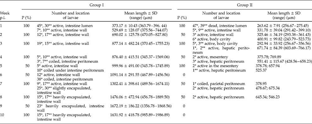
Table 1 summarizes data on the prevalence and intensity of infection, location of larvae in hosts and length of larvae.
Prevalence (P%), number and location of larvae, mean length±SD (the range of total length in parentheses) of C. rudolphii larvae in goldfish infected via copepods (Group I) or with free larvae (Group II).
p.i., postinfection.
* The first fish.
** The second fish.
Fish exposed to the previously infected cyclopoids (Group I) showed 90% prevalence. A week after the first dose of infected cyclopoids was administered, the intestine lumen and wall of the two fish autopsied yielded C. rudolphii larvae, motile and devoid of the old cuticle. The length of the larvae found in the intestine lumen and intestine wall averaged 373.17 ± 10.43 and 529.69 ± 128.07 μm, respectively. Two and 3 weeks p.i., only the intestine wall contained larvae, which moved within the muscle layer. All the larvae were motile after the intestine had been pepsin digested. Two and 3 weeks p.i., the larvae were 690.02 ± 125.78 and 877.14 ± 482.24 μm long, respectively. The larvae that were about 1000 μm long had a well-developed larval tooth; three primordial lips; a well-developed, 142.56 μm long ventricular appendix; and a 40 μm long intestinal caecum; the nerve ring was positioned 129.16 μm away from the anterior end of the body. Shorter larvae showed only a well-developed larval tooth, the remaining structures being poorly visible. Autopsies performed in subsequent weeks produced fewer and fewer larvae in the intestinal muscle layer; most larvae – slightly or tightly coiled – stayed underneath the intestine's serous membrane. Eight weeks later, almost all the larvae found in the intestine wall were surrounded by a thick light-brown sheath. After the sheath was digested away, most larvae – measuring from 1476.06 ± 472.94 μm (week 8) to 1631.92 ± 418.78 μm (week 10) – remained alive. Those larvae had the nerve ring 133.26 μm away from the anterior end; their ventricular appendix and intestinal caecum were 261.42 and 88.21 μm long, respectively. The mouth was surrounded by three lips. Throughout the experiment, goldfish exposed to the infected cyclopoids contained the larvae only in the intestine wall or underneath the intestine's serous membrane, no larvae being revealed either in the body cavity or in internal organs.
The prevalence of infection in fish infected with free-living larvae (Group II) was 50%, intensity of infection varying from two to nine larvae per host. On days two and three p.i., numerous C. rudolphii larvae were found in the fish tank sediment containing faeces. The larvae were mostly dead and had no cuticular sheath. Autopsies performed 1 week after infection showed the intestine lumen to contain only dead larvae, whereas live larvae measuring 331.78 ± 39.04 μm were found actively penetrating the intestine muscle layer. Two weeks p.i., the autopsy of one fish revealed three larvae (325.46 ± 34.19 μm long) in the intestine wall and six larvae (400.91 ± 99.82 μm long) in the body cavity. After 3 weeks, the larvae were found in the body cavity and in the hepatic peritoneum. Larvae isolated from the liver measured 671.74 ± 84.39 μm and showed a well-developed larval tooth and a ventricular appendix 85 μm long. After 4 and 5 weeks following infection, no larvae were found in the intestine wall and live larvae were found in the mesentery and liver peritoneum. In week six p.i., no autopsied fish contained any larvae, whereas in week seven, one fish contained a single larva, slightly coiled beneath the parietal peritoneum, and two larvae in the liver peritoneum. In week eight, one fish was found to host two larvae beneath the hepatic peritoneum. Autopsies performed in subsequent weeks produced no nematode larvae.
Discussion
The present results show that, under experimental conditions, goldfish may be infected by both newly hatched larvae and already infected cyclopoids. The type of infective material is important for subsequent development of the larvae in their hosts. Larval development was asynchronous in both groups of fish (table 1). For a few weeks p.i., fish contained live larvae which hardly differed in terms of length and developmental advancement of their internal organs, from larvae isolated from crustaceans (Group I) or from newly hatched larvae (Group II). A similar lack of synchronicity in C. rudolphii larval development in experimentally infected fish was also reported by Huizinga (Reference Huizinga1966) and Bartlett (Reference Bartlett1996). Larvae isolated by Bartlett (Reference Bartlett1996) from experimentally infected guppies were 0.8–2.9 mm long on day 87 p.i., the length range of 3.1–3.9 mm being recorded on day 159. Similarly, Huizinga (Reference Huizinga1966) reported 201.6–901.6 μm long larvae, isolated on day 18 p.i. from the experimentally infected mosquito fish. According to Moravec (Reference Moravec1994), third-stage larvae found in fish are 0.78–0.91 mm long when less developmentally advanced, and 15.0–24 mm when more advanced. Development of internal organs in the larvae isolated from goldfish in this study was similar to that described in larvae isolated from guppies and mosquito fish (Huizinga, Reference Huizinga1966; Bartlett, Reference Bartlett1996). Larvae, 1262.75 μm in mean length, isolated from mosquito fish infected with zooplankton (Huizinga, Reference Huizinga1966), had the nerve ring located, on the average, 105.09 μm away from the anterior end, the intestinal caecum averaging 65.64 μm in length. The nerve ring in larvae (860 μm in mean length on day 45) isolated from guppies experimentally infected with zooplankton (Bartlett, Reference Bartlett1996) was, on average, 100 μm away from the anterior end; the length of the ventricular appendix and intestinal caecum averaged 140 and 40 μm, respectively. Larvae measuring 2700 μm in mean length had the nerve ring 140 μm away from the anterior end; the ventricular appendix and intestinal caecum were 260 and 140 μm long, respectively.
In this study, the mean distance between the nerve ring and the anterior end was 129.16 μm in the 1000 μm long larvae isolated from zooplankton-infected goldfish; the ventricular appendix and intestinal caecum were 142.56 and 40.00 μm in mean length, respectively. The nerve ring in 1631.92 μm long larvae was 133.26 μm away from the anterior end; the ventricular appendix and intestinal caecum measured 261.42 and 88.21 μm, respectively. Internal organs of larvae isolated from goldfish infected with newly hatched larvae developed at a much slower rate. Mosgovoy et al. (Reference Mosgovoy, Shakhmatova and Semenova1968) reported C. rudolphii larvae in dragonfly larvae to develop much faster and the development to be more synchronous. In their study, nematode larvae isolated from a larval dragonfly on day 10 p.i. measured 0.69–0.88 mm, the range increasing to 2.2–3.27 mm on day 30. Larvae isolated on day 30 had three primordial lips surrounding the mouth as well as a well-developed ventriculus with a 0.42–0.49 mm long appendix, and a well-developed, 0.23–0.26 mm long intestinal caecum. The nerve ring was 0.14–0.15 mm away from the anterior end. Larvae, 3.0–4.0 mm long, isolated from experimentally infected fish, were not so advanced in their development. On day 30 p.i., they showed only a well-developed ventricular appendix. Similar body sizes and advancement in the internal organ development in larvae isolated from naturally infected trout (Salmo trutta) were reported by Torres & Cubillos (Reference Torres and Cubillos1987). Larvae measured 1.43–2.82 mm (mean length: 1.98 mm); their intestinal caecum and ventricular appendix averaged 0.149 and 0.274 mm in length, respectively. The mouth was surrounded by three lips, the buccal cavity showed a well-developed larval tooth, and the nerve ring was, on the average, 0.041 mm away from the anterior end.
The type of infective material was also important for prevalence and intensity of infection as well as for the location of larvae in the host (table 1). Only half of the individuals which had been force-fed with the larvae (Group II) became infected; the number and size of larvae found in them were lower than in fish infected via the already infected zooplankton. Clearly, most of the administered larvae were excreted; because of their small size, they might have not been able to penetrate the intestine wall efficiently. Køie & Fagerholm (Reference Køie and Fagerholm1995) argued for a clear relationship between the size of a larva and its ability to penetrate the host's intestine wall. In their study, newly hatched larval Contracaecum osculatum were able to penetrate the intestine wall of small fish only; the intestine wall of larger fish could be penetrated by larvae originating from infected crustaceans or from small infected fishes.
Larval resistance to the host's enzymes could be another cause of low invasion parameters in Group II fish. Digestive enzymes, particularly proteases, are known to be a barrier for a parasite to overcome before it can settle in the host's body. Parasites dwelling in the intestine lumen protect themselves from digestive enzymes by producing enzyme inhibitors (Hawley et al., Reference Hawley, Martzen and Peanasky1994; Morris & Sakanari, Reference Morris and Sakanari1994). Goldfish is a cyprinid whose alimentary tract lacks a defined stomach. Free-living larvae, administered to fish, ended up at a site affected primarily by trypsin and chymotrypsin. As adult and larval C. rudolphii dwell in the stomach of cormorant, they are doubtless resistant to pepsin. Nematode larvae were only found in the intestine lumen of fish naturally infected by Szostakowska & Fagerholm (Reference Szostakowska and Fagerholm2007); in all other cases, larvae were observed in the intestine wall and intestinal mesentery (Torres & Cubillos, Reference Torres and Cubillos1987; Kvach, Reference Kvach2005).
Too many larvae fed to the fish could be another reason why larvae were excreted, although – under natural conditions – intensity of infection is frequently very high. Szostakowska & Fagerholm (Reference Szostakowska and Fagerholm2007) found 500 larvae in a single goldfish, whereas intensity of infection reported from the Valdivia River (Chile) ranged from 8 to 1972 (Torres et al., Reference Torres, Valdiviesa, Schlatter, Montefusco, Revenga, Marin, Lamilla and Ramallo2000). In this study, larvae isolated from fish infected with them directly were only slightly coiled and were not encysted. The fish was then the first host; as in other animals serving as the first intermediate hosts (copepods, amphipods, larval dragonflies), larvae did not encyst. Køie & Fagerholm (Reference Køie and Fagerholm1995) reported a similar finding from their study on the life cycle of C. osculatum. Having administered newly hatched larvae to a number of fish species (Gasterosteus aculeatus, Pungitius pungitius, Pomatoschistus microps, Pleuronectes platessa, Zoarces viviparus), they found that only P. platessa and Z. viviparus became infected. Two weeks after infection, fish contained a few non-encysted larvae each, mainly in the hepatic sinusoids. On the other hand, strongly encysted larvae were found in cod (Gadus morhua) fed eelpout that had been infected earlier. Køie & Fagerholm (Reference Køie and Fagerholm1995) contended that, when a fish is infected with newly hatched nematode larvae, for these to encyst they have to be transmitted to a subsequent host, for example a predatory fish.
The present experiment showed that fish, fed the already infected cyclopoid, developed a much higher prevalence and intensity of infection. Larvae in those fish were found mostly in the intestine wall, where they were firmly encysted, a cyst looking like a light-brown nodule with a larva visible inside. Larvae found in the intestine wall were distinctly longer and much more developmentally advanced (as shown by the length of the ventricular appendix and intestinal caecum) than those isolated from fish that had been directly infected. In the first case, fish were the second hosts; larvae that penetrated the intestine wall had already moulted in the cyclopoid intestine. In her study on larval C. rudolphii morphogenesis in various hosts, Bartlett (Reference Bartlett1996) found the changes that the parasite undergoes in a copepod to be very important for development, as they greatly increase the parasite's chance of settling in a new host. While in the copepod, the larva grows in size and develops the ventricular appendix and the excretory system. There are no published descriptions of mechanisms enabling larval C. rudolphii to penetrate the fish intestine wall. Penetration enzymes (proteases, glycosidases, hyaluronidase and aminopeptidases) found in excretion–secretion (ES) products of numerous parasites (Morris & Sakanari, Reference Morris and Sakanari1994; Sajid & McKerrow, Reference Sajid and McKerrow2002; Irwin et al., Reference Irwin, Morrisey, Ryan, Walsche, O'Neill, Carrington, Matthews, Fitzpatrick, Muleahy, Corfirld and Dalton2004) facilitate the parasite's settling in the host's body. In our opinion, enzymes secreted to the external medium protect the parasite from a host's immunological response and facilitate migration in tissues by decomposing the tissue barrier. Higher invasion parameters in fish exposed to the already infected cyclopoids could have resulted not only from the larger larvae being involved, but also from their penetration enzymes, whereby the larvae were able to penetrate the intestine wall faster.
The present results show that development and location of larval C. rudolphii in fish depends greatly on the infection pathway. Goldfish studied were similar in age and individual weight. Most of the larvae found in fish directly exposed to free-living larvae (Group II) moved to the body cavity and then to other internal organs. All the larvae were motile and non-encysted. On the other hand, goldfish exposed to the infected cyclopoids showed larvae, coiled and surrounded by a light-brown sheath, to have remained in the intestine wall. Those larvae were most probably at a stage when they did not require transmission to another intermediate or paratenic host, but could stay in the present host until transmission to the definitive host.
Fish infected naturally also show some differences in the location of larval nematodes, which suggests different infection pathways. According to Moravec (Reference Moravec1994), encysted C. rudolphii larvae occur in the body cavity, intestinal mesentery and intestine wall. Torres & Cubillos (Reference Torres and Cubillos1987) found encysted larvae of C. rudolphii only in the intestine lumen of trout (Salmo trutta) in River Valdivia, whereas other Valdivia fish (Cauque mauleanum, Basilichthys australis, Galaxias maculatus, and Oncorhynchus mykiss) carried larvae also in the intestinal mesentery (Torres et al., Reference Torres, Valdiviesa, Schlatter, Montefusco, Revenga, Marin, Lamilla and Ramallo2000). In two Black Sea fish species (N. melanostomus and Mesogobius batrachocephalus) studied by Kvach (Reference Kvach2005), encysted larvae of C. rudolphii were found in the intestinal mesentery, whereas the golden grey mullet (Liza aurata) from the Black Sea, examined by Pronkina & Belofastova (2005), revealed non-encysted C. rudolphii larvae in bile ducts. Szostakowska & Fagerholm (Reference Szostakowska and Fagerholm2007) reported encysted larvae in the intestinal mesentery of the round goby (N. melanostomus) from the Gulf of Gdańsk, and the crucian carp (C. carassius) of Lake Selment Wielki carried larvae in the intestine lumen, wall, mesentery and near the liver. It seems that, under natural conditions, the probability that fish become infected by free-living larvae is very low because of the small size and low density of larvae, as shown in an earlier study (Dziekońska-Rynko et al., Reference Dziekońska-Rynko, Rokicki and Wziętek2008). However, differences in the location of larvae in fish and the presence of non-encysted larvae in fish infected under natural conditions may suggest that direct infection is indeed possible. According to Szostakowska & Fagerholm (Reference Szostakowska and Fagerholm2007), fish feeding close to the bottom may become infected by swallowing nematode eggs or ovigerous nematode females contained in cormorant faeces.
The present results, as well as data reported by other authors, confirm the opinion of Køie (Reference Køie1993) that life cycles of most anisakids are similar. The first two moults occur partly, or entirely, in eggs. The newly hatched second- or third-stage larvae, surrounded by membranes remaining from the preceding moult, are the dispersal stages. The subsequent moult occurs in the intermediate hosts, i.e. crustaceans. In such a life cycle, fish are paratenic hosts in which larvae become sturdily encysted. On the other hand, when infection is caused by free-living, newly hatched larvae, fish are the initial hosts in which larvae do not encyst.