Introduction
Extracranial internal carotid artery (ICA) pseudoaneurysms in children are extremely rare, with few reported cases in the literature. A pseudoaneurysm or false aneurysm represents a blood-filled cavity in contact with an artery, with a persistent connection with the arterial lumen.Reference Requejo, Sierre, Lipsich and Zuccaro 1 , 2 This occurs as a result of a localised disruption of the arterial wall. 2 The aneurysm wall is formed by haematoma and reactive connective tissue.Reference Requejo, Sierre, Lipsich and Zuccaro 1 It has a high mortality rate if left untreated because of the great potential for thromboembolic events and massive life-threatening haemorrhage.Reference Requejo, Sierre, Lipsich and Zuccaro 1 , Reference Garcia-Monaco, Kohan, Martinez-Corvalan, Cacchiarelli, Peralta and Wahren 3 , Reference Pourhassan, Grotemeyer, Fokou, Heinen, Balzer and Ramp 4 Therefore, a high index of suspicion and timely diagnosis remain very important in order to manage these children before the development of life-threatening complications.Reference Garcia-Monaco, Kohan, Martinez-Corvalan, Cacchiarelli, Peralta and Wahren 3 , Reference Pourhassan, Grotemeyer, Fokou, Heinen, Balzer and Ramp 4
We report on the case of a boy aged two years and six months with an extracranial ICA pseudoaneurysm, who presented with a large parapharyngeal space mass and bloody otorrhoea secondary to ear infection. A successful endovascular occlusion procedure of the ICA and pseudoaneurysm was performed without any neurological sequelae.
Case report
A previously healthy boy, aged two years and six months, with a one-month history of intermittent bloody otorrhoea from the right ear and a right-sided neck mass, was seen at the ENT Clinic of Universitas Hospital, Bloemfontein, South Africa.
On examination, the patient appeared acutely ill and irritable. A hard, tender, right-sided neck mass was noted in the infra-auricular area, which was non-fluctuant and non-pulsatile. Examination of the right ear revealed an inflamed, granulomatous mass on the inferior aspect of the right external auditory canal, with an intact tympanic membrane. Neurological examination findings were normal. On inspection of the oral cavity, the patient was noted to have mild trismus and narrowing of the oropharynx due to a large right-sided parapharyngeal mass. The airway was not imminently threatened. He had persistent hypertension, observed since admission.
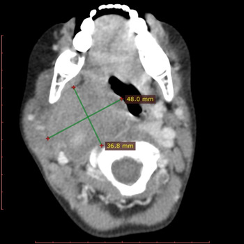
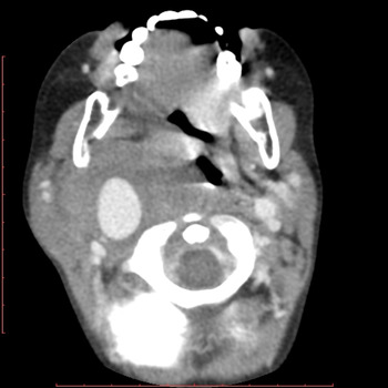
A neck ultrasound scan showed a poorly circumscribed mass on the side right, suspected to be of parotid gland origin. Computed tomography (CT) scans of the neck revealed a pseudoaneurysm of the right proximal internal carotid artery (ICA) measuring 4.8 × 3.6 × 4.4 cm, with mass effect on the pharynx and narrowing of the airway (Figures 1 and 2). Opacification of the right mastoid air cells and middle ear were observed, with an intact circle of Willis.
Axial computed tomography scan demonstrating internal carotid artery pseudoaneurysm.

Axial computed tomography scan with post-contrast enhancement in pseudoaneurysm.

With regard to laboratory results, white cell count was 7.69 × 10⁹/l, erythrocyte sedimentation rate was 62 mm/hour, C-reactive protein was 18 mg/l, enzyme-linked immunosorbent assay findings for human immunodeficiency virus (HIV) were negative, and renal functions and electrolytes were normal.
A biopsy of the mass in the right external auditory canal revealed ulcerated squamous epithelium and underlying inflamed granulation tissue, suggestive of chronic otitis externa. Auramine-O stain and tuberculosis cultures of the biopsy were negative, while a pus swab of the right ear cultured Klebsiella pneumoniae and beta-lactamase negative Staphylococcus aureus. Blood cultures were negative. A renal ultrasound scan was normal and autoimmune investigation findings were negative.
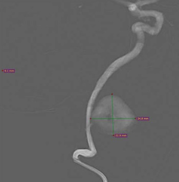
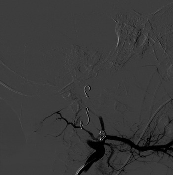
The patient underwent uneventful intubation, followed by endovascular occlusion of the ICA and pseudoaneurysm by coiling (Figures 3 and 4). Intubation was carried out by experienced personnel because of the potential for rupture during intubation. An endovascular approach was used because direct surgical repair would have required wide exposure of the neck in order to obtain proximal and distal control of the aneurysm, leading to higher morbidity and possible mortality.Reference Requejo, Sierre, Lipsich and Zuccaro 1
Arteriogram demonstrating pseudoaneurysm of right internal carotid artery.

Arteriogram post-coiling, demonstrating exclusion of internal carotid artery and pseudoaneurysm from circulation.

The patient was extubated 4 days after the procedure. He made a full recovery without any neurological sequelae. Post-procedural Doppler scans did not demonstrate any flow in the ICA or pseudoaneurysm. No anticoagulation was prescribed. The parapharyngeal mass gradually resolved and the right ear was successfully treated with systemic antibiotics and ofloxacin drops. Blood pressure gradually returned to normal after the resolution of the neck mass, and the assumption was made that the hypertension was secondary to the pressure on the carotid body caused by the pseudoaneurysm. At six months’ follow up, the ICA and pseudoaneurysm remained excluded from the circulation.
Discussion
Risk factors for extracranial internal carotid artery (ICA) pseudoaneurysm include prior carotid surgery or intervention, blunt and open trauma, primary or contiguous infection, spontaneous carotid dissection, connective tissue diseases, and radiation therapy.Reference Requejo, Sierre, Lipsich and Zuccaro 1 , 2 , Reference Pulli, Dorigo, Pratesi, Fargion and Pratesi 5 Contiguous infection, typically from meningitis, cervical lymphadenitis and ear infection, remains the most common cause of carotid artery aneurysm in children. 2 The infection typically spreads to involve periarterial lymphatics and vasa vasorum. 2 The incidence of infected aneurysms due to peritonsillar and middle-ear infections has declined with the use of antibiotics. 2 , Reference Garcia-Monaco, Kohan, Martinez-Corvalan, Cacchiarelli, Peralta and Wahren 3 The main reason for involvement of the vessels with infection of the pharynx and deep neck spaces is the close proximity of the ICA to these structures.Reference Requejo, Sierre, Lipsich and Zuccaro 1 , Reference Pourhassan, Grotemeyer, Fokou, Heinen, Balzer and Ramp 4 A case study published in 2012, on a five-year-old child, reported on an ICA pseudoaneurysm secondary to a throat infection, which was treated successfully with stent placement after thrombin injection failure.Reference Garcia-Monaco, Kohan, Martinez-Corvalan, Cacchiarelli, Peralta and Wahren 3
S aureus, salmonella, Streptococcus pyogenes, Escherichia coli and klebsiella are the most common pathogens associated with infected extracranial ICA pseudoaneurysm.Reference Requejo, Sierre, Lipsich and Zuccaro 1 , 2 The high incidence of HIV in our environment has led to an increase in HIV-associated carotid aneurysms.Reference Padayachy and Robbs 6 Carotid artery aneurysms represent 25 per cent of all HIV-related aneurysms and have been thought to occur as a result of HIV-related vasculopathy.Reference Padayachy and Robbs 6
Congenital aneurysms have also been described in children, and occur as a result of structural defects of the arterial wall secondary to developmental defects of the arterial muscular layer.Reference Pourhassan, Grotemeyer, Fokou, Heinen, Balzer and Ramp 4
Symptomatology of extracranial ICA pseudoaneurysm may be due to the local mass effect, embolism, thrombotic occlusion or aneurysm rupture. 2 Common symptoms include: a painful or painless mass, stroke or transient ischaemic attacks, visual disturbance, a protruding pharyngeal mass, epistaxis, and Horner's syndrome. Hoarseness, dysphagia, auricular pain and tongue weakness may occur as a result of cranial nerve palsies.Reference Requejo, Sierre, Lipsich and Zuccaro 1 , 2 , Reference Garcia-Monaco, Kohan, Martinez-Corvalan, Cacchiarelli, Peralta and Wahren 3 The condition may also be asymptomatic and discovered incidentally. 2 Rupture is a potentially life-threatening complication that may occur in as many as one in three children, and may present as pharyngeal haemorrhage, epistaxis or bloody otorrhoea.Reference Requejo, Sierre, Lipsich and Zuccaro 1 , 2 , Reference Pourhassan, Grotemeyer, Fokou, Heinen, Balzer and Ramp 4 Pourhassan et al. reported the highest risk of rupture to occur with traumatic and mycotic aneurysms.Reference Pourhassan, Grotemeyer, Fokou, Heinen, Balzer and Ramp 4
Diagnosis relies on the demonstration of an aneurysm on imaging studies. Ultrasound is the initial study of choice to evaluate a neck mass, but CT angiography and magnetic resonance angiography are useful confirmatory imaging studies that can determine the extent of dilatation and thrombus formation of the aneurysm, and demonstrate the relationship of the aneurysm with the surrounding structures. 2
Intervention is recommended for symptomatic aneurysms, aneurysms with intraluminal thrombus and small asymptomatic aneurysms that demonstrate expansion on imaging. 2 , Reference Pourhassan, Grotemeyer, Fokou, Heinen, Balzer and Ramp 4 Management was traditionally with open surgical repair, although it has recently been replaced by endovascular techniques, which seem to be a feasible and safe option.Reference Garcia-Monaco, Kohan, Martinez-Corvalan, Cacchiarelli, Peralta and Wahren 3 , Reference Pulli, Dorigo, Pratesi, Fargion and Pratesi 5 Open surgical repair is reported to have higher morbidity and mortality rates, and a higher risk of cranial nerve injury.Reference Pulli, Dorigo, Pratesi, Fargion and Pratesi 5 With free or impending rupture, it may be necessary to convert to an open surgical technique if there is continuous bleeding from the sac during endovascular procedures.Reference Pulli, Dorigo, Pratesi, Fargion and Pratesi 5
Options for open surgical repair include ligation of the carotid artery and excision of the aneurysm, with reconstruction using a saphenous vein graft or prosthetic material. 2 , Reference Pourhassan, Grotemeyer, Fokou, Heinen, Balzer and Ramp 4 In children younger than four years, the greater saphenous vein graft may be too small for supra-aortal interposition,Reference Pourhassan, Grotemeyer, Fokou, Heinen, Balzer and Ramp 4 with the theoretical risk of stenosis. In these cases, the use of the profunda femoral vein graft for interposition has been described.Reference Pourhassan, Grotemeyer, Fokou, Heinen, Balzer and Ramp 4 In children, the delicate anatomy of the small vessels makes open surgery challenging.Reference Pourhassan, Grotemeyer, Fokou, Heinen, Balzer and Ramp 4
Endovascular repair techniques include stent placement, trans-stent coil embolisation, stent-graft exclusion of the aneurysm or endovascular carotid artery occlusion. 2 In a reported case of a four-year-old child with ICA pseudoaneurysm secondary to pharyngitis, the use of a covered stent was reported to be an effective therapeutic option to exclude the pseudoaneurysm while keeping the parent vessel patent.Reference Requejo, Sierre, Lipsich and Zuccaro 1 It was reported that long-term patency, outcomes and best antithrombotic therapy still need to be evaluated in children with covered stents.Reference Requejo, Sierre, Lipsich and Zuccaro 1 There are also concerns regarding the adaptability of the growing carotid artery to the covered stent.Reference Garcia-Monaco, Kohan, Martinez-Corvalan, Cacchiarelli, Peralta and Wahren 3
-
• Extracranial internal carotid artery (ICA) pseudoaneurysms in children are rare
-
• Endovascular intervention remains the choice of treatment for this condition
-
• It has a high mortality rate if left untreated given the potential for development of life-threatening complications
-
• A high index of suspicion and timely diagnosis remain important
-
• This paper reports on a child successfully treated with endovascular occlusion of the ICA and pseudoaneurysm by coiling
Carotid sacrifice may be performed via an open ligation or via an endovascular approach. 2 Ligation of the ICA is associated with a substantial risk for neurological morbidity in the short and long term.Reference Rosset, Albertini, Magnan, Ede, Thomassin and Branchereau 7 Stroke and even death may result if the collateral circulation is not intact.Reference Pourhassan, Grotemeyer, Fokou, Heinen, Balzer and Ramp 4 McCann (as cited in Rosset et al.)Reference Rosset, Albertini, Magnan, Ede, Thomassin and Branchereau 7 reported a stroke risk of 25 per cent and mortality rate of 20 per cent with ICA ligation. Although endovascular exclusion techniques are usually safe and curative, there is a small risk for aneurysmal disease in the contralateral ICA due to chronic increase in flow.Reference Requejo, Sierre, Lipsich and Zuccaro 1 , Reference Garcia-Monaco, Kohan, Martinez-Corvalan, Cacchiarelli, Peralta and Wahren 3 Given the fragile nature of the pseudoaneurysm wall, selective embolisation of the pseudoaneurysm may be hazardous.Reference Garcia-Monaco, Kohan, Martinez-Corvalan, Cacchiarelli, Peralta and Wahren 3
Conclusion
Extracranial internal carotid artery pseudoaneurysm is rare in children, but has a high mortality rate if left untreated. The diagnosis should be kept in mind in a child presenting with a neck mass, especially in a patient with a concomitant protruding pharynx mass. This entity may rarely be caused by recent ear infection. Endovascular intervention remains the treatment of choice in children because of lower associated mortality rates compared to an open surgical approach. 2 , Reference Pulli, Dorigo, Pratesi, Fargion and Pratesi 5