Introduction
The description and identification of larval phase in Decapod crustaceans is a challenging task. The difficulty in finding morphological differences among specimens included in the same taxon increases when there is a scarcity of meticulous description of characteristics, limited illustrations available and few parental individuals identified to the species level (González-Gordillo et al., Reference González-Gordillo, Santos and Rodríguez2001; Negreiros-Fransozo et al., Reference Negreiros-Fransozo, González-Gordillo and Fransozo2002). Additionally, a limited supply of studies focused on this subject contributes to the obstacle in the descriptive and comparative process.
Among the Decapoda, the caridean shrimps present a relatively broad literature concerning the larval morphology (Guerao and Cuesta, Reference Guerao, Cuesta, Martin, Olesen and Hoeg2014), although, when the total number of species in an isolated taxon is computed it is possible to highlight the insufficiency of documented described larvae to meet some objectives. This is illustrated by some of the richest genera within Caridea. The Palaemonidae genus Macrobrachium Spence Bate, 1868, with approximately 17% out of the approximately 265 valid species (De Grave et al., Reference De Grave, Decock, Dekeyzer, Davie, Fransen, Boyko, Poor, Macpherson, Ahyong, Crandall, Mazancourt, Osawa, Chan, Ng, Lemaitre, van der Meij and Santos2023; WoRMS, Reference WoRMS2024) with described larvae (e.g. Magalhães, Reference Magalhães1985, Reference Magalhães1989; Jalihal et al., Reference Jalihal, Sankoli and Shenoy1993; Pereira and García, Reference Pereira and García1995, Reference Pereira, García, Escobar-Briones and Alvarez2002; Rodrigues and Bueno, Reference Rodrigues and Bueno1995; Ito et al., Reference Ito, Fujita and Shokita2006; Mejia-Ortiz et al., Reference Mejia-Ortiz, Hartnoll and Lopez-Mejia2010; Pantaleão et al., Reference Pantaleão, Gregati, Taddei and Costa2011; Vieira et al., Reference Vieira, Santos and Anker2017; Marco-Herrero et al., Reference Marco-Herrero, Anger and Hayd2019), and the Alpheidae genus Alpheus Fabricius, 1798, that presents approximately 335 valid species (Casaubon et al., Reference Casaubon, Hultgren, Murray, Hanscom and Hurt2023; WoRMS, Reference WoRMS2024), but not exceeding 10% comprising its entire species count (Pescinelli et al., Reference Pescinelli, Pantaleão, Mantelatto and Costa2017; Pantaleão et al., Reference Pantaleão, Pescinelli, Costa, Cuesta and Mantelatto2020; Pescinelli and Costa, Reference Pescinelli and Costa2022).
Within the Alpheidae, the genus Synalpheus Spence Bate, 1888 currently have 18 species with some information about first larval stage morphology (Santos et al., Reference Santos, Pescinelli and Costa2020, Reference Santos, Teles, Mantelatto and Costa2024), of the approximately 170 valid species, covering only about 10% of the total species (Ashrafi and Hultgren, Reference Ashrafi and Hultgren2023; Mantelatto et al., Reference Mantelatto, França, Cunha and Almeida2023; WoRMS, Reference WoRMS2024). Among those only eight of them present a detailed description of its zoeal stages appendages. If the subsequent larval stages are taken into consideration, the number of described species decreases even more, in which only six species present descriptions for the zoea II and zoea III, four for the zoea IV and only three for the zoea V (Brooks and Herrick, Reference Brooks and Herrick1892; Gurney, Reference Gurney1938; Weise, Reference Weise1975; Albornoz and Whertmann, Reference Albornoz and Whertmann1997; Yang, Reference Yang2009).
The limitation in detailed studies regarding the morphological larval descriptions of Synalpheus, in some cases, occurs due to a dearth of information, e.g., the species S. scaphoceris Coutière, 1910, S. longicarpus (Herrick, 1891), S. spinifrons (H. Milne Edwards, 1837), and S. williamsi Ríos and Duffy, Reference Ríos and Duffy1999 (Brooks and Herrick, Reference Brooks and Herrick1892; Dardeau, Reference Dardeau1986; Albornoz and Whertmann, Reference Albornoz and Whertmann1997; Ríos and Duffy, Reference Ríos and Duffy1999) in which any morphological characteristics are reported, or in cases where only a few description are provided, there is lack of specificity regarding the appendages or seta (Brooks and Herrick, Reference Brooks and Herrick1892; Gurney, Reference Gurney1938, Reference Gurney1949). Moreover, even in species that present a more extensive and all-encompassing description it is possible to observe that some structures have not been described, such as the first zoea of S. fritzmuelleri Coutière, 1909 (Weise, Reference Weise1975).
Apart from the lack of detailed structure descriptions, studies regarding Synalpheus larvae are in majority dated, hence, there is no standardized description framework that facilitates a comparative analysis, as has already been suggested for zoeae of Brachyura (Rice, Reference Rice1979; Clark et al., Reference Clark, Calazans and Pohle1998; Clark and Cuesta, Reference Clark, Cuesta, Castro, Davie, Guinot, Schram and Von Vaupel Klein2015). Not merely this, but within this genus there is a variable development feature, that may manifest an abbreviated, an extended or in some cases considered a direct post-embryonic development (Knowlton, Reference Knowlton1973). For this reason, there is adversity to establish morphological patterns among the Synalpheus larvae, which may result in the absence of certain details in new descriptive studies since there still is a difficulty to find conservative patterns in the larval structures (Santos et al., Reference Santos, Pescinelli and Costa2020).
Including the species with already described morphological characteristics, S. fritzmuelleri was first described by Weise (Reference Weise1975) (five zoeal stages), sampled in Florida's waters (USA). However, within this description there are a few lack of information along the development, such as the absence of morphological characteristics of the maxillule. Following this, is necessary to review and redescribe some species. Thus, the aim of the present study is to redescribe the three first zoeal stages of S. fritzmuelleri hatched under laboratory conditions and compare with the previous descriptions (Weise, Reference Weise1975).
Materials and Methods
Seven females with embryos of S. fritzmuelleri were captured in the subtidal region of five sampling areas: Tamanduá Island, Caraguatatuba, São Paulo (23°27′45″S; 45°02′48″W) in August 2018; Alcatrazes Archipelago, São Sebastião, São Paulo (24°05′43,69″S; 45°41′35,51″W) in October 2020 and October 2021; Cais do Porto, Itaguá beach, Ubatuba, São Paulo (23°27′05″S; 45°02′48″W) in October 2021; Figueira Isand, Cananéia, São Paulo (25°20′50″S; 48°01′40″W) in November 2020 and February 2022; and Ganchos Island, Paraty, Rio de Janeiro (23°10′29″S; 44°38′01″W), in November 2021 (Figure 1). Samples were performed by free and scuba diving using the sampling of biogenic substrate and the installation of artificial refuge substrate (ARS), following Moraes et al. (Reference Moraes, Almeida, Cobo, Davanso and Castilho2021). The specimens found in biogenic substrate were associated with the sponge Amphimedon viridis Duchassaing and Michelotti, 1864, the bryozoan Schizoporella sp. and the coral Madracis decactis (Lyman, 1859).
Location indicating the sampling areas of the parental female of Synalpheus fritzmuelleri Coutière, 1909.

Females with embryos were kept alive in individual water containers (3 l) containing natural water from the sampling location and were monitored daily until the larvae hatched. After they hatched, the females were removed from the containers and the larvae were maintained with constant temperature (25°C) and salinity (30), partially renewed every day. The larvae were served with nauplii of Artemia sp., although none of them fed during the cultivation. The larval stages were checked daily, and each obtained stage was removed from the recipients and conserved in a mixture (1:1) of ethyl alcohol (70%) and glycerin.
The carapace length (CL mm) of each female was measured and they were deposited at the Crustacean Collection of the Department of Biology, FFCLRP, University of São Paulo, Ribeirão Preto, Brazil (CCDB/FFCLRP/USP); CCDB – 6634, 6725, 6911, 7884, 7885, 7886, 7887. For each female and larval stage, ten larvae were randomly selected and had their CL measured from the margin of the ocular orbit to the posterior margin of the carapace, and the total length (TL mm), from the tip of the rostrum to the posterior margin of the telson, excluding setae. Appendages were dissected under a Zeiss Stemi 2000C trinocular stereomicroscope, and drawings and measurements were made using a Leica DM750 microscope equipped with a camera lucida. The larval description followed Clark et al. (Reference Clark, Calazans and Pohle1998) updated by Clark and Cuesta (Reference Clark, Cuesta, Castro, Davie, Guinot, Schram and Von Vaupel Klein2015), and the setal terminology proposed by Pohle and Telford (Reference Pohle G and Telford1981), Garm (Reference Garm2004) and Pantaleão et al. (Reference Pantaleão, Gregati, Costa, López-Greco and Negreiros-Fransozo2017) was used. For comparative analysis, the morphological characters described in the present study were compared with the first larval description accomplished by Weise (Reference Weise1975).
Results
The parental female CL varied from 5.03 to 5.47 mm, with an average size of 5.52 ± 0.80 mm. The larval morphology of zoea I, II, and III of S. fritzmuelleri are redescribed in detail.
Morphological description of early zoeal stages of Synalpheus fritzmuelleri Coutière 1909.
Zoea I
Dimensions: CL: 0.65 ± 0.04 mm; TL: 1.67 ± 0.08 mm – n = 10.
Carapace (Figure 2A, B): rostrum present and short; spines absent; eyes sessile.
Synalpheus fritzmuelleri Coutière, 1909, first larval stage. (A) Dorsal view; (B) lateral view; (C) antennule; (D) antenna; (E) maxillule; (F) maxilla; (G) first maxilliped; (H) second maxilliped; (I) third maxilliped; (J) pereiopod I; (K) pereiopod II; (L) pereiopod V; (M) telson. (Scale bars: a, b = 0.2 mm; c, j, k, l, m = 0.1 mm; d, f, g, h, i = 0.05 mm; e = 0.02 mm).

Antennule (Figure 2C): peduncle unarticled showing two annuli; endopod (accessory flagellum) as a plumose seta; exopod (primary flagellum) with four aesthetascs, one longer than others and one short plumose seta.
Antenna (Figure 2D): peduncle unarticled with an inner terminal short spine; endopod with one simple seta and one plumose seta; exopod (antennal scale) four-articled with 11 plumose setae, nine on the inner side (3, 1, 1, 4) and two on the outer side (1,1, 0, 0), and one simple seta on the outer side of the distal article.
Maxillule (Figure 2E): coxal endite with four simple setae; basial endite with two stout spines and one short spine; endopod with one simple seta and one denticulate seta (with denticules on medial to distal region).
Maxilla (Figure 2F): coxal endite with three (one proximal and two distal) plumose setae; basial endite bilobed with three simple setae each lobe; endopod bilobed with two simple and three (two simple and one plumose) setae, respectively; inner and outer margin with microtrichia; exopod (scaphognathite) with four (three terminal and one marginal) plumose setae, inner and outer margin with microtrichia.
First maxilliped (Figure 2G): coxa with one simple seta; basis with four spines and three simple setae; endopod unarticled with three (one short proximal and two distal) simple setae; exopod unarticled, bearing four long plumose natatory setae terminally.
Second maxilliped (Figure 2H): coxa unarmed; basis with one simple seta; endopod four-articled, with 0, 0, 1 terminal serrate seta, five (one marginal and three terminal simple plus one terminal serrate) setae; exopod unarticled, with one subterminal short simple seta and five (one subterminal and four terminal) long plumose natatory setae.
Third maxilliped (Figure 2I): coxa unarmed, basis with one simple seta; endopod five-articled, with 0, 0, 0, 2 terminal, four (one proximal and three terminal) simple setae; exopod unarticled with six (two subterminal and four terminal) long plumose natatory setae.
Pereiopods (Figure 2J–L): first and second as rudimentary biramous and fifth as rudimentary uniramous.
Pleon (Figure 2A, B): with five pleonites, without setae and rounded, pleopods absent.
Telson (Figure 2M): round, with deep postero-median concavity, margin with 7 + 7 plumose setae, outermost two pairs plumose only on inner margin, inner pair shorter; one row of minute spinules on distal margin between the four innermost pairs.
Zoea II
Dimensions: CL: 0.61 ± 0.01 mm TL: 2.11 ± 0.12 mm – n = 3.
Carapace (Figure 3A, B): rostrum longer than first zoea; pterogostomial spines present; eyes sessile.
Synalpheus fritzmuelleri Coutière, 1909, second larval stage. (A) dorsal view; (B) lateral view; (C) antennule; (D) antenna; (E) maxillule; (F) maxilla; (G) first maxilliped; (H) second maxilliped; (I) third maxilliped; (J) pereiopod I; (K) pereiopod II; (L) pereiopod V; (M) telson. (Scale bars: a, b = 0.4 mm; c, d, g, h, i, j, k, m, n = 0.1 mm; e, f, l = 0.05 mm).

Antennule (Figure 3C): peduncle three-articled, proximal article with eight (one marginal and seven terminal, with one longer than others) plumose setae, medial article with five plumose setae and distal article with seven (two marginal simple and five terminal plumose) setae; endopod (accessory flagellum) with a single plumose seta; exopod (primary flagellum) with five aesthetascs and one plumose seta.
Antenna (Figure 3D): peduncle unarticled; endopod as a slender spine at distal end with a small inner spine; exopod (antennal scale) four-articled with ten plumose setae, nine on the inner side (3, 1, 1, 4) and one on the outer side (0, 1, 0, 0) and two simple setae on the outer side (1, 0, 0, 1).
Maxillule (Figure 3E): coxal endite with six simple setae; basial endite with four (two short + two stout) spines; endopod unchanged.
Maxilla (Figure 3F): coxal endite with four (three plumose and one short simple) setae; basial endite bilobed with five and three simple setae, respectively; endopod unchanged; exopod (scaphognathite) with five (four terminal and one marginal) plumose setae; inner and outer margin with microtrichia.
First maxilliped (Figure 3G): coxa with two simple setae; basis unchanged; endopod unchanged; exopod unchanged.
Second maxilliped (Figure 3H): coxa unchanged; basis with two simple setae; endopod four-articled, with 0, 0, 1 terminal serrate, five (one marginal simple plus two terminal sparsely plumose plus two terminal serrate) setae; exopod unchanged.
Third maxilliped (Figure 3I): coxa unchanged, basis with two simple setae; endopod five-articled, with one terminal simple, one terminal simple, 0, 2 (one serrate and one sparsely plumose) terminal, four (one proximal and three terminal) simple setae, exopod unchanged.
Pereiopods (Figure 3J–L): first and second as rudimentary biramous, third as biramous bud, and fifth as rudimentary uniramous.
Pleon (Figure 3A, B): five pleonites, sixth somite starting to differentiate, without setae and rounded, pleopods absent.
Telson (Figure 3N): round, with deep postero-median concavity, margin with 8 + 8 (7 + 7 outer plumose setae plus 1 + 1 inner pair short and simple), outermost pair plumose only on inner margin; minute spinules on distal margin between the pair of setae two to five.
Zoea III
Dimensions: CL: 0.71 ± 0.01 mm TL: 2.22 ± 0.08 mm – n = 3.
Carapace (Figure 4A, B): rostrum longer than zoea II and exceeding the margin of the eyes; pterogostomial spines present; dorso-median papilla present; eyes sessile.
Synalpheus fritzmuelleri Coutière, 1909, third larval stage. (A) Dorsal view; (B) lateral view; (C) antennule; (D) antenna; (E) maxillule; (F) maxilla; (G) first maxilliped; (H) second maxilliped; (I) third maxilliped; (J) pereiopod I; (K) pereiopod II; (L) pereiopod III; (M) pereiopod V; (N) telson. (Scale bars: a, b = 0.4 mm; m, n = 0.2 mm; c, d, g, h, i, j, k = 0.1 mm; e, f, l = 0.05 mm).

Antennule (Figure 4C): peduncle three-articled, proximal article with six (one medial and five terminal) plumose setae, medial article with seven terminal plumose setae with one longer than others and distal article with nine (one marginal and five terminal short plumose and three long terminal plumose) setae; endopod (accessory flagellum) with a single simple seta; exopod (primary flagellum) with two aesthetascs and three (two short plumose and one simple) setae.
Antenna (Figure 4D): peduncle unchanged; endopod as a slender spine at distal end; exopod (antennal scale) two-articled with 11 plumose setae, ten on the inner side (7, 3) and one on the outer side (1, 0) and two simple setae on the outer side of the proximal article.
Maxillule (Figure 4E): coxal endite with six (one proximal simple and five terminal sparsely plumose) setae; basial endite with four stout spines (the two distal with two rows of spinules) and two simple setae; endopod unchanged.
Maxilla (Figure 4F): coxal endite with four (one simple and three sparsely plumose) setae; basial endite bilobed with five (two sparsely plumose and three simple) and five (two simple and three sparsely plumose) setae, respectively; endopod with two sparsely plumose setae plus three (two simple + one sparsely plumose) setae, inner and outer margin with microtrichia; exopod unchanged.
First maxilliped (Figure 4G): coxa unchanged; basis unchanged; endopod with four (one marginal and three terminal) simple setae, one terminal longer than others; exopod unchanged.
Second maxilliped (Figure 4H): coxa unchanged; basis unchanged; endopod four-articled, with 0, 0, 1 terminal plumose, five (one subterminal simple and one terminal sparsely plumose plus three terminal serrate) setae; exopod unarticled with one subterminal short plumose setae and five (one subterminal and four terminal) long plumose natatory setae.
Third maxilliped (Figure 4I): coxa unchanged; basis with three simple setae; endopod four-articled, with two (one proximal and one distal) simple setae, 0, 3 (two plumose + one simple) terminal setae, four terminal (one simple and three sparsely plumose) setae; exopod unchanged.
First Pereiopod (Figure 4J): coxa unarmed; basis with three (two simple and one sparsely plumose) setae; endopod four-articled, with two (one proximal and one distal) simple, two (one proximal simple and one distal serrate), two distal sparsely plumose, and three (two plumose and one simple) setae; exopod unarticled with six (two subterminal and four terminal) long plumose natatory setae.
Second Pereiopod (Figure 4K): coxa unarmed; basis with three (two simple and one sparsely plumose) setae; endopod four-articled, with two (one proximal and one distal) simple, two (one proximal simple and one distal serrate), two distal sparsely plumose, and three (two sparsely plumose and one simple) setae; exopod unarticled with six (two subterminal and four terminal) long plumose natatory setae.
Third–Fifth pereiopods (Figure 4L, M): third as a biramous bud, fourth absent, and fifth uniramous, five-articled unarmed with dactylus elongated, sharply pointed.
Pleon (Figure 4A, B): with six pleonites, unarmed, rounded; pleopods absent.
Uropods (Figure 4N): endopod rudimentary; exopod with six plumose setae on terminal margin and three dorsal short plumose setae, with microtrichia along the margin.
Telson (Figure 4N): with 7 + 7 setae, inner pair simple and two to seven pairs plumose; one row of minute spinules on distal margin between the pair of setae one to four.
Discussion
The morphological redescription of the early zoeal stages S. fritzmuelleri showed significant differences when compared with the first description (Weise, Reference Weise1975). Besides that, in both descriptions (Weise, Reference Weise1975 and the present study) the zoea I of S. fritzmuelleri shared some typical morphological characteristics among the first larva with extended development within the Synalpheus species (Knowlton, Reference Knowlton1973; Santos et al., Reference Santos, Pescinelli and Costa2020). Only a few of them are described with all (or most of it) morphological structures available to comparison (Gurney, Reference Gurney1927; Weise, Reference Weise1975; Yang, Reference Yang2009; Santos et al., Reference Santos, Pescinelli and Costa2020) and shared with both descriptions of the first larva of S. fritzmuelleri the carapace and pleon unarmed, eyes sessile, inner flagellum of antennule rudimentary as a plumose seta, exopod of first maxiliped with four plumose natatory setae, exopod of third maxiliped with six plumose natatory setae, pleon with five pleonites being the sixth undifferentiated from telson and a round telson with seven + seven plumose setae.
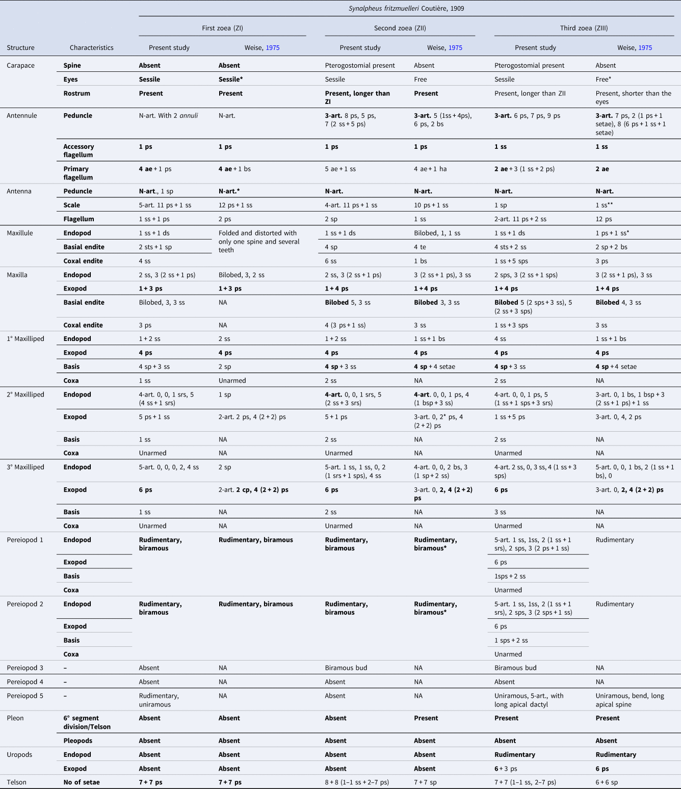
The comparative examination of the early larval stages of S. fritzmuelleri by Weise (Reference Weise1975) with the one presented here vary in the number and type of setae, and the presence or absence of articles, in which some appendages showed an expressive variation between the two descriptions (Table 1). In terms of articulation, for all zoeal stages compared, the antennal scale does not present any articles in the previous description, while in the present study it varies from 2 to 4, and in the exopod of the second and third maxillipeds, all stages described by Weise (Reference Weise1975) had articles, while in the current description these structures were unarticled. Moreover, some nomenclatural definitions of setae are not standardized, in which terms to designate them are used such as ‘bristled’, ‘hair’, and ‘teeth’, and in some cases there was only the mention of ‘setae’, without any determination of type.
Comparison of the described morphological characteristics of the early larval stage (ZI, ZII, and ZIII) of Synalpheus fritzmuelleri Coutière, 1909 between the present study and the first description by Weise (Reference Weise1975)

ss, simple setae; ps, plumose setae; sps, sparsely plumose setae; srs, serrate setae; ds, denticulate setae; sts, stout spine; sp, spine; ae, aesthetascs; art., article; NA, unavailable; bs, described in Weise, Reference Weise1975 as bristled setae; bsp, described in Weise, Reference Weise1975 as bristled spine; ha, described in Weise, Reference Weise1975 as hair; te, described in Weise, Reference Weise1975 as teeth; * = morphological characteristics observed through image boards. The highlight in bold demonstrate morphological similarities of each zoeal stage.
The non-standard terms used to describe some setae were found to approach the flagellum of antennule in the zoea I; penduncle and primary flagellum of antennule, basial and coxal endites of the maxillule, and endopod of the maxillipeds for the zoea II; and basial endite of maxillule, and endopod of maxillipeds of the zoea III. Through the observation of the described setae that receives these terms together with the figure boards provided by Weise (Reference Weise1975), it is possible to establish that the above-mentioned ‘teeth’ could be reporting spines, the ‘hair’ could be simple setae or microtrichia (in the mouth parts) and the ‘bristled setae’ or ‘bristled spines’ would probably be describing plumose setae, sparsely plumose setae, denticulate setae, or spiniform setae, if we follow the nomenclature proposed by Garm (Reference Garm2004) and Clark and Cuesta (Reference Clark, Cuesta, Castro, Davie, Guinot, Schram and Von Vaupel Klein2015), and by the observation of other contemporary studies with larval descriptions of Alpheidae (Bartilotti et al., Reference Bartilotti, Calado and Santos2005; Yang, Reference Yang2009; Pescinelli et al., Reference Pescinelli, Pantaleão, Mantelatto and Costa2017; Pantaleão et al., Reference Pantaleão, Pescinelli, Costa, Cuesta and Mantelatto2020; Pasinatto et al., Reference Pasinatto, Mantelatto and Terossi2020; Santos et al., Reference Santos, Pescinelli and Costa2020; Pescinelli and Costa, Reference Pescinelli and Costa2022; Pescinelli et al., Reference Pescinelli, Sanches and Costa2023; Sanches et al., Reference Sanches, Costa and Pescinelli2024).
Besides the variation in the nomenclature, the absence of information in the first description (Weise, Reference Weise1975) of the early S. frizmuelleri's zoeal stages makes it difficult for comparison. Some appendages do not present any information, as can be found in the coxa from the three maxillipeds and the basis of the second and third maxilliped for the first three zoeal stages; the basial and coxal endites of the maxilla for the zoea I; the pereiopods and pleopods of the zoea II; and the third and fourth pereiopods from the zoea III. Not only the complete lack of information, but also do not meticulously detail the morphology. This is noted in the maxillule of the zoea I of S. fritzmuelleri described by Weise (Reference Weise1975), in which is it folded and distorted with only one bristled spine and several teeth. All of this reinforces the importance of a redescription in order to complement the complete morphological characteristics of this species.
For the zoea I, a set of morphological characteristics were common for the two descriptions, as highlighted in bold in Table 1, most of them usually found in the first zoea of Synalpheus (Santos et al., Reference Santos, Pescinelli and Costa2020). Nonetheless, 18 differences were found, considering the appendages that were described by the two studies. Among those, some of the marked differences that can be mentioned are observed where the presence of a distal spine in the inner side of the antennal peduncle; the number of setae in the basis of the first maxilliped; the endopod of the second maxilliped; the endopod of third maxilliped; the number of pereiopods; and the minute spines and setuletes present in the margin of the telson were not mentioned in the first description. For more details about the differences among the first and the present description of the zoea I of S. fritzmuelleri, see Table 1.
A slightly but relevant difference in the zoea I descriptions is the presence of annuli that may be identified as markings of possible articles in the peduncle of the antenulle. This characteristic described here is shared only with the first zoea of S. ubatuba Mantelatto, França, Cunha & Almeida, 2023 (Santos et al., Reference Santos, Teles, Mantelatto and Costa2024). Commonly, the first zoeal stage in the caridean shrimps do not present articles in the peduncle of antenulle (Guerao and Cuesta, Reference Guerao, Cuesta, Martin, Olesen and Hoeg2014), although it may occur in some Alpheidae species. In the species Alpheus heterochaelis Say, 1818, A. simus Guérin-Méneville, 1855, S. brooksi Coutière, 1909, S. brevicarpus (Herrick, 1891), S. neptunus (Dana, 1852), and possibly S. biunguiculatus (Stimpson, 1860) these articulations occur due to an abbreviation of zoeal stages (Brooks and Herrick, Reference Brooks and Herrick1892; Dobkin, Reference Dobkin1965; Al Kholy and Mahmoud, Reference Al Kholy and Mahmoud1967; Knowlton, Reference Knowlton1973; Whertmann and Albornoz, Reference Whertmann and Albornoz2002; Pantaleão et al., Reference Pantaleão, Pescinelli, Costa, Cuesta and Mantelatto2020). Nevertheless, for Synalpheus species, the zoea I seems to present this characteristic in species of extended development, such as S. apioceros Coutière, 1909, S. neomeris (De Man, 1897), S. tumidomanus (Paulson, 1875), and S. williamsi (Gurney, Reference Gurney1927; Ríos and Duffy, Reference Ríos and Duffy1999; Yang, Reference Yang2009; Santos et al., Reference Santos, Pescinelli and Costa2020). Nonetheless, the articulation in the peduncle of antenulle must be examined carefully among the current and new descriptions of Synapheus first larvae. It is possible that the markings observed are associated with the rapid transition that larvae changes from the first to the second stage, in up to 2 h (Santos et al., Reference Santos, Pescinelli and Costa2020). These marks may represent the location where the articles will occur in the second zoea, but without being true articulation (Santos et al., Reference Santos, Teles, Mantelatto and Costa2024).
In the second zoea, it is possible to observe a low number of similar characteristics when comparing the first description with the one presented here. Only the rostrum, accessory flagellum of antennule, exopod of maxilla, first maxilliped and also first and second pereopods were equivalent. The variation in the number and type of setae, as well as for the presence/absence of articulation of some appendages (antennal scale and exopods of maxillipeds) remains constant as found for the first zoea. The most remarkable distinct morphological features are related to the presence of the pterogostomial spine in the zoea II in the present study; the eyes that are presented as free by Weise (Reference Weise1975), while they were described as sessile here; the absence of articulation in the antennal scale in the prior description; and the main difference found is related to the absence of information about the pereiopods in the first description, an important feature since in addition to the first two pairs of pereiopods already observed in the zoea I, the third pair appears as a rudimentary bud in the second stage as described in the present study. Additionally, during the review of the first description in the zoea II, some incongruences were found when comparing the text with the illustration, such as in the exopod of the second maxilliped in which is textually described as presenting one subterminal plumose seta (in the second article), while figure 24g of Weise (Reference Weise1975) presents two plumose natatory setae and no articulation was illustrated.
The available data in the literature for the second zoea description of Synalpheus species with extended development are exceedingly scarce. There is information about the morphology of the zoea II only for S. minus (Say, 1818), S. pectiniger (Coutière, 1907), S. spinifrons, S. triunguiculatus (Paulson, 1875), S. tumidomanus (Gurney, Reference Gurney1938; Weise, Reference Weise1975; Albornoz and Whertmann, Reference Albornoz and Whertmann1997; Yang, Reference Yang2009). Besides these, Al Kholy and Mahmoud (Reference Al Kholy and Mahmoud1967) also provide the description of S. biunguiculatus which presents the typical morphological characteristics of second zoea after hatch, presenting a possible abbreviation. It is difficult to find a pattern in the morphology of the second zoea within the genus, first due to the only species that presents all the appendages described is S. fritzmuelleri (present study) and S. tumidomanus (Yang, Reference Yang2009). Besides that, if we exclude the S. spinifrons and S. minus descriptions, since they lack key information about the appendage's morphology, the presence of a three articled antennular peduncle and six plumose setae in the exopod of third maxilliped is the only shared characteristics among the zoea II in Synalpheus.
In the third zoea, the number of shared characteristics decreases even more when compared with the previous stages, mainly zoea I (Table 1). The rostrum, the accessory flagellum of antennule, the peduncle of antenna, the exopod of maxilla and the exopod of first maxilliped were equally described in both studies (Weise, Reference Weise1975 and present study). Moreover, in terms of setae, the primary flagellum of the antennule presents the same number of aesthetascs, the basis of the first maxilliped presents the same number of spines and the exopod of the third maxilliped presents the same number of plumose natatory setae. Notably, the number of setae and the articulation are the most common variable characteristics within both zoea III descriptions of S. fritzmuelleri, following the same pattern found in the previous zoeal stages.
The differences that stand out the most in zoea III is related to the pereiopods, the first and the second above all. For the third and fourth pereiopods, no information is mentioned in Weise (Reference Weise1975), while in the present study, the third pereiopod is a rudimentary biramous bud and the fourth is absent. The fifth pereiopod is previously described as long with an apical spine, differing only in terms of articulation, described here as a five-articled structure. The first and the second pereiopods are extremely different, being related in the first description as rudimentary, the same description that occurs for the first and second zoea in both studies. Nonetheless, these structures were found here with a complex morphology (Table 1), while the second pereiopod differentiates from the first only by presenting three (two sparsely plumose and one simple) setae in the distal article of the endopod. This variation in the pereiopods must be cautiously investigated since the only described zoea III that shares this characteristic with the current description of S. fritzmuelleri is in the study by Yang (Reference Yang2009) with S. tumidomanus. Then, a review focusing in those appendages may clarify if there is any variation in the development of Synalpheus zoea among stages in which the pereopods present a more complex morphology, or if only these features were not thoroughly observed in other descriptions.
The current available zoea III descriptions of extended development of Synalpheus species available in the literature are for only four species, S. minus, S. pectiniger, S. triunguiculatus, and S. tumidomanus (Gurney, Reference Gurney1938; Weise, Reference Weise1975; Yang, Reference Yang2009). Those descriptions compared with the one of the present study shows a few shared morphological characteristics. Excepting the description of S. triunguiculatus that lacks information of several appendages, the following morphological characteristics are shared among the third zoea of Synalpheus: the rostrum present, the articulation of the antennular peduncle, the setae of the exopod of first and third maxilliped, the sixth pleonite separated from telson, and uropod present with a rudimentary endopod and an exopod with setae. Nevertheless, as far as new descriptions and reviews start to be carried out in more advanced stages of Synalpheus larvae it is possible that a greater number of shared characteristics could be found.
The results showed in the presented study evidenced the importance of review descriptions in the larvae morphology of Synalpheus. Regarding the variation of number and type of setae, highly present in the studied species and all zoeal stages compared, it is possible that a lack of observation due to the difficulty of detailing those structures, often very small and usually damaged, explain the variations observed. The lack of information that is common in the majority of the previous studies is one of the main problems to provide knowledge about this study subject, and a hindrance to accomplish comparative processes that help to develop new descriptions with accuracy. Thus, we reinforce the need to new reviews together with new descriptions of Synalpheus larvae to complement the insufficiency of characteristics that may establish a solid relationship in the larval morphology of the genus.
Data
The data that support the findings of this study are available from the corresponding author, R. C. S., upon reasonable request. The parental females and the their respective larvae were deposited in the Crustacean Collection of the Biology Department (CCDB) of the FFCLRP, University of São Paulo, available for consultation by the numbers: CCDB – 6634, 6725, 6911, 7884, 7885, 7886 and 7887.
Acknowledgements
We thank the ‘Instituto Chico Mendes de Biodiversidade’ (ICMBio) and the ‘Instituto Brasileiro de Meio Ambiente e dos Recursos Renováveis’ (IBAMA) for granting the permission to sampling and transport of in situ wild fauna for scientific purposes according to the State and Federal regulatory SISBIO number 69588-4/2020 and number 78386-1/2021. The authors are also thankful to the laboratory partners from Laboratory of Biology of Marine and Freshwater Shrimp (LABCAM) and Dr Isabela Ribeiro Rocha de Moraes for fieldwork, and to the Laboratory of Bioecology and Crustacean Systematics (LBSC) coordinated by Dr Fernando Luis Medina Mantelatto for the deposit of the parental females and larvae in the collection (CCDB).
Author Contributions
Rafael Carvalho Santos: Sampling, shrimp and larvae rearing, larvae description and illustration, main text writing, bibliography observation. Régis Augusto Pescinelli: Sampling, main text revision, English grammar correction, bibliography observation Matheus Sene: Larvae illustration, measurements, bibliography observation, formal manuscript revision. João Alberto Farinelli Pantaleão: Main text revision, English grammar correction, bibliography observation. Rogerio Caetano Costa: Sample's coordination, text writing, full text revision, main financial support.
Financial Support
This work was supported by the ‘Fundação de Amparo à Pesquisa do Estado de São Paulo-FAPESP/SP’ under grant number 2023/03694-5 (scholarship granted to MSO); number 2018/00739-0, (PhD scholarship granted to RCS); number 2020/14484-3 (post-doctoral scholarship granted to RAP); Regular Research Grant 2024/01176-0 to RAP and ‘Temático Biota INTERCRUSTA’ number 2018/13685-5 granted to RCC; ‘Coordenação de Aperfeiçoamento de Pessoal de Nível Superior – CAPES Ciências do Mar II’ number 2005/2014 – 23038.004308/2014-14); ‘Conselho Nacional de Desenvolvimento Científico e Tecnológico – CNPq’ number 155813/2018-8 (post-doctoral scholarship granted to RAP); 302253/2019-0 and 304368/2022-9 (research scholarships PQ to RCC) and PPBio 2023-07/2023 – Linha 8: Redes Costeiras Marinhas – 442421/2023-0, to RCC.
Competing interest
The authors declare no conflict of interests.
Ethical Standards
All ethical standards were applied according to the Brazilian laws and licenses, even though the work was performed with invertebrates.