Introduction
Intrauterine or fetal growth restriction (IUGR) is a major complication of pregnancy, affecting up to 9% of pregnancies in high resource countries, and considerably more in low resource countries.Reference Miller, Huppi and Mallard 1 It is the leading identifiable contributor toward perinatal morbidity and stillbirth,Reference Gardosi, Madurasinghe, Williams, Malik and Francis 2 and despite significant advances in obstetric and newborn care perinatal mortality rates remain four to eight times higher for growth-restricted infants. There is no current intervention for IUGR other than premature delivery, which itself carries short- and long-term health risks for the infant,Reference Blencowe, Cousens and Chou 3 especially the IUGR preterm infant.Reference Garite, Clark and Thorp 4
Placental insufficiency is the most readily identifiable cause of IUGR.Reference Nardozza, Júnior and Barbosa 5 As a result of the reduced transfer of oxygen and nutrients fetal growth is reduced, but this is often asymmetric where brain (and therefore head size) and heart growth are preserved relative to overall body growth, lung maturation is retarded, and other peripheral organs such as the kidney and liver are reduced in size.Reference McMillen, Adams and Ross 6 – Reference Morrison, Botting and Soo 9 As such, assessment of multiple organs, including but not limited to the brain, heart, lung, liver and kidney is essential to further our understanding of the pathophysiology of IUGR, and to develop treatment strategies that protect multi-organ development.
Clinical interest in the adaptations that occur in utero as a result of placental insufficiency has been the basis for developing animal models of IUGR, to further both understanding of the detailed effects on fetal growth, and also to test therapeutic interventions.Reference Swanson and David 10 Methods used in the majority of existing large and small animal models of IUGR employ either sudden reduction or permanent reduction of umbilical or uterine blood flow. These models have provided valuable insight, but are typically associated with high fetal loss, presumably due to the sudden and sustained loss of blood flow which may not replicate the gradual reduction of uteroplacental perfusion that underlies the majority of IUGR cases in the clinical setting.Reference Jansson and Persson 11 , Reference Ergaz, Avgil and Ornoy 12
Herrera et al. recently developed a model of progressive uterine artery occlusion in the guinea pig that resulted in reduced fetal growth with minimal fetal loss.Reference Herrera, Alegria and Farias 13 In the current study we examined the effect of limiting the gestation-dependent increase of uterine blood flow by placing a non-expandable silastic cuff around one uterine artery at 0.7 of gestation in the spiny mouse (Acomys cahirinus). Thus, this new approach examined the effects of preventing the normal increase of uterine blood flow on fetal growth. We used the spiny mouse because it is a species that menstruates,Reference Bellofiore, Ellery and Mamrot 14 and therefore early embryo implantation and placental formation are likely to be more comparable to humans unlike all other rodents. Spiny mice give birth to highly developed, and relatively few young (one to four, usually three) after a long (for rodents) gestation of 38–39 days.Reference Brunjes 15 The placenta is hemochorial,Reference O’Connell, Moritz, Walker and Dickinson 16 cortisol is the primary circulating glucocorticoid,Reference Quinn, Ratnayake and Dickinson 17 and both the fetal adrenal gland and brain synthesize the androgen dehydroepiandosterone, which is characteristic of the human feto-placental unit.Reference Quinn, Ratnayake and Dickinson 17 In addition, previous studies have identified that organogenesis of the brain,Reference Fleiss, Parkington and Coleman 18 kidney,Reference Dickinson, Walker, Cullen-McEwen, Wintour and Moritz 19 liverReference Lamers, Mooren, De Graaf and Charles 20 and lungReference Oosterhuis, Mooren, Charles and Lamers 21 are essentially completed during the fetal period in the spiny mouse, unlike other rodents where organ development continues after birth. The advanced stage of development at birth renders the spiny mouse ideal for fetal and perinatal studies, in particular for studying the short- and long-term effects of an abnormal intrauterine environment, including that caused by low uterine blood flow.
Accordingly, our aim was to determine if IUGR in the spiny mouse could be induced by reducing (but not completely abolishing) blood flow to one side of the uterus throughout the last third of gestation. We proposed that a reduction in uterine blood flow by the placement of a silastic cuff around the left uterine artery would lead to poor fetal growth in those fetuses in the left side of the uterus, but without significant fetal loss. Here we provide the descriptive methodology for this technique, which we show to produce two distinct growth phenotypes depending on the side of the uterus occupied by the fetus. We present data on the weight parameters of fetuses collected at 37 days gestational age, that is 10 days after reducing the blood flow and 1 day before term in the spiny mouse.
Methods
All experiments were approved in advance by Monash University Animal Ethics Committee (MMCA 2015/56) and conducted in accordance with the Australian Code of Practice for the Care and Use of Animals for Scientific Purposes.
Surgical procedures
On day 26 of gestation spiny mice (n=17) were weighed to confirm pregnancy and prophylactic analgesia provided (paracetamol; 3 mg/ml in drinking water). On day 27 of pregnancy (0.7 gestation) dams were anesthetized for aseptic surgery (induction: 3–4% isoflurane in room air for ~2 min; maintenance: ~2.5% isoflurane in room air; delivered by nose cone). Sufficient depth of anesthesia was confirmed by absence of toe-pinch reflex and regular, steady respiration. Hair was clipped from the maternal abdomen and 70% ethanol used to sterilize the skin. A sterile plastic drape, with a small hole in the center was placed over the maternal abdomen. An incision (1–1.5 cm) was made in the midline of the maternal abdomen for visualization of the uterus. After infiltration of the incision with bupivacaine (50 μl), the position of each fetus, placenta and amniotic sac was identified and recorded. Using a blunt probe the left side of the pregnant uterus was removed from the abdominal cavity. A piece of gauze moistened with warmed sterile saline was placed over the uterus with only the upper third of the left side of the uterus exposed. Care was taken to minimize handling and movement of the pregnant uterus.
For IUGR surgeries (n=10) the left uterine artery was identified and gently separated from the surrounding connective tissue inferior to the ovarian artery. Care was taken to not disturb the ovary or the ovarian vasculature. Just before placement, the silastic tubing (0.8 mm internal diameter, 4 mm long) was cut longitudinally to open the tube to a flat segment of silastic. This flattened tube was wrapped around the exposed left side of the uterine artery, immediately inferior to the ovarian artery. The tube was allowed to naturally fold around the uterine artery, forming a cuff. Supporting the uterine artery independently of all other tissues, the cut edge of the silastic cuff was sealed with tissue glue and the glue allowed to set for at least 2 min.Footnote a With the cuff secured in place and the tissue glue dried, the uterus was gently returned to the maternal abdomen. The maternal abdomen was sutured closed in two layers. First, the internal abdominal wall was closed with size 6-0 synthetic monofilament suture; this was followed by closure of the skin. Sham operated dams (n=7) underwent all surgical procedures, but without placement of the silastic cuff around the left uterine artery.
Upon withdrawal of isoflurane, spiny mice recovered rapidly (<2 min). Once regular breathing and activity were re-established, the dam was returned to the home cage placed on a warmed mat. Dams were monitored regularly (every 5–10 min) until fully recovered from the anesthesia, and then closely observed for a further hour, after which time the warmed mat was removed and the dam left undisturbed. Based on our experience the entire surgical procedure takes 15–20 min. Surgery time for the sham and IUGR dams were consistent ensuring equivalent periods of anesthetic exposure for all fetuses. Paracetamol (3 mg/ml) was supplied via drinking water as an analgesic for 72 h post-operatively. Spiny mice were weighed daily for 3 days after surgery, then every 3 days until the end of the experiment.
Post-mortem and tissue weights
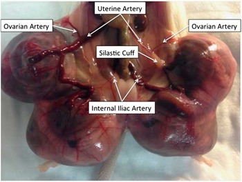
At 37 days gestation, (10 days post-surgery and 1 day before term) dams and their fetuses were humanely killed (cervical dislocation of dams; decapitation of fetuses) for sterile collection of fetal and gestational tissues. Figure 1 shows an example of a uterus 10 days after cuff placement. All tissues were weighed and collected for subsequent analysis. Whole fetal body weights were recorded. All fetal organs were removed and weighed, before sections of these organs were either fixed or snap frozen for future analysis. At the completion of the postmortem fetal carcass weight (muscle, skin, skull and bone) was recorded.
Representative image of anesthetized pregnant spiny mouse dam, 10 days after occlusion of the left uterine artery, with a silastic cuff. Image shows differential vasculature in the uterine artery, ovarian artery and internal iliac artery between the left and right sides of the uterus.

Statistical analysis
Data were assessed for normality. Organ to body weight ratios were calculated by dividing the organ weight by the total fetal weight. Descriptive statistics were tabulated. The general linear model was used to compare fetal body, organ and organ to body weight ratios between sham and occluded offspring. Dam was included in all models to account for litter effects. The estimated marginal means were also adjusted for fetal location within each uterine horn (1 is closest to ovary, 4 is closest to cervix), uterus side (left or right), dam weight and fetal sex (male or female). A P value of less than 0.05 (two-tailed) was regarded as statistically significant. The potential interactions between the interventions (sham, occluded), the uterus side and location of offspring within the uterine horn on total fetal weight were also assessed. Where an interaction was identified, we undertook a simple main effects analysis. Pairwise comparison with SIDAK adjustment for multiple comparisons was also performed. All analyses were performed using the SPSS statistical package (standard version 14.0, SPSS, Chicago, IL, USA).
Results
There was no dam mortality associated with the placement of the uterine artery cuff or sham surgical procedures in this study. Of the 43 fetuses counted at the time of surgery, 41 (95.3%) were carried to 37 days gestational age. The two fetuses lost (dead at postmortem; 37 days gestational age) were in the left side of the uterus from two pregnancies where the left uterine artery had been occluded. No adverse signs were noted after surgery in pregnant spiny mice; that is, activity levels and weight gain were normal, and no signs of wound infection or discomfort after surgery. The success rate for maintaining the pregnancy until 37 days gestational age was 90% (9/10) in the occluded group and 85.7% (6/7) in the sham operated group. Two dams, one each from the occluded and sham groups, delivered at 36 and 37 days gestation (term 38–39 days gestation), and in each case the offspring survived and appeared healthy despite their slight prematurity.
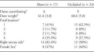
Analyses were conducted on a total of 41 offspring, born to 15 dams. Offspring were in 1 of 4 positions within either side of the uterus (1 representing closest to the ovary, 4 representing closest to the cervix). A total of 17 offspring contributed to the sham group and 24 offspring contributed to the occluded group (placement of a silastic cuff around the left uterine artery); all offspring from these pregnancies are included in the analysis. The characteristics of offspring in the two groups are presented in Table 1.
Characteristics of sham and occluded fetuses and dams

Fetus location: 1 denotes the fetus closest to the ovary, 4 denotes the fetus closest to the cervix within a litter. We have not compared the characteristics of these two populations statistically as individual offspring in each arm are not all independent of each other.
a Dams did not contribute to both arms.
b Mean (s.e.).
c Number (%).
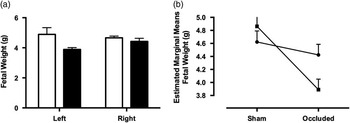
We hypothesized that the side and position of the offspring relative to the cuff placement would be associated with offspring weight. Overall, we did not identify a significant interaction between cuff placement, fetal position and uterus side with fetal weight (F=2.1, P=0.14), or between cuff placement and fetal position with fetal weight (F=1.8, P=0.19). However, we did identify a significant interaction between the uterus side and cuff placement with fetal weight (F=5.4 P=0.004). The effect of uterus side and cuff placement on fetal weight was then determined using simple main effects. There was no effect of cuff placement for right uterus side (F=0.8 (1,30), P=0.38), whereas there was for the left side of the uterus (F=7.5 (1,30), P=0.01). The mean difference in fetal body weight between occluded and sham was −1.02 g (95% CI −1.78 to 0.26), P=0.01 for left uterus side and −0.22 g (95% CI −0.72 to 0.28), P=0.28 for right uterus side. Raw fetal body weights and the adjusted estimated marginal mean plot for the mean differences with the uterus side, by treatment, are presented in Fig. 2.
(a) Body weight data for fetuses in the left and right uterine horn from sham (open) and occluded (closed) dams. (b) Estimated marginal means of fetal weight by uterus side and group show fetuses in the left side (squares) of the uterus (occluded) are 1 g smaller than right side (circles) fetuses (F=5.4, P=0.004). No significant difference is noted in shams between the left and right side of the uterus.

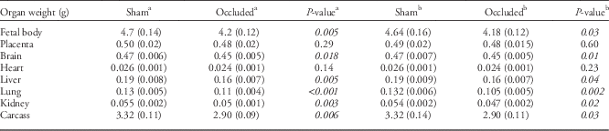
The estimated marginal mean organ weights for occluded and sham fetuses are presented in Table 2. Offspring from dams exposed to placement of a silastic cuff around the left uterine artery had significantly lower body, brain, liver, lung, kidney and carcass weights at 37 days of gestational age, compared with sham operated controls. After additional adjustment with the uterus side (left or right), fetal location within the uterus (ovarian, middle, cervical), sex and dam weight all differences remained statistically significant.
Estimated marginal means of organ weights for sham and occluded fetuses

Data is Mean (s.e.).
Italic values indicate P-values that are statistically significant.
a Model 1 corrects only for mother.
b Model 2 also adjusts for estimated marginal means after adjustment for uterus side, fetal location, sex and dam weight.
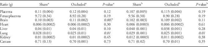
Differences in organ to body weight ratios of offspring from dams exposed to placement of a silastic cuff around the left uterine artery compared with shams are presented in Table 3. Offspring from dams exposed to the cuff had significantly higher brain and lung to body weight ratios, compared with sham fetuses. After additional adjustment with uterus side (left or right), fetal location within the uterus, sex and dam weight, only the lung to body weight ratio remained significant.
Estimated marginal means of organ to body weight ratios for sham and occluded fetuses.

Data is Mean (s.e.).
Italic values indicate P-values that are statistically significant.
a Estimated marginal means after adjustment for mother.
b Estimated marginal means after adjustment for, mother, uterus side, fetal location, sex and dam weight.
Discussion
In this study we show that limiting the increase of blood flow in one uterine artery for the last third of gestation is sufficient to induce fetal growth restriction in the spiny mouse, without significant fetal loss. We placed a silastic cuff around the left uterine artery at 27 days gestation (~0.7 gestation) to prevent an increase in size of the left uterine artery, which in turn would prevent the gestation-dependent increase of blood flow for the last third of pregnancy. In most mammals blood is delivered to the uterus via a dual arterial anastomotic loop, in which one end (the two ovarian arteries) originates from the aorta and the other (uterine arteries) from the internal iliac arteries. Unlike the linear, branching pattern of many vascular networks where blockage leads to downstream ischemia, this bilateral anatomical arrangement provides the uterus with a dual source of blood and considerable redundancy in case of loss of one or other of the main arterial supplies. At postmortem at 37 days gestation, we noted that the left uterine artery of the occluded dams was restricted in size beyond the cuff, and the left internal iliac artery size was larger. This alteration in blood flow to one side of the uterus produced a distinct phenotype of fetal growth restriction. Fetuses in the left side of the uterus were ~21% smaller than sham fetuses in the left side of the uterus, and abdominal organ weights were smaller in these fetuses, consistent with the growth restriction being asymmetric. Our gross measures of placental function (fetal/placental ratio, as a measure of placental efficiency) do not support placental insufficiency in this model. We will undertake comprehensive analysis of placentas in subsequent studies to elucidate the mechanism of growth restriction in this model.
The relatively precocial nature of spiny mouse fetal development means that we have targeted late gestation fetal growth and organ maturation, and therefore our model is relevant to late onset human IUGR.Reference Basilious, Yager and Fehlings 22 As such, this model could be successfully utilized to study both the immediate and long-term outcomes following IUGR, and also for evaluating treatments given prenatally to ameliorate the effects on fetal growth.
There are several established and published techniques for reducing fetal growth in animal models. Generally, fetal growth is altered via surgical, environmental or genetic manipulations, with each method closely relating to placental, maternal or fetal etiologies of IUGR.Reference Ergaz, Avgil and Ornoy 12 Ligation of the uterine artery has been the most common method for inducing IUGR in small animals, but this is associated with a high rate of fetal loss. In the rat, ligation of the uterine artery results in ~30% fetal loss, with fetuses located closest to the cervix most at risk of dying in utero.Reference Ergaz, Avgil and Ornoy 12 In the guinea pig, uterine ligation is performed at mid-gestation (at 28–30 days, a stage of gestation chosen to coincide with the best outcome for fetal survival), and while reducing blood supply to the uterus it results in fetal loss of ~20%.Reference Jansson and Persson 11 Herrera et al., recently developed a gradual occlusion model, like that used in this study, in the guinea pig, reducing fetal loss to 8%.Reference Herrera, Alegria and Farias 13 The method of uterine artery blood flow restriction described here in the spiny mouse reports a fetal loss of only 5%. Both methods rely on restriction of expected gestational increases in uterine artery diameter. Thus, uterine blood flow and subsequently fetal growth are gradually reduced over of time in late gestation, which is likely to better reflect the slow progression of growth restriction in human IUGR pregnancies. We suggest that this approach provides a more economical and realistic means of inducing fetal growth restriction in a small laboratory animal with a high relevance to the most notable clinical features of human IUGR.
Experimental limitations
A current limitation of this experimental technique is that we do not yet know the degree of uterine blood flow reduction over time in response to the cuff placement. Also, we have not yet assessed the physiological and metabolic parameters, or the endocrine status (e.g. hypoxemia, glucose and cortisol levels, hormone profile) that can reflect the degree of fetal compromise following induction of IUGR. These studies are now underway, together with characterization of uteroplacental vascular structure before and after uterine cuff placement using a vascular casting technique. In addition, we have not yet characterized the structure and function of the placenta in this model of IUGR.
Future directions
It is pertinent to ask whether a new animal model of reduced fetal growth is necessary, given there are numerous animal models of placental insufficiency, chronic hypoxia and IUGR in precocial and altricial species. The primary aim was to determine if fetal growth restriction occurred when only the gestation-related increase of uterine blood flow was prevented in the spiny mouse, and if so, to take advantage of the in utero development of this small laboratory rodent and eventually to use a number of validated physiological and behavioral tests available for this species.Reference Dickinson, Moritz and Kett 23 – Reference LaRosa, Ellery and Parkington 26 Our secondary aim was to improve on existing rodent models of IUGR and induce a more subtle insult that might improve fetal survival. Studies of reduced fetal growth in spiny mice will now proceed toward characterization of the multi-organ pathology, the assessment of antenatal or postnatal strategies to improve multi-organ growth and development, and the potential for promoting long-term health in IUGR babies.
Conclusion
By reducing blood flow in the left uterine artery with placement of a silastic cuff at 0.7 gestation we reduced fetal growth in the spiny mouse at term. In response to left uterine artery cuff placement, fetal loss was minimal. This technique could be readily adapted to reduce fetal growth in other small animal species by varying the internal diameter of the silastic tubing. Our ongoing work will establish the use of this model to characterize the short- and long-term physiological and molecular changes associated with human IUGR, and to test therapies for improving organ and body growth.
Acknowledgments
The authors acknowledge the technical support of Meghan Boomgardt. H.D. is an NHMRC Career Development Fellow; S.E. is funded by a career development fellowship from Cerebral Palsy Alliance to H.D; M.D.T is an NHMRC Early Career Fellow; S.M. is an ARC Future Fellow. A grant from the Victorian Government Infrastructure Support Fund to the Hudson Institute of Medical Research was received.
Conflicts of Interest
None.