Introduction
The yearly incidence of syphilis has risen markedly in Japan and worldwide. This increase in incidence is expected to continue. Considering disease severity, it is imperative to prevent the progress of infection through early diagnosis and treatment.
Moreover, there has also been an increased incidence of human papilloma virus (HPV) associated oropharyngeal cancer, which presents with clinical features similar to those of syphilis, making it difficult to distinguish between them. Herein, we report a case of syphilis with a clinical manifestation resembling that of HPV-associated oropharyngeal cancer. A literature review of similar cases was also conducted.
Case report
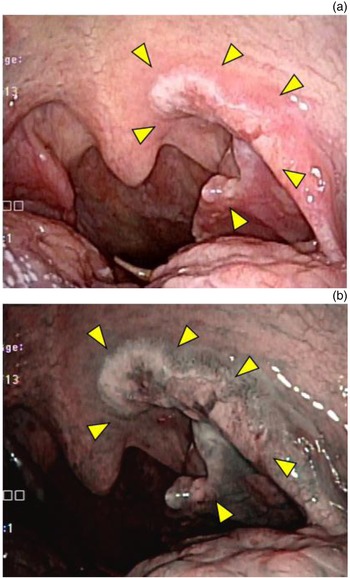
A 59-year-old man presented to our department with the chief complaint of swelling on the left side of his neck with an unknown history of onset. Intra-oral examination revealed a shallow, ulcerated mucosal lesion on the left side, extending from the palatine arch anteriorly to the palatine tonsil posteriorly (Figure 1). Extra-oral examination revealed a palpable lymph node on the left side of the neck.
Endoscopic view of the palatine tonsil under: (a) normal white light; and (b) narrow-band imaging. A shallow and ulcerated mucosal lesion (arrowheads) on the left side extending from the palatine arch anteriorly to the palatine tonsil posteriorly (a). Narrow-band imaging enhances an abnormality of small vessels within the lesion (b).

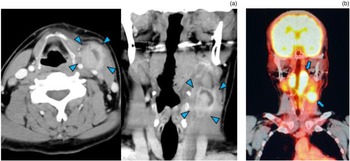
Contrast-enhanced computed tomography (CT) revealed an enlarged cervical lymph node, with a maximum diameter of 27 mm, and possible cystic changes or central necrosis. Positron emission tomography-CT showed uptake in the left palatine tonsil and an enlarged cervical lymph node (Figure 2).
Imaging. (a) Contrast-enhanced computed tomography (CT); and (b) positron emission tomography – CT. Contrast-enhanced CT (a) reveals an increase in the size of the lymph node, with a maximum diameter of 27 mm, and suspected cystic changes or internal necrosis (arrowheads). Positron emission tomography – CT (b) showed uptake in the left palatine tonsil and the enlarged cervical lymph node (arrows).

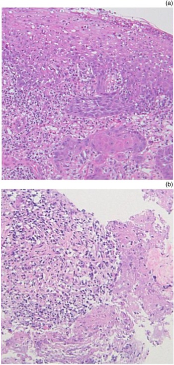
These findings were suggestive of cervical lymph node metastasis of oropharyngeal cancer. However, histological examination of a biopsy specimen from the left palatine tonsil revealed inflammatory changes with no sign of malignant features (Figure 3a). Needle biopsy of the lymph node was also performed, which revealed inflammatory cell infiltration of fibrous connective tissue (Figure 3b). In order to rule out an error related to insufficient biopsy material obtained from the out-patient clinic, an open biopsy of the left cervical lymph node taken under local anaesthesia was planned, to make a definitive diagnosis.
Histopathological findings of the left palatine tonsil (a) and the enlarged lymph node on the left side (b). Diffuse infiltration of inflammatory cells with hyperplastic squamous epithelium is evident in the tonsil (a), and lymphoplasmacytic infiltration with capillary proliferation is seen in a needle biopsy sample of the lymph node (b) (H&E; ×100)

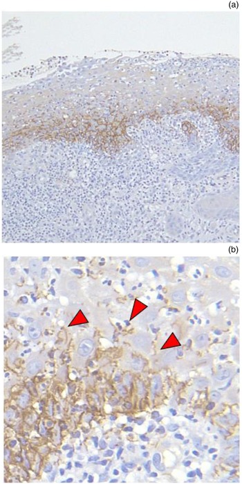
The Treponema pallidum latex agglutination test (anti-T pallidum antibody test), performed as a part of pre-operative screening for syphilis, revealed positive results. A subsequent quantitative rapid plasma reagin test revealed elevated values of up to 62.0 rapid plasma reagin units. The results of immunostaining with T pallidum antibodies were consistent with a syphilis infection (Figure 4).
Immunostaining (with Treponema pallidum antibody) of the left palatine tonsil shows infiltration of spirochaetes (arrowheads): (a) ×100 magnification; and (b) ×400 magnification.

The patient confirmed he had visited an adult entertainment establishment six months before presenting at our hospital. Thus, the patient was treated for secondary syphilis with a two-week course of amoxicillin and probenecid. Three weeks after the start of treatment, both the oropharyngeal lesion and the cervical lymph node had decreased in size. Three months after treatment, the clinical signs had subsided and the rapid plasma reagin test result was negative.
Discussion
Syphilis is a chronic and gradually progressing Treponema infection that manifests as lesions in multiple organs, including the oral mucosa, lymph nodes, central nervous system and cardiovascular system. As the clinical features of primary syphilis are non-specific, it is often difficult to diagnose. Its incidence reportedly decreased after the discovery of penicillin. However, in recent years, its incidence has increased worldwide.Reference Cerchione, Maraolo, Marano, Pugliese, Nappi and Tosone1 In Japan, the number of new cases of syphilis reported per year remained below 1000 after 1993. However, it has shown a rapid increase since 2010, with approximately 7000 cases reported in 2018.2
Meanwhile, HPV-associated oropharyngeal cancer has also become increasingly common.Reference Chaturvedi, Engels, Pfeiffer, Hernandez, Xiao and Kim3,Reference Näsman, Attner, Hammarstedt, Du, Eriksson and Giraud4 In Japan, approximately 50 per cent of reported cases of oropharyngeal cancer are associated with HPV, and this incidence is expected to increase in the future.Reference Hama, Tokumaru, Fujii, Yane, Okami and Kato5
The clinical findings in patients with syphilis are sometimes suggestive of malignancy. This resulted in a delayed diagnosis in a number of previously reported cases.Reference Hamlyn, Marriott and Gallagher6–Reference Sato, Tsubota and Himi11
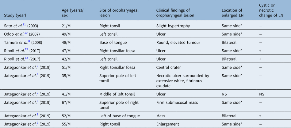
A PubMed search result for the key words ‘syphilis’ and ‘oropharyngeal’ revealed 40 publications published from 1999 to 2019. Five of those articles were written in English language and described 11 cases of syphilis in which oropharyngeal cancer was suspected, as in our patient (Table 1).Reference Tamura, Takimoto, Hoshida, Okada, Yoshimura and Uji8–Reference Ripoll, Montironi, Alos, Pujol, Berenguer and Oleaga12 Amongst these cases, syphilis was diagnosed comparatively early in: male homosexuals, individuals testing positive for human immunodeficiency virus infection and sex workers. In 10 of these 11 cases, lymph node enlargement was evident on either the same side as the oropharyngeal lesion or on both sides. However, there were no enlarged lymph nodes in areas other than the neck, or any oral or cutaneous symptoms. These clinical findings were suggestive of oropharyngeal cancer and further delayed the diagnosis of syphilis. In some of the cases, histopathological examination of biopsied tissue revealed non-specific inflammation, and several biopsies were conducted.Reference Jategaonkar, Klimczak, Agarwal, Badhey, Portnoy and Damiano9,Reference Ripoll, Montironi, Alos, Pujol, Berenguer and Oleaga12 In other cases, a diagnosis was only reached after consulting an infectious disease specialist,Reference Jategaonkar, Klimczak, Agarwal, Badhey, Portnoy and Damiano9 or following the discovery of syphilitic lesions in the stomach during upper gastrointestinal endoscopy intended to exclude multiple cancers,Reference Tamura, Takimoto, Hoshida, Okada, Yoshimura and Uji8 or syphilis was diagnosed accidentally during screening tests, as was the case for our patient.Reference Sato, Tsubota and Himi11 Enlarged lymph nodes with cystic or necrotic changes were present in 3 of the 11 cases,Reference Jategaonkar, Klimczak, Agarwal, Badhey, Portnoy and Damiano9,Reference Ripoll, Montironi, Alos, Pujol, Berenguer and Oleaga12 and in 1 of those cases, HPV-associated oropharyngeal cancer was suspected.Reference Jategaonkar, Klimczak, Agarwal, Badhey, Portnoy and Damiano9
Published syphilis case reports where oropharyngeal cancer was suspected

*Same side as the oropharyngeal lesion. ‘−’ = absent; ‘+’ = present. LN = lymph node; M = male; NS = not specified
The oropharyngeal mucous patches that present in secondary syphilis are initially erythematous, and gradually turn cream-coloured as they spread and merge, with marginal erythema. They have a ‘butterfly appearance’, which is a typical feature of pharyngeal syphilis. In our patient, oropharyngeal cancer was initially suspected in light of the presence of a shallow ulceration in the left oropharynx and cervical lymph node enlargement. In hindsight, however, cream-coloured patches were present within the shallow ulceration from the left anterior palatine arch to the palatine tonsil, an important finding suggestive of syphilis. Human papilloma virus associated oropharyngeal cancer may be difficult to diagnose given the small size of the primary lesion;Reference Ebisumoto, Okami, Sakai, Sugimoto and Iida13 thus, a biopsy may not always lead to a diagnosis. Considering this, oropharyngeal cancer could not be immediately ruled out, despite the lack of malignant findings in the biopsy.
Lymph node enlargement occurs in patients with secondary syphilis, along with occasional cystic changes.Reference Jategaonkar, Klimczak, Agarwal, Badhey, Portnoy and Damiano9,Reference Ripoll, Montironi, Alos, Pujol, Berenguer and Oleaga12 Similar clinical features can be seen in HPV-associated oropharyngeal cancer because of metastasis.Reference Goldenberg, Begum, Westra, Khan, Sciubba and Pai14 In our patient, contrast-enhanced CT revealed cystic changes that were suggestive of cervical lymph node metastasis, similar to HPV-associated oropharyngeal cancer. Therefore, it is important to consider syphilis as a differential diagnosis.
The diagnosis of syphilis can be confirmed by the demonstration of T pallidum spirochaetes using Warthin–Starry silver impregnation staining or T pallidum antibodies.Reference Ripoll, Montironi, Alos, Pujol, Berenguer and Oleaga12,Reference Komeno, Ota, Koibuchi, Imai, Iihara and Ryu15 Some of the previously reported cases were initially diagnosed as non-specific inflammation owing to the absence of clinical features suggestive of syphilis.Reference Cerchione, Maraolo, Marano, Pugliese, Nappi and Tosone1,Reference Ikenberg, Springer, Bräuninger, Kerl, Mihic and Schmid7,Reference Oddo, Carrasco, Capdeville and Ayala10,Reference Ripoll, Montironi, Alos, Pujol, Berenguer and Oleaga12 Similarly, in our patient, the initial histological analysis of both the palatine tonsil and the cervical lymph node indicated inflammatory changes. However, the immunostaining results confirmed syphilis. Therefore, with the increasing incidence of both HPV-associated oropharyngeal cancer and syphilis, differential diagnoses that include both HPV-related oropharyngeal cancer and syphilis, and their subsequent distinction, may be very important for early diagnosis and treatment.
• Syphilis may cause irregular mucosal lesions of the oropharynx and cystic lymphadenopathy
• In human papilloma virus related oropharyngeal cancer, pharyngeal mucosal lesions may be small and difficult to identify, and are characterised by cystic changes in lymph node metastasis
• Biopsy may not result in definitive diagnosis of oropharyngeal cancer because of the primary lesion's small size, and diagnosis may not be possible until palatine tonsil removal
• A biopsy of the palatine tonsil showed no malignant findings, but the possibility of oropharyngeal cancer could not be ruled out
• It is difficult to diagnose syphilis by only examining pathological specimens, without clinical information such as T pallidum antibody findings
• It is necessary to understand the characteristics of both diseases to ensure prompt diagnosis and treatment
Conclusion
We treated a patient with syphilis who was initially suspected to have cervical lymph node metastasis of HPV-associated oropharyngeal cancer, owing to the presence of both oropharyngeal mucosal lesions and an enlarged lymph node with cystic changes. Syphilis, which is becoming increasingly common, should be considered as a differential diagnosis in suspected oropharyngeal cancer cases, when oropharyngeal mucosal lesions and cervical lymph node enlargement are present. Collaboration with pathologists is also important for the correct diagnosis of syphilis.
Acknowledgements
We would like to thank Ayumi Tsuda, Saya Miyahara, Naoya Nakamura and Satomi Asai for their clinical support and comments. We would also like to thank Editage (www.editage.com) for editing and reviewing the English language of this manuscript. Daisuke Maki would also like to express his sincere gratitude to his brother for his insightful comments and suggestions.
Competing interests
None declared