Introduction
About 60 years ago, researchers in Kenya described Stephanofilaria dinniki Round, Reference Round1964 (Nematoda: Spiruridae) from the black rhinoceros (Round Reference Round1964; Tremlett Reference Tremlett1964). Stephanofilaria Ihle & Ihle-Landenberg has received little attention in Africa since the 1960s but has now come under increased scrutiny in Kenya (Chiyo and King’ori Reference Chiyo and King’ori2025; King’ori et al. Reference King’ori, Waiguchu, Ruoro, Muriithi, Mumbi, Omondi, Aminga, Angwenyi, Mijele and Chiyo2024). This is at least partly due to unexpected mortality in translocated rhinos both with and without filarial sores (Mutinda et al. Reference Mutinda, Otiende, Gakuya, Kariuki, Obanda, Ndeere, Ndambiri, Kariuki, Lekolool, Soriguer, Rossi and Alasaad2012). The goal of this study was to investigate the prevalence of filarial nematodes in rhinos and to explore possible vectors in rhino conservation areas. The vectors of Stephanofilaria have never been documented in Africa; in other parts of the world, they are Musca L. or Haematobia Lepeletier & Serville (Johnson Reference Johnson1987).
Exploratory work involved the dissection of possible vectors during ecological studies of Stomoxyinae and other biting flies, including tsetse (Mihok and Clausen Reference Mihok and Clausen1996; Mihok, Maramba, et al. Reference Mihok, Maramba, Munyoki and Saleh1996; Mihok, Moloo, et al. Reference Mihok, Moloo, Oden’y, Brett, Rakwar, Munyoki, Kiilu and Kyorku1996). Rhino blood was also examined for the detection of any circulating nematodes, and some limited work was done to attempt transmission of the parasites found to a few available vectors (tsetse, mosquitoes). Although filaria such as Stephanofilaria are unlikely to invade the bloodstream (Solismaa et al. Reference Solismaa, Laaksonen, Nylund, Pitkanen, Airakorpi and Oksanen2008), checks on their presence in rhino blood were done as all samples were routinely checked for trypanosomes. Immunosuppression appears to occur in rhinos during translocation due to elevated stress hormones; hence it was deemed prudent to consider this possibility. This work was facilitated by routine access to rhinos during translocations in the 1990s during investigations of the role of trypanosomiasis in rhino health (Gakuya et al. Reference Gakuya, Kock, Lekolool and Mihok2024; Kock et al. Reference Kock, Mihok, Wambua, Mwanzia and Saigawa1999). The focus of our work was on Nairobi National Park. This is the one site in Kenya that was well protected from poaching and had an in situ population of rhinos and hence was likely representative of the natural state of parasite and vector biology. Other sites in Kenya had devastating declines from poaching or were effectively ex situ populations set up on sanctuaries on cattle ranches or within fenced enclosures.
Here, we report on the patterns of infection in Stomoxyinae from thousands of fly dissections and the prevalence of parasites matching Stephanofilaria in rhino blood. Only Stomoxys Geoffroy and Haematobosca Bezzi were infected with Stephanofilaria.
Materials and methods
Rhino sampling
Blood was collected from rhinos at 10 locations in Kenya before or after translocations, typically from the medial radial vein, occasionally from ear veins. Data consisted of 88 black and 27 white rhino samples. The locations were Ngulia (27), Solio (24), Nairobi (16), Mara (18), Tsavo East (13), Lewa Downs (5), Ol Jogi (4), Aberdares (1), Samburu (1), and Marsabit (1). Five white rhinos were also sampled on arrival from South Africa. The focus was on obtaining several repeat samples over two months from 14 black rhinos before and after they were moved to a lowland fenced sanctuary (Ngulia Rhino Sanctuary, -2.965° S, 38.239° E, elevation, 715 m) from highland populations such as Nairobi National Park. Samples were also obtained from 10 sick white rhinos after introduction from South Africa to the Ol Choro Oirua Wildlife Conservancy (–1.111° S, 35.253° E, 1850 m) adjacent to the Maasai Mara National Reserve. Routine Coulter Counter ZM haematology and specific examination for filaria were done by the senior author, supplemented by occasional manual haematology conducted by the Kenya Wildlife Service (KWS) (Kock et al. Reference Kock, Wambua, Mwanzia and Saigwa1995). Additional tests in this study included measurements of the osmotic fragility of erythrocytes and the frequency of Heinz bodies in a subset of rhinos. This was done to check for erythrocyte anomalies sometimes found in captive rhinos (Chaplin et al. Reference Chaplin, Malecek, Miller, Bell, Gray and Hunter1986).
Microfilariae were searched for in the buffy coat and on thin and thick smears. A random subset from 14 animals (3–5 mL) was also lysed with 1% sodium dodecyl sulphate (Ndao et al. Reference Ndao, Pandey, Zinsstag, Pfister and Van Meirvenn1995) or was passed through a 3 μm Nuclepore filter (5–10 mL) to concentrate microfilariae. Laboratory-reared tsetse were routinely fed on rhinos in the field or through membrane feeding at the laboratory later in the day for xenodiagnoses. Fresh blood was also injected into cyclophosphamide-immunosuppressed mice (often in the field) to detect any parasite growth (and occasionally into rabbits).
Fly sampling
Flies for dissection were captured in Vavoua traps typically baited with dry ice and 1-octen-3-ol. Traps were set at the edge of clearings in semi-evergreen Croton/Oliea forest by Nairobi National Park headquarters (–1.363° S, 36.772° E, elevation 1774 m) and within Acacia xanthophloea thickets/woodland (–1.412° S, 36.904° E, 1533 m) near the Mbagathi River in Kenya (Lesilau et al. Reference Lesilau, Verschueren, van’t Zelfde, Musters, de Snoo and de Iongh2021). Most flies were caught during rainy periods in April–May and November–December of 1993–1994 (Mihok et al. Reference Mihok, Kang’ethe and Kamau1995; Mihok, Maramba, et al. Reference Mihok, Maramba, Munyoki and Saleh1996). These two habitats were about 16 km apart with lightly treed grassland and bushland in between.
In the early 1990s, the park contained about 60 black rhinos (Okita-Ouma Reference Okita-Ouma2004). A sample of 146 flies from Solio Ranch (–0.237° S, 36.885° E, 1940 m) in Laikipia (King’ori et al. Reference King’ori, Waiguchu, Ruoro, Muriithi, Mumbi, Omondi, Aminga, Angwenyi, Mijele and Chiyo2024) was also dissected in 1994, along with 57 flies netted from black rhinos at capture at Solio (Mihok, Moloo, et al. Reference Mihok, Moloo, Oden’y, Brett, Rakwar, Munyoki, Kiilu and Kyorku1996). Solio Ranch had roughly 60 black rhinos in the 1990s (Patton et al. Reference Patton, Campbell and Parfet2007), along with roughly 70 white rhinos (KWS 2007, 2022). Solio Ranch was overstocked and hence the understory was heavily browsed relative to similar habitat in Nairobi National Park (Figure 1). No flies were dissected from the other eight areas where blood was collected.
Habitats with Stomoxyinae in two rhino sanctuaries in Kenya. A Nairobi forest clearing near park headquarters with abundant Stomoxys, B Far view of Acacia woodland at the Mbagathi River, C Close-up view of a riverine thicket at the Mbagathi River, D Acacia woodland at Solio Ranch.

Figure 1. Long description
A four-panel set of photos labeled A through D.
* Panel A, top-left: A forest clearing in Nairobi featuring tall, dry yellowish grass in the foreground and a dense wall of green leafy trees and shrubs in the background.
* Panel B, top-right: An elevated wide-angle view of Acacia woodland at the Mbagathi River. The landscape shows rolling hills with patches of green trees interspersed with dry, light-colored grassland.
* Panel C, bottom-left: A close-up of a riverine thicket at the Mbagathi River. A small stream is visible at the bottom, bordered by thick green bushes and tall Acacia trees with wide, flat canopies reaching toward the sky.
* Panel D, bottom-right: A view from inside an Acacia woodland at Solio Ranch. The scene is dominated by the slender, light-colored trunks and tangled branches of Acacia trees, with green foliage and grass visible through the gaps in the trunks.
Fly nomenclature follows Zumpt (Reference Zumpt1973), which was generally applicable (Duvallet and Hogsette Reference Duvallet and Hogsette2023), but with the discovery of one new species of Haematobosca (Pont and Mihok Reference Pont and Mihok2000). The ecology of most African Stomoxyinae remains poorly documented with some taxonomic revisions likely needed. The subspecies status of S. niger bilineatus Grünberg, 1906, S. taeniatus f. brunnipes Grünberg, 1906, and the species status of S. boueti Roubaud, 1911 likely need to be assessed following DNA sequencing (Dsouli et al. Reference Dsouli, Delsuc, Michaux, de Stordeur, Couloux, Veuille and Duvallet2011; Moyaba et al. Reference Moyaba, Modise, Esterhuizen, Suganuma, Inoue, Thekisoe and Taioe2025; Muita et al. Reference Muita, Bargul, Makwatta, Ngatia, Tawich, Masiga and Getahun2025).
Photographs of key species are provided here to assist in fly identification (Figures 2 and 3); Zumpt (Reference Zumpt1973) should be used for definitive identification. In practical terms for field sorting in East Africa, one can often sort flies based on size, colour, and a few features available at low magnification. Stomoxys calcitrans with its spotted abdomen is familiar to most biologists but should be checked against the possible presence of S. sitiens Rondani, 1873 (northern arid environments). Stomoxys pallidus with its bright yellow abdomen stands out but can easily be confused with non-biting muscids if one does not check the proboscis. Stomoxys transvittatus also stands out as a particularly large fly with a well-demarcated white and black pattern of the thorax that is different from the rather shiny S. inornatus. Large yellowish species with mainly yellow legs are typically S. taeniatus or S. taeniatus f. brunnipes, with the latter having dark brown femora. Stomoxys varipes females are readily recognised by the distinctive legs, but males are very similar to S. niger niger. Males have to be identified by the long and dense hairs on the mesonotum. The three remaining species we have illustrated are similar but are easy to identify with experience. Stomoxys niger bilineatus is distinctly brown with crisper thorax and abdominal markings than S. niger niger and can be very abundant. S. boueti is easily confused with S. niger niger but is smaller, with more well-defined abdominal bands and a distinctive curve in the media of the wing (only slightly curved upwards). Stomoxys niger niger is sometimes very abundant and hence identification of other species present can be tedious in areas with high Stomoxys diversity (forests vs savannah).
Dorsal views of common Stomoxys from Nairobi National Park (not to scale across panels). A S. taeniatus female 7 mm, B S. varipes female 7 mm, C S. inornatus male 7 mm, D S. transvittatus male 9 mm, E S. niger bilineatus female 7 mm, F S. niger niger female 7 mm.

Figure 2. Long description
A collection of six photos arranged in a two-column by three-row grid.
* Panel A at the top-left shows S. taeniatus with a light tan body, clear wings, and distinct dark longitudinal stripes on the thorax.
* Panel B at the top-right shows S. varipes with a darker, reddish-brown thorax and abdomen, and wings that have a noticeable amber tint.
* Panel C in the middle-left shows S. inornatus with a very dark, almost black thorax featuring two prominent white patches at the top and clear wings.
* Panel D in the middle-right shows S. transvittatus with a greyish thorax marked by thick black vertical stripes and a rounded dark abdomen.
* Panel E at the bottom-left shows S. niger bilineatus with a yellowish-tan thorax featuring thin dark lines and an abdomen with horizontal dark bands.
* Panel F at the bottom-right shows S. niger niger with a grey-toned thorax, dark stripes, and a more elongated dark abdomen compared to panel E.
All specimens are shown with wings spread and legs extended, viewed from directly above.
Dorsal views of other Stomoxys from Nairobi (not to scale across panels). A S. calcitrans female 7 mm, B S. taeniatus f. brunnipes female 9 mm, C S. pallidus male 7 mm, D S. boueti male 5 mm.

Figure 3. Long description
Panel A, top-left, shows a female S. calcitrans with a grey and black patterned thorax and a broad abdomen featuring dark transverse bands. Panel B, top-right, shows a female S. taeniatus f. brunnipes with a more elongated, yellowish-brown body and slender legs. Panel C, bottom-left, shows a male S. pallidus with a dark, metallic thorax and a bright orange-yellow abdomen. Panel D, bottom-right, shows a male S. boueti with a dark, uniform purple-black coloration across the thorax and abdomen. All specimens are shown with wings spread, revealing translucent membranes and distinct venation patterns. The heads feature prominent compound eyes and forward-pointing proboscises.
Dissections
Fly proboscides were dissected from 19 species of Stomoxyinae identified according to the keys of Zumpt (Reference Zumpt1973). Fly parts were teased apart and suspended in insect physiological saline, then lightly pressed between a microscope slide and a cover slip. These preparations were examined at 250x magnification on a Leica compound microscope to detect filaria, which were typically mobile. We also dissected several non-biting muscids (Musca spp. and Morellia abdominalis Stein, 1918), as well as a smaller number of Tabanidae [mostly Haematopota vulnerans (Surcouf, 1909)] and Sarcophagidae. Stomoxys calcitrans (L.) and Musca domestica L. were only minor components of the fly fauna in the park but are common in peridomestic settings in Nairobi. The use of dry ice as a bait facilitated the capture of diverse Stomoxyinae, but traps caught hardly any Haematobia.
Fly proboscides were dissected fresh after brief freezing with large catches frozen for later sorting. Larval filaria were still motile after weeks in a chest freezer. Twenty-four females of Stomoxys transvittatus Villeneuve, 1916 were dissected for ovarian ageing to scope the age of females caught in traps (Moobola and Cupp Reference Moobola and Cupp1978). The guts and thoraces of a small sample of Stomoxys were also dissected to look for developing larvae. Statistics in text for body landmarks are presented as the arithmetic mean and standard deviation. Parasites were photographed as is without use of a clearing agent.
Attempts at transmission
Rhinos were screened by xenodiagnosis for transmission to various species of tsetse as part of the routine trypanosomiasis protocol during rhino sampling (Mihok et al. Reference Mihok, Munyoki, Brett, Jonyo, Röttcher, Majiwa, Kang’ethe, Kaburia and Zweygarth1992). Tsetse are found throughout the traditional range of rhino in lowland areas but are not present in Nairobi except on the very southern edge of the park (just one G. longipennis caught in many years of work). Blood was also injected into cyclophosphamide-immunosuppressed mice. The one sample from a rhino with many filaria was fed to particularly large numbers of tsetse only a few hours after collection, as well as to 50 Aedes aegypti the day after collection. Insects were dissected over a month to detect any parasite growth and blood was examined regularly over this same time period from mice.
Results
Rhino sampling
Microfilariae with the morphology of Stephanofilaria were found in 3 of 89 blood samples from three black rhinos (Figure 4); none were found in white rhinos. Parasites were unsheathed and had a short headspace with a long and thin tapering, clear (mostly anucleate) sharp tail. Morphology did not resemble other common microfilariae transmitted by mosquitoes, midges, or black flies (Mathison et al. Reference Mathison, Couturier and Pritt2019). Unfortunately, there is little information on filaria in wildlife (Abo-Aziza et al. Reference Abo-Aziza, Hendawy, Abdullah, El Namaky, Laidoudi and Mediannikov2022); hence, there may be some uncertainty in the identity of the parasites found (Remya et al. Reference Remya, Manju and Prasanta2025). In Asian rhinos, DNA evidence exists for the presence of Onchocerca (larger microfilariae) and Mansonella (similar small microfilariae) in a few animals (Pandey et al. Reference Pandey, Manandhar, Shrestha, Sadaula, Hayashi, Abdelbaset, Silwal, Tsubota, Kwak, Nonaka and Nakao2024).
Stephanofilaria dinniki in a buffy coat smear in a black rhino from Nairobi National Park in February 1992 (Giemsa stain). Mean length of eight parasites was 183 μm.

Reference material was limited to Setaria Viborg from buffalo (longer and sheathed), which was dissimilar. The rhino microfilariae were 183 ± 20 μm (N = 8, range 141–207) in length, which is within the size range of Mansonella. Mansonella is present in Africa and is also unsheathed. However, Mansonella differs in terms of having a blunt, curved tail with many nuclei densely packed to the tip of the tail (Perles et al. Reference Perles, Otranto, Barreto, de Macedo, Lia, Mendoza-Roldan, Herrera, de Oliveira, Machado and André2023; Uni et al. Reference Uni, Bain and Takaoka2004). The position of the nerve ring was distinct in four microfilariae at 26% of body length. These samples were from the late dry season and early rainy season: one from Nairobi at first capture, and two from Ngulia, four and eight weeks after translocation. All three animals were free of trypanosomes and healthy.
The presence of ulcerative sores in rhinos was not systematically recorded as in King’ori et al. (Reference King’ori, Waiguchu, Ruoro, Muriithi, Mumbi, Omondi, Aminga, Angwenyi, Mijele and Chiyo2024), but animals at Nairobi and Solio were often noted to have sores (Figure 5). These healed gradually once animals were moved to the lowlands. For example, repeat photographs of deep sores from an animal on first capture at Nairobi in August 1990 showed considerable healing of these sores two weeks later at Ngulia. Flies were abundant on the sores in Nairobi but absent at Ngulia.
Black rhino ulcerative sores indicative of Stephanofilaria. A Animal from Solio where flies were captured by hand net, B Animal from Nairobi at first capture with unidentified muscids on August 30, 1990, C The same animal two weeks later after translocation to Ngulia with healing wounds and no flies.

Figure 5. Long description
The three panels are arranged vertically.
* Panel A at the top shows a wide-angle photo of a sedated black rhino lying on its side in dry grass, restrained with ropes around its legs and head. Large, bloody ulcerative sores are visible on its shoulder and flank. Several people in green uniforms stand around the animal.
* Panel B in the middle is a close-up photo of a deep, linear skin lesion on a rhino. The wound is raw, red, and wet, with numerous small black flies clustered around the edges and inside the laceration.
* Panel C at the bottom is a close-up photo of the same anatomical area on the rhino two weeks later. The previously raw wounds have closed and formed dry, dark scabs. The skin appears thickened and greyish around the healing site, and no flies are present on the surface.
Osmotic fragility of erythrocytes and the frequency of Heinz bodies did not reveal any differences related to the presence of filaria. Complete blood counts were within expected ranges, except for the animal from Nairobi with the highest numbers of filaria at first capture (1 per 10–20 fields in a smear of the buffy coat). This subadult female had a high leukocyte count (16.3 × 10⁹/L, > 95th percentile) consisting of an even number of lymphocytes and neutrophils. It also had a high eosinophil count (1.25 × 10⁹/L, > 90th percentile), typical of helminth infections and hence consistent with a Stephanofilaria infection. No filaria were detected in the other two positive animals when first captured in Nairobi. Filaria were rare (1–2 per entire buffy coat smear) when present on just two of seven occasions at Ngulia when blood was collected.
Repeat measurements of erythrocyte size distributions were available (Coulter ZM 256 Channelyzer) for the two animals that had detectable filaria after translocation to Ngulia (Linda subadult female, Observation old adult male) and for the animal with high filaria at capture in Nairobi (Bahegwa subadult female). Repeat counts were available at six and eight weeks for Linda and Observation; Linda was also sampled 3.5 years later (when recovering from a poacher’s arrow after living free near Mzima Springs). Erythrocyte size distributions gave no clear indication of the presence of filaria (Figure 6). There was only a hint of minor disturbance in haematopoiesis in terms of a more peaked distribution of cells for Bahegwa with many filaria, and for Linda during recovery from an injury.
Erythrocyte size distributions (log scale) for the three animals in which Stephanofilaria was detected. Bahegwa was positive at capture but never sampled again. Linda was positive only at 8 weeks. Observation was positive only at 4 weeks. The sample for Linda at 3.5 years was when she was recovering from an injury; it is almost identical to the sample from Bahegwa.

Figure 6. Long description
The X-axis represents Erythrocyte size in f l on a log scale ranging from 40 to 180. The Y-axis represents the percentage in Channel ranging from 0.0 to 1.4.
Eight data series are plotted as bell-shaped curves peaking between 70 and 80 f l.
* Linda: Black dotted line peaking at approximately 1.2.
* Linda 6 wk: Blue dashed line peaking at approximately 1.2.
* Linda 8 wk: Solid blue line peaking at approximately 1.2.
* Observation: Red dotted line peaking at approximately 1.2.
* Observation 6 wk: Red dashed line peaking at approximately 1.25.
* Observation 8 wk: Solid red line peaking at approximately 1.1.
* Bahegwa: Solid black line peaking at approximately 1.2.
* Linda 3.5 yr: Blue line with diamond markers peaking highest at 1.4.
The curves for Linda at 3.5 years and Bahegwa show the highest concentration of smaller cells, while the Observation 8 week curve is shifted furthest to the right, indicating a higher proportion of larger erythrocytes.
Attempts at transmission
All tsetse and mice fed on or injected with blood from rhino did not show evidence of filaria over a month of blood and health monitoring. Importantly, blood from the positive rhino on first capture in Nairobi with many filaria (Bahegwa) was negative, despite feeding to large numbers of three species of tsetse. Examination of tsetse guts a few days after feeding showed that microfilariae were ingested but the parasites were dead. Filaria were also not detected in Aedes aegypti fed on this rhino’s blood, but these mosquitoes were not examined till several weeks after feeding with few still alive.
Fly sampling
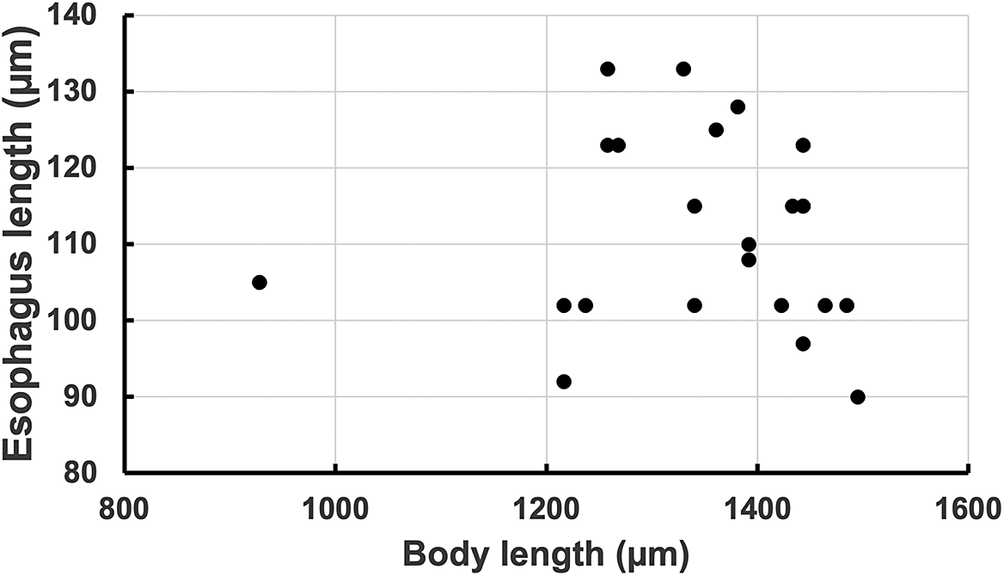
Altogether, proboscides from 4,819 Diptera were dissected and examined for filaria (Table 1). Eighteen examples of Stephanofilaria larvae were obtained in seven species of Stomoxys and from one Haematobosca latifrons Malloch, 1932 and one H. squalida Grünberg, 1913. Stephanofilaria parasites were similar in size only to a small form of Habronema in Rhinomusca (Mihok et al. Reference Mihok, Kock, Krüger and Giordano2026), and could be readily differentiated at medium magnification by differences in the oesophagus. Stephanofilaria had a uniformly short and undifferentiated oesophagus regardless of body length (Figure 7, Table 2), whereas the small form of Habronema had a longer oesophagus with distinct muscular and glandular portions. A single H. hirtifrons harboured 15 slender, short filaria (642 ± 28 μm), but material was too degraded for identification.
Occurrence of Stephanofilaria larvae in proboscides of flies caught in traps at Nairobi National Park and Solio Ranch along with flies caught with a hand net from sedated black rhinos at Solio Ranch

Table 1. Long description
The table consists of five columns: Fly species or group, Traps Nairobi, Traps Solio, Rhino Solio, and Positive.
* Stomoxys varipes: 433 in Traps Nairobi, 6 in Traps Solio, 2 Positive.
* S. niger bilineatus: 426 in Traps Nairobi, 13 in Traps Solio.
* S. inornatus: 343 in Traps Nairobi, 5 Positive.
* S. transvittatus: 309 in Traps Nairobi, 5 Positive.
* S. taeniatus: 244 in Traps Nairobi, 4 Positive.
* S. taeniatus f. brunnipes: 244 in Traps Nairobi, 1 Positive.
* S. niger niger: 228 in Traps Nairobi, 64 in Traps Solio, 1 Positive (noted as being from Solio).
* S. boueti: 222 in Traps Nairobi, 2 in Traps Solio, 1 Positive.
* S. calcitrans: 168 in Traps Nairobi, 16 in Traps Solio.
* S. pallidus: 145 in Traps Nairobi.
* S. ochrosoma: 3 in Traps Nairobi.
* Haematobosca latifrons: 244 in Traps Nairobi, 4 in Traps Solio, 1 Positive.
* H. aurata: 128 in Traps Nairobi.
* H. squalida: 128 in Traps Nairobi, 1 in Traps Solio, 1 Positive.
* H. hirtifrons: 117 in Traps Nairobi.
* Rhinomusca dutoiti: 203 in Traps Nairobi, 40 in Traps Solio, 6 in Rhino Solio.
* Prostomoxys saegerae: 131 in Traps Nairobi.
* Stygeromyia sanguinaria: 115 in Traps Nairobi.
* Stygeromyia maculosa: 47 in Traps Nairobi.
* Haematobia: 1 in Rhino Solio.
* Non-biting muscids: 631 in Traps Nairobi, 50 in Rhino Solio.
* Tabanidae (5 species): 71 in Traps Nairobi.
* Sarcophagidae: 36 in Traps Nairobi.
a All positive flies were from Nairobi except for one S. niger niger at Solio.
Total length versus oesophagus length in Stephanofilaria in fly proboscides in Nairobi National Park, Kenya.

Figure 7. Long description
A scatter plot with a horizontal X axis labeled Body length in micrometers mu m and a vertical Y axis labeled Esophagus length in micrometers mu m. The X axis ranges from 800 to 1600 in increments of 200. The Y axis ranges from 80 to 140 in increments of 10.
Data points are represented by solid black circles.
- A single outlier is located on the far left at approximately 930 on the X axis and 105 on the Y axis.
- The main cluster of data points is concentrated between 1200 and 1500 on the X axis.
- Within this cluster, esophagus lengths vary widely between 90 and 135 micrometers.
- There is no clear linear correlation between the two variables, as the points are scattered vertically across the 1200 to 1500 body length range.
Measurements in μm of Stephanofilaria larvae in the proboscis of Stomoxys in Kenya

Table 2. Long description
The table presents Mean, Standard Deviation, and Sample Size (N) for various anatomical features.
Length from tip of head:
* Body: Mean 1306, Std Dev 202, N 76.
* Oral cavity: Mean 6, Std Dev 1, N 26.
* Total esophagus: Mean 112, Std Dev 13, N 23.
* Muscular esophagus: Mean 112, Std Dev 13, N 22.
* Nerve ring: Mean 81, Std Dev 14, N 24.
Body width at various landmarks:
* Mouth: Mean 6, Std Dev 1, N 28.
* Head: Mean 17, Std Dev 1, N 28.
* Nerve ring: Mean 33, Std Dev 2, N 24.
* Junction (esophagus and intestine): Mean 34, Std Dev 3, N 25.
Width of internal structures:
* Typical esophagus: Mean 10, Std Dev 2, N 26.
* Posterior esophagus: Mean 15, Std Dev 2, N 6.
* Intestine: Mean 16, Std Dev 3, N 43.
Tail and Anus:
* Width tail: Mean 14, Std Dev 2, N 30.
* Anus from tip tail: Mean 29, Std Dev 4, N 35.
Notes: Junction – body width at intersection of oesophagus and intestine; s.d. – Standard deviation.
a This measurement is for six Stephanofilaria with a wider posterior section; other parasites with a uniform width.
Stephanofilaria was never found outside of the proboscis in 41 guts and 21 thoraces examined, mostly from S. transvittatus. In the 24 females that were aged by ovarian development, half of the females had undifferentiated cells or were in age class I, indicating an age up to about three days. Only one of these flies had a Stephanofilaria infection and it was the only parous female. It had oocytes in early development, indicating an age of about ten or more days. Counts of larval forms of Stephanofilaria in the proboscis were 6.2 ± 8.5 (N = 18, range 1–26). Given the very low infection rates (1.6% at best), some vectors could have been missed. For example, Stephanofilaria was detected in S. niger niger Macquart, 1851 at Solio but was absent in 228 dissections at Nairobi. It was not, however, found in S. niger bilineatus, an abundant species in the East African savannah with many dissections.
Stephanofilaria had a rather plain head with a ring of six tiny cephalic spines around a smooth buccal capsule, just visible at 1000× magnification (Figure 8). These cephalic spines are illustrated in drawings but are difficult to count with light microscopy (Chiyo and King’ori Reference Chiyo and King’ori2025). The oesophagus of Stephanofilaria was just 8.5% of body length and was typically undifferentiated (Figure 8). Stephanofilaria also had a ring of small “teeth” at the front of the oesophagus, which is occasionally mentioned in the literature. This ring was difficult to discern without clearing. The tail of Stephanofilaria was plain and often lightly curved. The anus was near the tip of the tail (29 μm, 2.2% of body length) and was occasionally everted. The cuticle was lightly striated, but this was not obvious in all specimens. It was not possible to assess Haematobia as a potential vector as it was hardly caught in traps even with dry ice or both dry ice and 1-octen-3-ol (67 vs 25,916 Stomoxys).
Stephanofilaria larval forms in two species of Stomoxys. A S. varipes showing ring of tiny cephalic spines with four in sharp focus and two others barely visible; ring at entrance to oesophagus is visible but associated small teeth are not, differential interference contrast (DIC), 1000× magnification (6 μm width at the tip). B S. transvittatus showing nerve ring and undifferentiated oesophagus leading to the wider intestine; tiny cephalic spines are out of focus, 400× magnification (junction of the intestine is 120 μm from the tip).

Discussion
The microfilariae found in black rhinos matched Stephanofilaria best and were only slightly shorter than the 205 μm length of three S. dinniki “micro larvae” isolated from sores in Chiyo and King’ori (Reference Chiyo and King’ori2025). Larval filaria also had numerous features in common with other Stephanofilaria spp. (Johnson Reference Johnson1987). They were just slightly shorter than the two 1.6 mm larvae obtained from skin lesions in Chiyo and King’ori (Reference Chiyo and King’ori2025) but were longer than the few measurements available for L3 larvae of four other Stephanofilaria spp. (range 928–1,425 vs 640–1,123 μm) (Dutt Reference Dutt1970; Hibler Reference Hibler1966; Kono and Fukoyoshi Reference Kono and Fukoyoshi1967; Saparov et al. Reference Saparov, Akramova, Azimov and Golovanov2014; Srivastava and Dutt Reference Srivastava and Dutt1963). Larval filaria had an undifferentiated oesophagus, a diagnostic feature of the genus (Anderson et al. Reference Anderson, Chabaud and Willmott2009). Some larvae had a protruding buccal capsule as in other Stephanofilaria (Hibler Reference Hibler1966; Saparov et al. Reference Saparov, Akramova, Azimov and Golovanov2014), and a greenish-brown anal spot as in Parafilaria bovicola Tubangui, 1934 from Musca autumnalis De Geer, 1776 (Bech-Nielsen et al. Reference Bech-Nielsen, Bornstein, Christensson, Wallgren, Zakrisson and Chirico1982). Cuticular spines were never noticed with light microscopy (Naseem, Raza, Allavena, et al. Reference Naseem, Raza, Allavena, McGowan, Morgan, Constantinoiu, Tabor and James2021).
The rarity of infections in Stomoxyinae was at least partly a function of the characteristics of flies caught in traps, which are presumed to be host seeking. Most Stomoxys in traps had fed on nectar (Mihok and Carlson Reference Mihok and Carlson2021) and were likely quite young. The only ovarian-aged female with a Stephanofilaria infection was about two weeks old and hence could have developed a mature infection from blood feeding. There was no apparent pattern in the frequency of infections in the various species. Stomoxys varipes, S. taeniatus and S. transvittatus, the species with the most infections, varied from abundant to uncommon in Nairobi Park. All Stomoxys are particularly active around sunset; other genera without Stephanofilaria infections are more active in the afternoon (Mihok, Maramba, et al. Reference Mihok, Maramba, Munyoki and Saleh1996). This could correlate with the observation that wildlife in general often visit waterholes around sunset.
The rarity of microfilariae in white versus black rhino was also consistent with the pattern of lesions in Kenya (King’ori et al. Reference King’ori, Waiguchu, Ruoro, Muriithi, Mumbi, Omondi, Aminga, Angwenyi, Mijele and Chiyo2024). Stephanofilaria parasites have also been isolated from black rhinos in Kruger National Park (Hitchins and Keep Reference Hitchins and Keep1970) and in Zimbabwe (Kock and Kock Reference Kock and Kock1990), but only from lesions. This is typical for Stephanofilaria, although there are a few reports of isolation from blood in cattle (Johnson Reference Johnson1989).
The assumption that Stephanofilaria in Stomoxys is S. dinniki is logical but there is a possibility that it could have originated from another wildlife species, of which there are many in the park (Ogutu et al. Reference Ogutu, Owen-Smith, Piepho, Said, Kifugo, Reid, Gichohi, Kahumbu and Andanje2013). Only larval Habronema Diesing were common in Rhinomusca in other dissections (Mihok et al. Reference Mihok, Kock, Krüger and Giordano2026). Most Stomoxys caught in traps fed on bushbuck (Mihok and Clausen Reference Mihok and Clausen1996), with meals from rhino uncommon (7%). Stephanofilaria has yet to be documented in bushbuck (Boomker et al. Reference Boomker, Horak and De Vos1986; Boomker et al. Reference Boomker, Keep and Horak1987). The ELISA (enzyme-linked immunosorbent assay) system was able to differentiate all common species but few alternative hosts other than bushbuck were detected. Stomoxys has rarely been reported as a pest of rhino, with only one observation of harassment of wildlife during an unusual outbreak in the Ngorongoro Crater in Tanzania (Fosbrooke Reference Fosbrooke1963). A second event occurred in the crater in 2000–2001 when large numbers of buffalo died (R. Kock, unpublished data). This was at a time when few rhinos were still present in the crater. Hippopotamus were severely harassed and lions even fell out of trees. It was extraordinary to observe. There was a distemper outbreak also coincident with this unusual event.
Stomoxys occasionally fed on warthogs (5%), which are a hypothetical wildlife reservoir for S. boomkeri, a disease detected in pigs in the former Zaire, now the Democratic Republic of Congo (Bain et al. Reference Bain, Van der Lugt and Kazadi1996). No blood meals were identified from giraffes, which have filarial skin lesions that are still being characterised (Han et al. Reference Han, Dadone, Ferguson, Bapodra-Villaverde, Dennis, Aruho, Sadar, Fennessy, Driciru, Muneza, Brown, Johnston and Lahmers2022). Lastly, no blood meals were identified from hippopotamus, which harbour Stephanofilaria thelazioides (Boomker et al. Reference Boomker, Bain, Chabaud and Kriek1995).
It is also unlikely that this parasite was a new form of Parafilaria, which causes bleeding sores and is found in cattle in East Africa (Getu et al. Reference Getu, Maru and Alemu2025) and in buffalo in South Africa (Keet et al. Reference Keet, Boomker, Kriek, Zakrisson and Meltzer1997). L3 larvae of P. bovicola Tubangui, 1934 in Musca spp. have been documented and are much larger (1,934–3,721 μm) (Nevill Reference Nevill1981), as are L3 larvae of P. multipapillosa (Condamine & Drouilly, 1878) in Haematobia (1,670–2,670 μm) (Gnedina and Osnov Reference Gnedina and Osnov1960). Parafilaria larvae also differ in morphology (Bain Reference Bain1981; Nevill Reference Nevill1981).
A few other Stomoxyinae were found to be uncommon vectors of Stephanofilaria (H. latifrons, H. squalida), with the status of Haematobia still to be properly investigated. Haematobia, Musca and Stomoxys are vectors of other Stephanofilaria outside Africa where infection rates in vectors are also very low (Johnson Reference Johnson1989; Naseem, Raza, Fordyce, et al. Reference Naseem, Raza, Fordyce, McGowan, Constantinoiu, Turni, Allavena, Tabor and James2021). Haematobia is an occasional pest of cattle in Africa (Newsholme et al. Reference Newsholme, Erster and Jacobs1983) and was commonly observed on black rhinos in Tsavo in the 1960s (Parsons and Sheldrick Reference Parsons and Sheldrick1964) but not in our study. Very few Haematobia were captured by hand net on three freshly sedated rhinos at Solio (9 of 242 flies).
Ulcerative skin lesions seem to be tolerated by black rhino, but there is one report of unexpected mortality in translocated white rhino. This mortality occurred at Meru National Park, a lowland sanctuary with high numbers of tsetse. A definitive interpretation of any role of Stephanofilaria in mortality was complicated by fatal trypanosome infections that occurred in animals both with and without sores (Gakuya et al. Reference Gakuya, Kock, Lekolool and Mihok2024; Mutinda et al. Reference Mutinda, Otiende, Gakuya, Kariuki, Obanda, Ndeere, Ndambiri, Kariuki, Lekolool, Soriguer, Rossi and Alasaad2012; Obanda et al. Reference Obanda, Kagira, Chege, Okita-Ouma and Gakuya2011). The park now contains a thriving population of rhinos following tsetse control measures and restriction to an 80–84 km2 fenced sanctuary. Skin lesions are also now quite rare in white rhinos at Meru Park [3 of 32, King’ori et al. (Reference King’ori, Waiguchu, Ruoro, Muriithi, Mumbi, Omondi, Aminga, Angwenyi, Mijele and Chiyo2024)] versus black rhinos (7 of 13). Unfortunately, there is no information on Stomoxys at Meru, as only tsetse fly surveys have been conducted.
Hopefully, this study will now facilitate more work on the epidemiology and life cycle of Stephanofilaria dinniki at rhino sanctuaries in Africa. More refined studies are now possible through PCR for specific DNA typing of filaria to rule out the possibility of a misidentification of Mansonella, for example (Chiyo and King’ori Reference Chiyo and King’ori2025). Successful laboratory rearing of various Stomoxys spp. should also be considered for xenodiagnosis and for definitive studies of larval development in vectors to confirm parasite identities (Muita et al. Reference Muita, Bargul, Makwatta, Ngatia, Tawich, Masiga and Getahun2025; Sumba et al. Reference Sumba, Mihok and Oyieke1998).
Acknowledgements
The authors thank K. McHugh for editorial assistance, the Kenya Wildlife Service (KWS) for permission to work in conservation areas, and A. Pont for his helpful assistance with muscid taxonomy. Numerous KWS and ICIPE staff provided field and logistical support in Kenya.
Financial support
This study was primarily funded by a European Union grant to the International Centre of Insect Physiology and Ecology (ICIPE), while the senior author was a research scientist with ICIPE. R. Kruger is supported by a research fellowship from the National Council for Scientific and Technological Development (CNPq – process no. 300899/2025–4).
Competing interests
The authors declare no competing interests.