INTRODUCTION
The avian Haemosporidia (phylum Apicomplexa; Levine, 1988) are ecologically successful parasites, exploiting hosts of most bird taxonomic families over an almost cosmopolitan distribution (Valkiunas, 2005). Parasitologists have described more than 200 species of haemosporidians infecting birds, and have placed these into 3 genera, Plasmodium, Haemoproteus, and Leucocytozoon (Valkiunas, 2005). Characters that have been used to define these genera, and species within each genus, include morphology seen under the light microscope, ultrastructure, the course and details of the life-cycle, and host range (Garnham, 1966; Laird, 1998; Valkiunas, 2005). Some species are differentiated based only on host species or subtle morphological features. Morphological characters within a parasite species can vary among infections, especially when the parasite is sampled in different hosts. The validity of some taxa can be questioned (Laird, 1998), but even apparently slight character differences such as the shape of hemozoin crystals (which differentiates P. cathemerium and P. relictum [Laird, 1998]) may signal important physiological specialization (Noland et al. 2003). A very similar morphological appearance can also mask important differences in life-cycle among species (Khan and Fallis, 1970). Nonetheless, identification of haemosporidian parasites to species when only stained blood smears are available is often vexing, and investigators may be stymied by questionable use of subtle features or host taxon to define species.
The advent of rapid DNA sequencing now allows a new perspective on the diversity of haemosporidians. Gene sequencing has become a common method in studies of avian haemosporidian diversity, biogeography, and host range (Bensch et al. 2000; Bensch and Akesson, 2003; Ricklefs and Fallon, 2002; Fallon et al. 2005). A striking result in these studies is the finding of possible distinct species with very similar morphology (cryptic species); indeed, benchmark results suggest there is a substantial cryptic diversity of Plasmodium and Haemoproteus in bird hosts, with perhaps as many parasite taxa as bird species (Bensch et al. 2004). This pattern may apply also to Leucocytozoon (Hellgren, 2005).
Two problems thus emerge in studies of the systematic diversity of avian haemosporidian parasites (and for studies on parasite diversity in general [Poulin and Morand, 2004]). The characters used to describe species may not be phylogenetically valid, leading to an inflated estimate of species diversity. The contrasting problem is the acute possibility of a cryptic diversity of species that share a similar, or even identical, morphology when examined in stained blood smears. Identification of species would then be possible only from diagnostic gene sequences. We explore these issues with a study of the 3 genera of avian haemosporidian parasites of birds sampled at 1 site in Israel. We compared identification of parasites based on morphological characters and gene sequence data. Three species concepts were compared for these parasites (Mayden, 1997; Perkins, 2000). The morphological species concept defines species based on similarity and differences in morphology as seen in blood smears. This is the classical method used to define species of haemosporidians. The related genetic species concept seeks to define taxa based on genetic similarity (or divergence). Last, the phylogenetic species concept requires species (or any named taxon) to be a monophyletic group. We seek to determine if results for these 3 methods of species recognition concord, and evaluate the 3 concepts for use in identification of parasite species. This effort was provoked by publication of a major monograph on avian haemosporidia that reviews all the relevant literature, including the original species descriptions (Valkiunas, 2005). Only a few publications have previously presented comparisons of morphological and gene sequence data for avian haemosporidian parasites, and these include only 1 or few species in the analysis (Bensch et al. 2000; Bensch et al. 2004; Kissinger et al. 2002). Our research tactic was to have the morphological identifications done in one laboratory, and the gene sequence analysis done in another, thus eliminating any biasing of the identifications to known species.
MATERIALS AND METHODS
The avian community was sampled from sites around Kfar Ruppin Bird Watching Center (www.birdwatching.org.il) in the mid Jordan Valley, south of Lake Kinereth, Israel, during the spring 2002 migration season. Birds were collected using mist-nets, and species determined by the bird-ringing officer (K. Merom) and by consulting Heinzel et al. (1972) and Svensson et al. (1999, 2003 Hebrew Edition). Each bird was ringed, and various data recorded for other ecological studies. Blood was taken by puncture of the brachial vein, and smears made to be fixed in absolute methanol and later stained (60–90 min, 12% Giemsa, pH 7·4). Duplicate smears were made, 1 for complete microscopical examination, and 1 stored as a reference. Blood drops were dried on filter paper, and stored with silica gel beads. Collection and processing of birds was conducted under the appropriate government permits.
The entire area of each smear was examined at 1000×. Measurements of each parasite and infected host cell encountered were made using a digital camera and video screen or from digital prints enlarged and printed at 400× additional magnification. Parasites were identified by consulting Garnham (1966), Valkiunas (the 1997 Russian edition, now Valkiunas, 2005), Laird (1998), Landau et al. (2003), and many original species descriptions by Bennett and his collaborators (see Bennett et al. 1981). Specific morphological traits used in identifications differed among the 3 genera. For Haemoproteus, diagnostic characters included the shape and size of the gametocyte, its nucleus size and position in the cell, the size and number of pigment granules, the size and shape of its host cell, the gametocyte position relative to the host cell nucleus, and the degree of the host cell nucleus displacement (Nuclear Displacement Ratio of Bennett and Campbell, 1972; see Valkiunas, 2005). For Leucocytozoon, important characters included shape and size of the parasite relative to the host cell, intracytoplasmic structures, and the percentage cover of the gametocyte by the host cell nucleus (Bennett et al. 1992). Identification of Plasmodium was more difficult because many literature descriptions lack measurements, the described features are often based on infections in experimental unnatural hosts such as canaries, and diagnostic characters include extra-erythrocytic developmental traits obtained in experimental infections. However, infections encountered could be identified readily to subgenus (see Garnham, 1966), and most were consistent with a single species, P. relictum. All identifications were made by a single author without reference to the gene sequence results (IP).
A 607 bp region of the cytochrome b gene (comprising the first half of the gene) was sequenced for infections with parasitaemia sufficient for identification by morphology as described above. Only infections containing a single species within a genus were processed. For mixed-genus infections (for example, Plasmodium mixed with Haemoproteus), genus-specific primers allowed clear sequencing of each parasite. DNA was extracted from dried blood dots using the Qiagen DNeasy kit (Qiagen, USA) and the supplier's protocol. Amplification of the gene segment was achieved with a nested PCR design. Outer reactions were carried out with the primers DW2 (5′-TAA TGC CTA GAC GTA TTC CTG ATT ATC CAG-3′) and DW4 (5′-TGT TTG CTT GGG AGC TGT AAT CAT AAT GTG-3′) (Escalante et al. 1998, Perkins and Schall, 2002). The reaction mixed 2 μl of extraction product, 1 μl of each 10 μm primer, and a Ready-to-Go PCR bead (Amersham, USA) which includes a complete dNTP, polymerase, and buffer mix for optimal PCR conditions. After an initial denaturation period of 4 min at 94 °C, conditions were 35 cycles of 94 °C for 20 sec, 60 °C for 20 sec, and 72 °C for 1·5 min. For all of the samples, an inner reaction was performed using 1 μl of the outer product. For samples for which a single infection was found, an inner PCR reaction was carried out using the primers DW1 (5′-CAT ATC CTA AAG GAT TAG AGC TAC CTT GTA A-3′) and DW3 (5′-TGC TGT ATC ATA CCC TAA AG-3′) (Perkins and Schall, 2002). For infections with more than 1 genus of parasite present, we used genus-specific primers designed in our laboratory, including FH3 (5′-GAT TRA ACT CAT TTT TTG TTT TTA CT-3′) and RH3 (5′-ACA ATT GCA TTA TCA GGA TGA GC-3′) for Haemoproteus, FP3 (5′-TAT ATA ACT TAT TTT TTG TTT ATA TG-3′) and RP3 (5′-GTR ATW GCA TTA TCT GGA TGT GA-3′) for Plasmodium, and FL4 (5′-GGT TTG TTT GYR YGA ATT WTT AYG TA-3′) and RL6 (5′-ACA CAT TAR AGC ATA GAA TGT G-3′) for Leucocytozoon. For all inner reactions, the PCR conditions were 40 cycles of 94 °C for 20 sec, 52 °C for 20 sec, and 72 °C for 30 sec, following an initial temperature of 94 °C for 1 min. For each outer and inner PCR, a negative control was used. In no case was contamination detected by gel electrophoresis of the PCR products.
PCR products were purified using ExoSAP-It (USB, USA) and sequenced directly using Big Dye Terminator v3.0 Cycle Sequencing Kit and an ABI Genetic Analyzer (ABI, USA) at the University of Vermont Cancer Research Center Facility. The cytochrome b gene segment was sequenced using the forward inner reaction primer (DW1, FH3, RP3, or FL4). If any polymorphisms were detected, the PCR and sequencing reactions were repeated.
Sequences were edited using Sequencher (Genecodes Corp., USA) and aligned by eye in PAUP*, version 4b10 (Swofford, 2002). For our cytochrome b gene region, no indels were observed among isolates of haemosporidians. However, to align the sequences with the chosen outgroup, Toxoplasma gondii, 3 indels were required at base pair position 304. T. gondii was chosen as the appropriate outgroup as it is the most closely related Apicomplexan parasite available and differs from the ingroup taxa by 35–38%. MacClade version 4.02 (Maddison and Maddison, 2001) was used to search for stop codons and assure proper alignment. Pairwise genetic distances were calculated in PAUP* for identification of samples with identical sequences. Identical sequences were combined into 1 lineage for all analyses. Phylogenetic reconstruction was conducted under parsimony and likelihood frameworks. Uncorrected pair-wise genetic distances between sequences were calculated using PAUP*, version 4b10.
Maximum parsimony analysis was conducted using the heuristic search option with 1000 random stepwise addition replicates in PAUP. The region of the cytochrome b gene analysed here is not saturated at the levels of sequence divergence observed between ingroup taxa in the present data set (data not shown). Nodal support values were generated by 10 bootstrap replicates per random stepwise addition heuristic replicate search for a total of 10000 bootstrap replicates.
For selection of an appropriate evolutionary model, the Akaike Information Criterion implemented in ModelTest 3.06 was used (Posada and Crandall, 1998). Maximum Likelihood and Bayesian analysis were performed with the general-time-reversible model with a gamma distribution of rates at variable sites and a proportion of the sites as invariable (GTR+Γ+I). For the Bayesian analysis, a Markov chain Monte Carlo sampling regime was run for 1000000 generations with sampling every 100th generation using MrBayes v3.0b4 (Huelsenbeck and Ronquist, 2001). A total of 10001 trees were produced, with the first 1000 trees discarded as burn-in, or suboptimal trees. Posterior probability values were calculated from the remaining 9001 trees. PAUP was used for Maximum Likelihood analysis, which involved a heuristic search of 100 random stepwise addition replicates. Nodal support was calculated by bootstrap analysis including 100 random addition replicates.
To compare the observed topology to one in which species are monophyletic, the same likelihood analysis was performed enforcing the topological constraint of monophyly for each morphological species. Thus, for species represented by more than 1 haplotype, haplotypes were constrained to a clade. This alternative tree topology was developed using MacClade version 4.02 (Maddison and Maddison, 2001). As a statistical test of the observed and constrained phylogenies, the Shimodaira-Hasegawa test was employed using RELL approximation and 1000 bootstrap replicates (Shimodaira and Hasegawa, 1999).
Quality control for the sequence data was critical because many of the isolates differed by only a few sites of the 607 examined, and previous studies suggest that a single substitution may represent a unique species (Bensch et al. 2004). Therefore each sequence was examined, site-by-site with the Sequencher software at least twice. If even a single ambiguous site was detected, the sample was amplified and sequenced again. Any samples that retained ambiguous sites were discarded from the analysis. All remaining sequences revealed a high signal-to-noise ratio. Quality analysis of all sequences was performed by a single observer (ESM). All sequences are deposited in Genbank (Accession numbers DQ451403 to DQ451439).
The goal of these procedures was to associate unambiguously a parasite identified to species by morphology with a specific gene sequence. Therefore, only infections containing a single species within a genus were included in the study. The genus-specific primers allowed separation of species from 2 or 3 genera in a single infection. A few congeneric mixed-species infections were detected by visual examination of chromatograms. Even if PCR products from mixed infections were cloned, it would not be possible to link the sequence data with the species identified by morphology. Therefore, all mixed species infections (within a genus) were discarded from the study.
RESULTS
During the spring 2002 migration season, a total of 357 birds of 26 species (migrants and residents) from 13 families were sampled, with 153 (43%) found infected (27% with Haemoproteus; 9% Leucocytozoon; 13% Plasmodium) using microscopical examination. Positive infections with parasitaemia sufficient to allow morphological measurements and identification to species, and which also met the criterion of containing a single species within a genus (a single sequence) were obtained from 73 birds of 19 species from 4 orders.
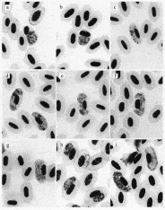
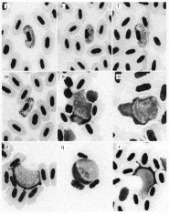
A total of 15 parasite species was identified by morphology, 14 of these to described species, and 1 distinctive Leucocytozoon that could not be identified to a known species. Additionally, 1 sample was not identified to species but only to the Plasmodium subgenus Haemamoeba. Figures 1 and 2 present photographs of each of the parasite species. These photographs should not be assumed as exemplars of the species because most parasite identification required a series of infected cells and measurements.

Fig. 1. Photomicrographs of haemosporidian parasites isolated from the bird community at 1 site in Israel. Each photograph matches a parasite indicated in Fig. 4.

Fig. 2. Photomicrographs of haemosporidian parasites isolated from the bird community at 1 site in Israel. Each photograph matches a parasite indicated in Fig. 4.
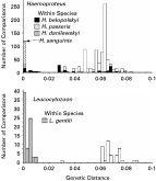
Each of the 9 parasite species identified by morphology from a single infection was represented by a unique gene sequence. Some morphological species represented by more than 1 sample either were identical in sequence or differed by only a single synonymous base-pair substitution (a genetic distance of 0·16%: P. relictum, H. sanguinis, H. danilewskyii, H. passeris). Two species identified by morphology revealed greater genetic distances among the sampled infections. Parasites identified by morphology as L. gentili were found in 13 infections; these differed from 0 to 0·89%. More extreme is the case for H. belopolskyi, which differed among infections by 0 to 7·7%. Fig. 3 presents all pair-wise genetic distance comparisons for infections, both within and between morphological species, for Haemoproteus and Leucocytozoon. Overall, within species distances ranged from 0 to 7·7%, and between species from 2·9 to 8·9% (Mann-Whitney U-test, P<0·0001). H. belopolskyi appears to be the outlier for the within-species comparisons (comparing only within-species divergence for Haemoproteus, Kruskal-Wallis test, P<0·0001). Excluding H. belopolskyi results in a range of 0–0·89% for the within-species comparisons.

Fig. 3. Pair-wise uncorrected genetic distances (cytochrome b gene, 607 bp) for Haemoproteus and Leucocytozoon species identified from a sample of birds from a single site in Israel. To determine genetic distances, each pair of sequences was examined for the proportion of bases that differed. Thus, every pair of infections identified as Haemoproteus or Leucocytozoon were compared, and comparisons for samples within a morphological species are indicated by different fill patterns. Open bars are for comparisons between morphological species. Results show genetic distance within species of parasite identified from different infections is slight compared to between-species distances except for a single morphological species, H. belopolskyi.
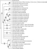
A total of 32 unique sequences obtained from 73 infections were incorporated into a phylogenetic analysis, using Toxoplasma gondii as the outgroup taxon. All 3 analyses produced the same topology. Maximum parsimony analysis produced 53 equally parsimonious trees, with the strict consensus shown in Fig. 4. The figure includes nodal support values from parsimony and likelihood bootstrap analyses (posterior probability values not shown). All phylogenetic analyses support the 3 genera as monophyletic groups, although support was weakest for Haemoproteus. There is support for monophyly for most morphological species sampled multiple times, but generally weak nodal support for more basal relationships for all methods of analysis.

Fig. 4. Strict consensus tree of 53 equally parsimonious trees obtained from maximum parsimony analysis of 607 bp cytochrome b sequences from haemosporidian parasites pictured in Fig. 1. Bootstrap support values are provided by both maximum parsimony (above each branch) and likelihood methods (below each branch). Bootstrap values below 50% are not shown. Letters refer to species shown in Figs 1 and 2.
Of the morphological species represented by more than 1 sample, P. relictum, H. sanguinis, H. danilewskyii, H. passeris, and L. gentili all displayed monophyly. Only a single morphological species consisted of more than 1 clade. The main H. belopolskyi clade consisting of 15 infections and 8 sequences was found primarily in 3 Acrocephalus species and 1 species of Hippolais. The other 3 H. belopolskyi haplotypes were from 2 species of Sylvia, and only 2 of these sequences formed a well-supported clade. Enforcing the constraint of monophyly for H. belopolskyi resulted in a significantly different topology than that observed as determined using the Shimodaira-Hasegawa test (P=0·009).
As noted above, each of the 9 morphological species represented by a single sample had a unique sequence, and these species were not contained within any of the clades containing species with multiple samples. Two pairs of these morphological species, however, H. syrnii and H. turtur, and L. squamatus and the new unidentified species of Leucocytozoon, did fall together into well-supported groups, with a nodal support value of over 90% by one or both bootstrap analyses. Both of these pairs of morphological species differ from one another by a sequence divergence of 2·9%, well outside the range of intraspecific values noted for each genus.
Some parasites were quite host specific; H. passeris was found infecting 15 birds of 3 species of Passer. P. relictum, in contrast, was identified from birds of 3 families, Old Word Sparrows (Passeridae), Crows (Corvidae), and Old World Buntings (Emberizidae).
DISCUSSION
The study compared identification of parasites by morphological and genetic means. The results can be used to explore 3 species concepts (Mayden, 1997; Perkins, 2000). The morphological and genetic species concepts require grouping the parasites by similarity as seen under the microscope (morphology) or by genetic distance. The phylogenetic species concept recognizes species or higher-level taxa based on monophyly; we used 3 methods of phylogenetic analysis, maximum parsimony, maximum likelihood, and Bayesian methods. The results supported the validity of most of the morphologically identified species of haemosproidian parasites infecting birds from the study site. Every parasite identified as a morphological species from a single infection presented a unique cytochrome b gene sequence. Some parasite species identified from many samples had either a unique sequence or differed by only a few synonymous sites (P. relictum, H. sanguinis, H. danilewskyi, H. passeris, L. gentili). Additionally, intraspecific and interspecific genetic distances were significantly different. These results demonstrate that the sequences for the recognized species are significantly more similar to each other than are the interspecific sequence comparisons. Only 1 morphological species, H. belopolskyi did not concord with species limits as defined by genetic and phylogenetic analysis.
A total of 10 species of Haemoproteus were recognized based on morphology. Six of these species occurred only once in the dataset and are distinct from the other Haemoproteus species by both genetic distance and phylogeny. The other 4 species are represented by 2, 6, 14 and 17 samples, respectively. For 3 of these species, H. sanguinis, H. danilewskyii, and H. passeris, morphological identification corresponds to small intraspecific sequence divergence (0 to 0·49%), and strong support of a monophyletic relationship (97 to 100% nodal support). Results for H. belopolskyi, as noted above, were less clear. While most infections identified to H. belopolskyi fell into one clade (14 out of 17 samples), 3 other infections resulted in quite different sequences that placed these samples in 2 other locations in the phylogeny. The H. belopolskyi within the main clade all infected Old World warblers, Sylviidae, but not the genus Sylvia. The infections of H. belopolskyi outside that clade were all found in Sylvia. The alternative topology test demonstrated a significant difference between the morphological and phylogenetic species designation for H. belopolskyi. These results agree with those of Bensch et al. (2004) who also found substantial genetic variation in morphological H. belopolskyi that suggests this taxon represents 2 or more genetic or phylogenetic species.
P. relictum has been reported from a very broad host and geographical range (Atkinson et al. 1995) which suggests this described species masks a cryptic diversity of taxa. We identified P. relictum from 11 birds of 3 families, and found all sequences identical (10 infections) or differing by only a single synonymous substitution (1 infection). Monophyly was supported for all morphological P. relictum samples. Thus, for the birds sampled in Israel, P. relictum appears to be a single species by morphological, genetic, and phylogenetic criteria, with a broad host range. The results for Leucocytozoon reveal that those samples identified as L. gentili fall into a single clade with very little sequence divergence. Thus, all 3 species concepts agree on the designation of this Leucocytozoon species.
Some studies conclude that a single or very few base pair differences in the cytochrome b gene observed among infections of avian haemosporidian parasites may reveal cryptic species (Bensch et al. 2004; Ricklefs et al. 2005). Many other studies treat a single base difference among parasite isolates as presumably non-recombining ‘lineages’ (for example, Bensch et al. (2000), Waldenstrom et al. (2002), Fallon et al. (2005)). However, such small genetic distances found among infections could represent variation that is intraspecific, interspecific, or both. The nature of genetic variation within species of haemosporidian parasites has not been well explored. Joy et al. (2003) found 6 bases differing in the cytochrome b gene of P. falciparum of humans, but these samples were taken over a wide geographical range. Variation in the cytochrome b gene is typically observed for avian haemosporidian parasites even at local sites as described here (Bensch et al. 2000; Ricklefs and Fallon, 2002; Waldenstrom et al. 2002; Fallon et al. 2003a,b; Schrenzel et al. 2003; Beadell et al. 2004; Bensch et al. 2004; Fallon et al. 2005; Ricklefs et al. 2005). A ‘single base-pair rule’ to delimit species would lead to the conclusion that there is a substantial cryptic diversity of the parasites. Our results suggest a more cautious perspective. Every pair of sequences that differed at 1 or 2 sites represented changes that would not result in an amino acid substitution on the cytochrome b protein (synonymous substitutions), and these were always identified as being the same morphological species. A genetic distance of only 1 or 2 substitutions could well represent non-recombining taxa (species), but in most cases we found these to belong to well-supported clades for the phylogenetic analyses. A resolution of this issue will require examination of several genes to detect the kind of covariation expected for reproductively isolated species. That is, an appropriate rule for defining species of haemosporidians will emerge only with studies of several genes. In a unique example of this approach, Bensch et al. (2004) found that parasite isolates that differed by 1 base pair for cytochrome b, also differed for a nuclear gene (DHFR-RS), suggesting there may be a genome-wide difference in these forms.
Although genetic distance studies are useful measures in modern systematic studies, we favour a combined genetic distance/phylogenetic species concept; again this would best be based on a study of the parasite sequence data for at least 2 genes. Nonetheless, the general concordance of our phylogenetic analysis based on a single gene (which provides a gene tree rather than species tree) with the identification of parasites by classical morphological study argues that study of the cytochrome b gene can provide valuable insight into the validity of classical morphological species.
Many researchers and veterinarians must identify avian haemosporidia based on their appearance in stained blood smears, including studies in wildlife epizootiology, parasite virulence, conservation biology, and captive animal care in zoos. Our results are the first broad-scale comparison of morphological data with gene sequence data for avian haemosporidians, but the findings must be taken now as only tentative. The results presented here suggest detailed study of morphology, taking many measurements to be compared with published descriptions (such as in Valkiunas, 2005), will usually allow sound identifications for Plasmodium, Haemoproteus, and Leucocytozoon.
We thank Kobi Meron, ringing officer, Kfar Ruppin Bird Watching Center, Nature Protection Society, Israel, for assistance in collecting the birds, and Hagit Gil, Faculty of Agriculture, Food and Environmental Quality, the Hebrew University of Jerusalem, for giving a hand in collecting and processing birds. Assistance with the molecular studies came from C. William Kilpatrick, Patrick O'Grady, and Susan Perkins. G. Valkiunas and W. Barnard offered important guidance and encouragement early in the project. The research was funded by grants from the Morris Animal Foundation to J.J.S. and Vermont Genetics Network through the NIH BRIN programme of the NCRR to E.S.M.