Kawasaki disease is an acute vaculitis syndrome of unknown aetiology that mainly affects the small- and medium-sized arteries, particularly the coronary arteries.Reference Kawasaki, Kosaki and Okawa 1 The prognosis of the disease depends on the extent of coronary artery lesions and associated ischaemia. The most commonly seen lesions are transient coronary ectasias, which are self-resolving. Transient coronary aneurysms are less common during the acute stage of untreated Kawasaki disease, but clinicians need to have a higher level of awareness, because aneurysms have a different healing pattern. However, the use of intravenous immunoglobulins reduces their incidence.Reference Newburger, Takahashi and Burns 2 The coronary aneurysms regress spontaneously within 1–2 years after onset in more than half of the patients, but some may progress to obstructive coronary lesions.Reference Takahashi, Mason and Lewis 3 , Reference Akagi, Rose and Benson 4 Whereas coronary aneurysms are usually diagnosed by cross-sectional echocardiography, coronary stenosis is invasively assessed by coronary angiography.Reference Takahashi, Mason and Lewis 3 Although in adults angiography is the gold standard of diagnosis of coronary artery lesions, it cannot be repeated very often, especially in infants and children. Moreover, it is sometimes difficult to monitor progressive changes from aneurysms to stenotic lesions; moreover, coronary angiography may not correlate well with ischaemic risk.Reference Kondo, Hiroe and Nakanishi 5
Coronary artery lesions with aneurysmic nature can lead to perfusion defects distal to the dilated area that can be very unpredictable. They may predispose to thrombus formation, resolve with time, or become stenotic.Reference Kato, Sugimura and Akagi 6 In severely affected vessels, the media develops inflammation and necrosis of the smooth muscle cells. The internal and external elastic laminae can split, leading to aneurysms.Reference Burns, Shimizu and Shike 7 The vessel lumen eventually becomes narrowed or occluded owing to stenosis or a thrombus. Cardiovascular death may occur from a myocardial infarction secondary to thrombosis of a coronary aneurysm or from rupture of a large coronary aneurysm.Reference Lee, Chun, Yeon, Shin and Kim 8 , Reference Wang, Wu, Liu, Kuo and Yang 9
The myocardial perfusion imaging is a diagnostic procedure performed to evaluate the blood flow to the myocardium. Using dipyridamole stress technetium-99 m tetrofosmin single-photon emission computed tomography may identify the hemodynamic significance of coronary aneurysm. Therefore, it may help in making decisions about coronary artery bypass surgery and evaluation of prognosis.Reference Kondo 10
The objective of this study was to assess the diagnostic accuracy of technetium-99 m tetrofosmin single-photon emission computed tomography as a possible alternative method to invasive coronary angiography for detection and follow-up of myocardial ischaemia in patients with Kawasaki disease, pre-, and post-coronary bypass graft operation.
Patients and methods
The study group included 21 children with a previous history of Kawasaki disease (13 boys, 8 girls), with age ranging from 5 to 9 years and mean age 7.3±1.6 years. They were diagnosed based on established clinical criteria.Reference Dajani, Taubert and Gerber 11 The mean age at the time of presentation with acute Kawasaki disease was 2.6±2.2 years.
Coronary lesions were detected in all patients on two-dimensional echocardiography and they were referred to the department of nuclear medicine, chest diseases hospital. Therefore, the knowledge of perfusion deficits and viability of the myocardium will allow adequate decision regarding revascularisation surgery, from January 2010 to September 2013.
The interval between the onset of Kawasaki and both selective coronary angiography and myocardial perfusion evaluation by technetium-99 m tetrofosmin single-photon emission computed tomography was 1.8±0.4 years. There were no children under treatment for active bronchial asthma included in this study. After giving informed consent, the following protocol was performed:
Coronary angiography
Coronary angiography was performed on all patients few weeks before stress myocardial perfusion by technetium-99 m tetrofosmin single-photon emission computed tomography to assess of the presence of coronary aneurysms and/or stenosis. The percentage of coronary narrowing was calculated with the nearest normal-appearing portion of the coronary artery – either distal or proximal to stenosis – as a standard; coronary narrowing was considered significant if ⩾75%.Reference Nakanishi, Takao and Nakazawa 12
Coronary aneurysms were classified according to the American Heart Association statement, as small, <5 mm internal diameter; medium, 5- to 8-mm internal diameter; or giant (>8 mm internal diameter).Reference Dajani, Taubert and Gerber 11 , Reference Kondo, Hiroe and Nakanishi 13
Stress myocardial perfusion using single-photon emission computed tomography technetium-99 m tetrofosmin
With the patient in the supine position, while electrocardiography and blood pressure were being monitored, dipyridamole as a pharmacological stress agent: 0.56 mg/kg was administered by continuous intravenous infusion over 4-minute period through the antecubital vein.Reference Kondo, Hiroe and Nakanishi 13 Then every subject was administered I.V. technetium-99 m tetrofosmin at a dose of 0.34 mCi/kg, followed by intake of a fatty meal to minimise hepatic activity. Parenteral aminophylline was available for treatment of any adverse effects.
After 1 hour, myocardial uptake of technetium-99 m tetrofosmin was assessed with acquisition by gamma camera. Chest leads were placed, and through electrophysiology synchroniser, the heart beats were accumulated using software protocols of the gamma camera system. Acquisition started from right 45 to 135° left side as step and shots every 3°, for technetium-99 m tetrofosmin accumulation. All defects were graded as either fixed – if present in both rest and stress images – or reversible – if present only with exercise. Single-photon emission computed tomography images were interpreted by a consensus of two experienced blinded independent observers; analysis was determined according to Emory cardiac tool box protocol.
In groups I and II, 16 patients with myocardial perfusion defects, who underwent coronary artery bypass grafting, were followed up by single-photon emission computed tomography.
Stress myocardial perfusion using single-photon emission computed tomography technetium-99 m tetrofosmin was followed up by resting for 1 hour within the 72-hour period.
Statistical analysis
Data were expressed as percentages, mean±standard deviation. A χ2 analysis was used to test for concordance of coronary angiographic and technetium-99 m tetrofosmin single-photon emission computed tomography perfusion results among the three groups of Kawasaki disease patients. Diagnostic accuracy of technetium-99 m tetrofosmin single-photon emission computed tomography for identifying coronary lesions due to Kawasaki disease is given in terms of sensitivity, specificity, positive predictive value, negative predictive value, and accuracy. A p-value of <0.05 was considered significant.
Results
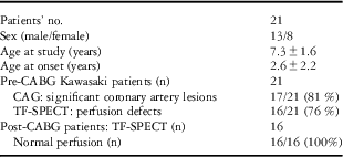
Summary of demographic, angiographic, and SPECT shown in Table 1. Of the 21 Kawasaki disease, pre-coronary bypass graft patients, 17 patients (81%) showed coronary angiographic lesions (stenosis ⩾75% or giant aneurysms >8 mm), and tetrofosmin single-photon emission computed tomography defects were positive in 16 Kawasaki disease patients (76%). Follow-up on 16 patients, who had undergone tetrofosmin single-photon emission computed tomography, of groups I and II, with myocardial perfusion defects after coronary bypass graft surgery, demonstrated that pre-coronary bypass graft myocardial ischaemic defects had disappeared.
Summary of demographic, angiographic, and SPECT resultsFootnote *.

CABG=post-coronary bypass graft; CAG=coronary angiography; Tf-SPECT=tetrofosmin single-photon emission computed tomography
* Data expressed as mean±standard deviation
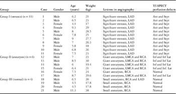
The findings of coronary angiography and tetrofosmin single-photon emission computed tomography myocardial perfusion defects in patients with Kawasaki disease are shown in Table 2. According to angiographic findings and coronary artery status, in the study, 21 patients were classified into three groups. In group I (n=11), significant coronary stenosis (>75%) was observed in the left anterior descending coronary artery, and all patients (100%) showed anterior and septal wall perfusion defects on technetium-99 m tetrofosmin single-photon emission computed tomography. Group II (n=6) patients had giant aneurysms (>8 mm) found in the right coronary artery and left main coronary artery, and five of six patients (83%) showed perfusion defect in the inferior and inferolateral myocardial walls. Group III (n=4) patients had small coronary artery aneurysms, and normal myocardial perfusion scan was performed in all patients. All perfusion deficits in patients of groups I and II were reversible. No immediate complications were shown in all patients, such as myocardial infarction or sustained angina related to this stress test.
Findings of coronary angiography and Tc-99 m Tf-SPECT myocardial perfusion defects in pre-CABG Kawasaki disease patients.

Ant and septal=anterior and septal walls; CABG=coronary bypass graft; Inf and Inf Lat=inferior and inferolateral walls; LAD=left anterior descending coronary artery; LMCA=left main coronary artery; RCA=right coronary artery; giant aneurysm (>8 mm); small aneurysm (<3 mm); significant stenosis (⩾75%)
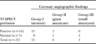
Results of tetrofosmin single-photon emission computed tomography perfusion were concordant with coronary angiography results among the three groups of Kawasaki disease patients, that is, no discordance could be detected by χ2 test; χ2 value=0.067, p=0.967 (non-significant), Table 3.
Concordance of coronary angiographic and Tc 99 m Tf-SPECT perfusion results among three groups of Kawasaki disease patients.

Tc 99 m Tf-SPECT=tetrofosmin single-photon emission computed tomography; χ2=0.067, p=0.967
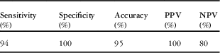
As shown in Table 4, overall sensitivity and specificity of technetium-99 m tetrofosmin single-photon emission computed tomography for the detection of coronary artery lesions with myocardial ischemia were 94 and 100%, respectively, whereas positive predictive value, negative predictive value, and accuracy were 100, 95, and 83%, respectively, as compared with coronary angiography.
Diagnostic accuracy of Tc 99 m Tf-SPECT for identifying coronary lesions in Kawasaki disease patients.

NPV=negative predictive value; PPV=positive predictive value
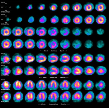
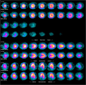
The changes in perfusion before and after coronary bypass surgery are demonstrated in Figs 1 and 2. Pre-coronary bypass graft image showing anterior and anteroseptal myocardial perfusion defects owing to occlusion of the left anterior descending coronary artery are displayed in Fig1. Follow-up post-coronary bypass grafting surgery of the same patients showed normal myocardial perfusion (Fig 2).
Myocardial perfusion technetium-99 m tetrofosmin, single-photon emission computed tomography (Tc-99 m-Tf SPECT) in patients with Kawasaki disease. Pre-coronary bypass graft (CABG) operation image showing anterior and anteroseptal myocardial perfusion defects owing to occlusion of the left anterior descending coronary artery (LAD).

Myocardial perfusion technetium-99 m tetrofosmin, single-photon emission computed tomography (Tc-99 m-Tf SPECT) in the same patient. Post-coronary bypass graft (CABG) operation. Image showing normal myocardial perfusion defect.

Discussion
Coronary artery lesions are critical complications of the Kawasaki disease; their long-term outcome is still unclear. It is sometimes difficult to monitor progressive changes from the aneurysms to stenotic lesions, because coronary angiography cannot be repeated very often owing to it being an invasive technique, especially in infants and children.Reference Chiewvit and Durongpisitkul 14 About 4% of the children with the Kawasaki disease ultimately develop ischaemic heart disease. Therefore, early detection, non-invasive monitoring, and follow-up of myocardial ischaemia are essential.Reference Jan, Hwang and Fu 15
In this study, the ischaemic areas with perfusion defects detected by tetrofosmin single-photon emission computed tomography have a good a concordance with coronary artery lesions identified by coronary angiography. In addition, the sensitivity and specificity of tetrofosmin single-photon emission computed tomography for the detection of the coronary artery lesions were 94 and 100%, respectively, whereas accuracy, positive predictive value, and negative predictive value were 95, 100, and 80%, respectively.
Although there are few published studies in relation to myocardial perfusion scans in children with Kawasaki disease, this study agrees with Fukuda et alReference Fukuda, Ishibashi and Yokoyama 16 , who demonstrated that regional myocardial hypoperfusion was detected by tetrofosmin single-photon emission computed tomography in 18 patients who had coronary artery stenosis of 75% or greater – sensitivity, 90%, and specificity, 100%. Another Japanese studyReference Karasawa, Ayusawa and Noto 17 has similarly found that the sensitivity for the detection of stress-induced perfusion defects by this method was 90%, and the specificity was 85%.
Contrary to this study, Fu et alReference Fu, Shiau and Tsai 18 in his study on 29 children with Kawasaki disease reported a poor agreement between coronary angiography and tetrofosmin single-photon emission computed tomography finding. The difference between the results of the studies in assessing myocardial ischaemia by the stress myocardial perfusion imaging could be partly explained by the presence of different coronary lesions among patients with Kawasaki disease, particularly lesions of the right coronary artery or multi-vessels.Reference Ono, Kohata and Iwatani 19 In addition, Onouchi et al,Reference Onouchi, Hamaoka and Kamiya 20 concluded in his study that the occluded vessels and vessels with segmental stenosis may be not associated with ischaemia or an episode of acute myocardial infarction as there is sufficient perfusion via collateral channels.
On one hand, coronary angiography has the advantage of providing a more detailed definition of the coronary artery than cardiac ultrasound, making it possible to detect coronary artery stenosis or thrombotic occlusion.Reference Miyagawa, Mochizuki and Murase 21 However, it adds to the risk for arterial occlusion or pseudoaneurysm formation at the catheterisation site, especially if repetitive follow-up cardiac catheterisation examinations are conducted.Reference Naoe, Takahashi and Masuda 22
On the other hand, according to Kondo et al,Reference Kondo, Hiroe and Nakanishi 13 tetrofosmin single-photon emission computed tomography has a number of advantages over coronary angiography. First, patients with Kawasaki disease can be evaluated serially, and the aggravation from the aneurysms to stenosis may be detected in a long-term follow-up of patients. Second, this method can detect the severity of myocardial damage. Third, myocardial perfusion state can be evaluated in patients with Kawasaki disease with no coronary stenosis on angiography.
Interestingly, some studies reported that even in asymptomatic children with previous history of Kawasaki disease and normal coronary arteries, reversible perfusion abnormalities were detected by technetium-99 m or thallium-201 single-photon emission computed tomographic imaging.Reference Paridon, Fnlioto and Vincent 23 , Reference Fukazawa, Fukushige and Takeuchi 24 This is will further highlight the value of myocardial perfusion imaging as a non-invasive method for the prediction of cardiac events during the chronic stage of Kawasaki disease.
In this study, 16 patients in groups I and II underwent coronary bypass graft operations and followed up by tetrofosmin single-photon emission computed tomography, which revealed a normal myocardial perfusion in all cases. The information from myocardial perfusion imaging is necessary to make a decision about coronary artery bypass surgery and to evaluate prognosis. Our results were comparable to Fukuda et al,Reference Fukuda, Ishibashi and Shinohara 25 who reported that patients who underwent coronary bypass graft had pre-coronary bypass graft ischaemic changes that disappeared after coronary bypass graft. Thus, coronary bypass graft is a useful tool in monitoring the progressive changes of myocardial ischaemia in Kawasaki disease after such operations.
In this study, there was no technetium-99 m tetrofosmin injection-related immediate complications, as well as in the other studiesReference Fukuda, Ishibashi and Yokoyama 16 , Reference Karasawa, Ayusawa and Noto 17 that used the paediatric field of myocardial perfusion imaging. The use of technetium-99 m compounds have been the radionuclide of choice for the last decade in myocardial perfusion assessment owing to its higher physical energy and shorter half-life, which allows better image resolution, onsite availability, and radiation dosimetry.Reference Chiewvit and Durongpisitkul 14 Moreover, biokinetic studies demonstrated that rapid clearance from the blood, with excellent cardiac uptake, relatively slow clearance and no significant redistribution.Reference Kelley, Forster and Highley 26 – Reference Jain, FJTh and McMahon 28 New other alternative non-invasive imaging modalities such as cardiac magnetic resonance imaging has recently emerged with no risk of radiation. It has the ability to evaluate the coronary arteries, cardiac function, and myocardial perfusion. Moreover, it facilitates both pharmacological stresses testing to assess reversible ischaemia and delayed contrast enhancement to visualise myocardial scar.Reference Lee, Simonetti and Harris 29 – Reference Schwitter, Wacker and van Rossum 30 However, such specialised magnetic resonance imaging scanning is not available in every hospital, and further studies for validation of its results are still recommended to incorporate. Cardiac magnetic resonance imaging in the guidelines for follow-up of patients with Kawasaki disease.Reference Tacke, Kuipers and Groenink 31
Study limitations
The 21 patients with Kawasaki disease in this study were selected on the basis of aneurysms detected by cross-sectional echocardiography. The data revealed there were perfusion defects detected by tetrofosmin single-photon emission computed tomography and a good concordance with coronary artery lesions detected by coronary angiography. However, tetrofosmin single-photon emission computed tomography data were not available on other patients with Kawasaki disease who did not have coronary aneurysms on echocardiography. Thus, we could not identify the utility and the false-positive rate of tetrofosmin single-photon emission computed tomography in the unselected patients with Kawasaki disease.
Conclusion
It has been shown that technetium-99 m tetrofosmin single-photon emission computed tomography can be applied as an accurate non-invasive diagnostic technique in the detection of myocardial perfusion defects with coronary artery lesions, and to show improved or even normalised perfusion of the myocardium in patients after surgical revascularisation. It was also found to be a useful technique without short-term complications and can be used to avoid unnecessary coronary catheterisation. Further studies with larger patients’ number are required to confirm this study finding.
Acknowledgement
The authors thank the staff of the departments of Pediatrics and Nuclear Medicine for their enthusiastic cooperation and technical assistance.
Financial Support
This research received no specific grant from any funding agency, commercial, or not-for-profit sectors.
Conflicts of Interest
None.