Introduction
The linker histone H1 maintains the higher-order chromatin and plays an essential role in the development and stabilization of the chromatin structure (Izzo et al., Reference Izzo, Kamieniarz and Schneider2008; Happel & Doenecke, Reference Happel and Doenecke2009; Medrzycki et al., Reference Medrzycki, Zhang, Cao and Fan2012). In mammals, at least eight H1 subtypes have been identified: the somatic variants H1a, H1b, H1c, H1d, H1e, and H10; the testis-specific variant H1t; and the oocyte-specific variant H1foo (Tanaka et al., Reference Tanaka, Hennebold, Macfarlane and Adashi2001; Happel & Doenecke, Reference Happel and Doenecke2009). It has been reported that the composition of the linker histone is tissue specific, as well as developmentally regulated (Khochbin & Wolffe, Reference Khochbin and Wolffe1994; Terme et al., Reference Terme, Sese, Millan-Arino, Mayor, Belmonte, Barrero and Jordan2011).
During oogenesis and embryogenesis, the type and expression level of linker-histone composition undergoes a dramatic change in many organisms (Godde & Ura, Reference Godde and Ura2009; Terme et al., Reference Terme, Sese, Millan-Arino, Mayor, Belmonte, Barrero and Jordan2011). In Xenopus, oocyte-specific linker histone B4 is the predominant subunit during the first division, however B4 is replaced by the somatic linker histones at midblastula transition (Dworkin-Rastl et al., Reference Dworkin-Rastl, Kandolf and Smith1994), demonstrating the tight developmental regulation of linker-histone expression and composition. In contrast, knockout mice studies have shown that any individual somatic linker histone H1, or even two, is dispensable for mouse development – as different subunits can compensate for each other's absence (Fan et al., Reference Fan, Sirotkin, Russell, Ayala and Skoultchi2001).
Oocyte-specific linker histone, H1foo in mammals, is the homolog of cs-H1 in sea urchins and B4 in Xenopus laevis (Tanaka et al., Reference Tanaka, Hennebold, Macfarlane and Adashi2001, Reference Tanaka, Kato, Tanaka, Kuji and Yoshimura2003a; McGraw et al., Reference McGraw, Vigneault, Tremblay and Sirard2006). H1foo is localized to the nucleus of germinal vesicle (GV) stage oocytes and binds to chromosomes in the subsequent stages (Tanaka et al., Reference Tanaka, Hennebold, Macfarlane and Adashi2001). H1foo is synthesized and accumulates during oogenesis, and its presence is maintained until some stage of the early embryo, at which rapid degradation follows (Tanaka et al., Reference Tanaka, Hennebold, Macfarlane and Adashi2001, Reference Tanaka, Kihara, Hennebold, Eppig, Viveiros, Emery, Carrell, Kirkman, Meczekalski, Zhou, Bondy, Becker, Schultz, Misteli, De La Fuente, King and Adashi2005; Fu et al., Reference Fu, Ghadam, Sirotkin, Khochbin, Skoultchi and Clarke2003). Unlike somatic linker histones, H1foo has been proven to be indispensable for meiotic maturation of mouse GV oocytes (Furuya et al., Reference Furuya, Tanaka, Teranishi, Matsumoto, Hosoi, Saeki, Ishimoto, Minegishi, Iritani and Yoshimura2007). Here, up-regulation and down-regulation of H1foo expression in bovine oocytes were performed to examine the function of H1foo during oocyte maturation.
Materials and methods
Construction of the expression vector for H1foo and preparation of siRNA
Bovine H1foo (NM001035372) cDNA was synthesized by reverse transcription polymerase chain reaction (RT-PCR; Fermentas-China, Shenzhen, China) from GV-stage oocytes, and then inserted into pMD19-T (Takara, Dalian, China) vectors. The desired recombinants were sent to the Shanghai Sangon Biotechnology Co. (Shanghai, China) for DNA sequencing. pH1foo–Venus was constructed by inserting H1foo cDNA into pVenus. The vector pVenus is adapted from eukaryotic expression vector pcDNA3.1, inserted coding sequence (CDS) region of the fluorescent protein Venus between BamHI and EcoRI.
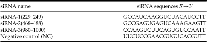
Three siRNA sequences that targeted bovine H1foo mRNA were designed and synthesized by GenePharma-a siRNA, and the siRNA stock solutions were diluted to a concentration of 20 μM. The siRNA sequences are listed in Table 1.
Details of siRNA sequences that target bovine H1foo coding sequence

Construction of the expression vector for H1e
Bovine H1e (NM_001098989) cDNA was synthesized by RT-PCR (Fermentas-China, Shenzhen, China) from cattle skin fibroblasts, and then inserted into pMD19-T vectors (TaKaRa, Dalian, China). The desired recombinants were sent to the Shanghai Sangon Biotechnology Co. (Shanghai, China) for DNA sequencing. As for pH1foo-Venus, pH1e-Venus was constructed by inserting H1e cDNA into pVenus.
Cotransfection of siRNA and plasmids into Hela cells
HeLa cells were cultured in Dulbecco's modified Eagle's medium (DMEM; Gibco) supplemented with 10% fetal bovine serum (FBS; HyClone) and 2 mM l-glutamine (Sigma) until 70% confluency. Three H1foo siRNAs and the negative control were cotransfected with plasmids into HeLa cells, using Lipofectamine™ 2000 (Invitrogen) in accordance with the manufacturer's procedure. The medium was changed with fresh DMEM at 5 h after transfection and cells were harvested at 48 h to determine the interference efficiency on RNA and protein expression, based on semi-quantitative RT-PCR and western blotting.
Effect of the different siRNA on H1foo mRNA and protein expression
RNA was isolated from about 1.0 × 10⁶ HeLa cells. Extraction of RNA was performed using Trizol (Invitrogen) in accordance with the manufacturer's protocol. Total RNA was dissolved in diethylpyrocarbonate (DEPC)-treated water, and quantified by means of ultraviolet (UV) light spectrophotometry.
Reverse transcription of purified RNA (1μ g) was performed using oligo(dT) priming and Moloney murine leukemia virus (M-MLV) in accordance with the kit procedure (TaKaRa). The PCR reaction was conducted in accordance with the manufacturer's instructions (Fermentas). The sequences of the bovine H1foo and β-actin primers are listed in Table 2; PCR products were separated by electrophoresis on 2.0% agarose gels, which were run in Tris–acetic acid–EDTA (TAE) buffer.
Details of primers used for RNA expression detection

Protein was extracted from about 1.0 × 10⁶ HeLa cells in lysis buffer. The samples were boiled and loaded onto a 12% sodium dodecyl sulphate polyacrylamide gel electrophoresis (SDS–PAGE) gel, and were transferred onto a polyvinylidene difluoride (PVDF) membrane (0.45-μm, Millipore) using semi-dry transfer cells (Bio-Rad). The membranes were stained with Ponceau S and blocked with Tris-buffered saline–Tween 20 (TTBS) that contained 10% skimmed milk, then the membranes were incubated with horseradish peroxidase (HRP)-conjugated goat anti-green fluorescent protein (GFP) antibody (1:10,000 dilution, Abcam). The immunoreactive bands were detected by the ECL Plus Western Blotting Detection System (Amersham Bioscience) in accordance with the manufacturer's instructions and visualized using Kodak BioMax XAR film. Quantitative analysis was performed by densitometric measurement (Gene).
Expression profiles of H1foo mRNA during bovine oocyte maturation
Before overexpressing or inhibiting H1foo expression in bovine oocytes, endogenous mRNA expression was identified by semi-quantitative RT-PCR. Specifically, bovine ovaries were obtained from a local slaughterhouse and transported to the laboratory within 6–10 h after slaughter in a thermo flask that contained 0.9% saline solution supplemented with penicillin and streptomycin. The cumulus oocyte complexes (COCs) were aspirated from follicles (2–8 mm in diameter) with a 12-gauge needle and COCs with multiple layers of cumulus cells were selected for in vitro maturation. The COCs were cultured in maturation medium that consisted of M199 (Earle's salts, Gibco) supplemented with 10% FBS, human menopausal gonadotrophins (HMG) (follicle stimulating hormone (FSH):leuteinizing hormone (LH) = 1:1) (0.1 IU/ml), estradiol (1.0 μg/ml), epidermal growth factor (EGF; 50 ng/ml), uracil (50 μg/ml), and insulin–transferrin–selenium (ITS) in groups of 50–100 in four-well dishes (Nunc) at 38.5°C under a humidified atmosphere of 5% CO2. The naked oocyte samples (zona pellucida free) in pools of five were collected at 0 h, 4 h, 8 h, 12 h and 24 h, and added in 5 μl lysis buffer (5 mM dithiothreitol (DTT), 20 U/ml RNase inhibitor, 1% NP-40), and stored at –80°C until RNA extraction.
H1foo-Venus, M-H1foo-Venus and H1e-Venus mRNA constructs
To construct pH1foo-Venus and pH1e-Venus, full-length coding sequence for bovine H1foo and H1e was amplified from GV-stage oocytes or cattle skin fibroblasts and cloned into the plasmid pVenus. Using linearised plasmids as templates, capped mRNAs were synthesized using the T7 mMESSAGE mMACHINE® Kit (Ambion) and dissolved in nuclease-free water to a final concentration of 1 μg/μl.
H1foo and H1e overexpression and H1foo-siRNA knockdown in bovine oocytes
Prior to microinjection, the COCs were cultured in maturation media at 38.5°C for 8 h. Then the COCs were completely denuded by gently pipetting in 0.3% hyaluronidase. The naked oocytes were incubated for 15 min in M-199 supplemented with 10% FBS and 7.5 μg/ml cytochalasin B before microinjection in order to reduce mechanical damage during injection (Paradis et al., Reference Paradis, Vigneault, Robert and Sirard2005). During microinjection, a group of 20–30 oocytes were placed in a 10 μl droplet of M-199 supplemented with 10% FBS and 7.5 μg/ml cytochalasin B under mineral oil (Sigma). Oocytes were either injected with mRNAs only or mix of siRNA and mRNA using Eppendorf Femtojet with EggJek injection needles (Eppendorf) and Precision Micro Devices holding pipets (Eppendorf). In total, 40–50 oocytes were injected per group. Before being transferred to maturation medium for further culture, the injected oocytes were incubated for 1 h to remove the oocytes with damaged plasma membranes following microinjection. Oocytes without microinjection were matured as controls. The rate of meiotic maturation was calculated based on first polar body extrusion at 22–24 h post oocyte maturation.
Oocyte imaging
Live oocytes were incubated with 10 μg/ml Hoechst 33342 stain in M-199 to stain chromatin. Brightfield, Hoechst 33342 and Venus fluorescence images were recorded with a charge coupled device (CCD) camera (Axiocam MRm; Carl Zeiss, Shanghai, China) attached to a Zeiss microscope.
Oocyte RNA isolation and semi-quantitative RT-PCR
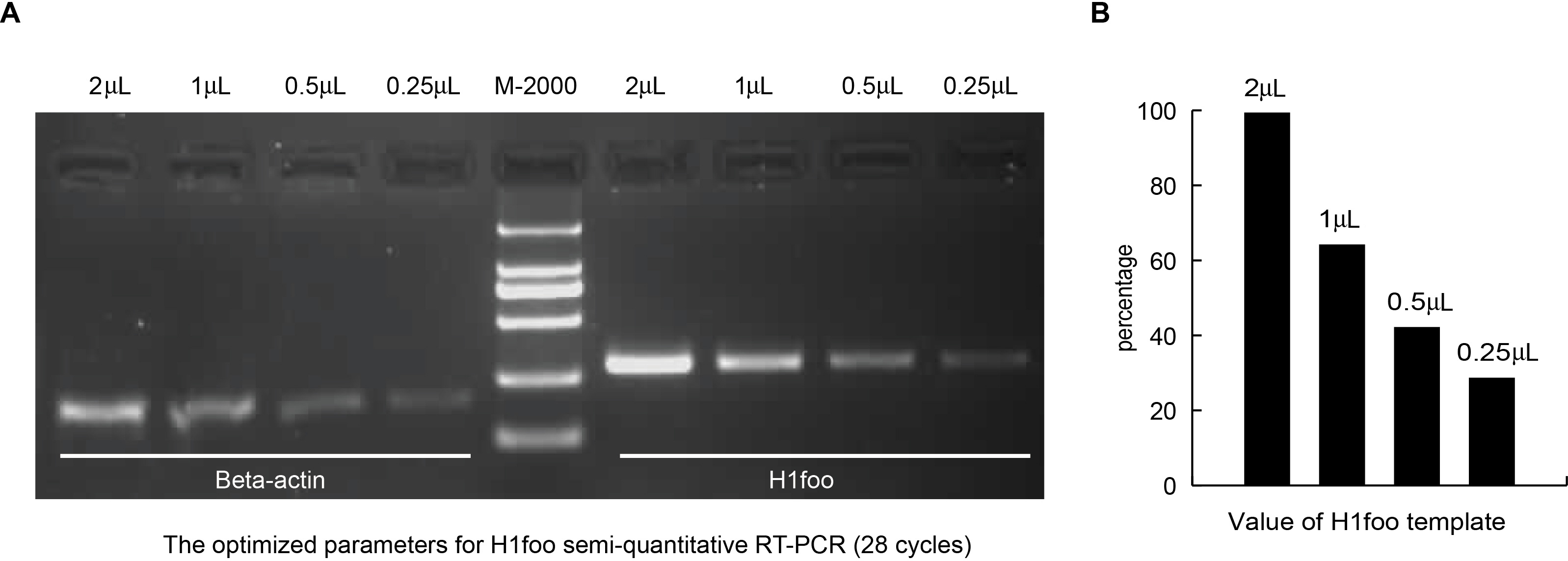
At 14–16 h after injection, total RNA was acquired by directly adding oocytes in 5 μl lysis buffer, followed by treatment with RNase-free DNase I (Fermentas) and RT-PCR of H1foo and β-actin. Every pool contained five oocytes from each group. The linear range of the amplification curve was validated using different numbers of PCR cycles and different amounts of RNA template. The optimized parameters were used for semi-quantitative RT-PCR.
Statistical analysis
To avoid the variable oocyte quality from different batch of collection, side-by-side experiments were always performed among groups. All experiments were repeated at least three times independently. Student's t-test and chi-squared test were used to evaluate the results. For all analyses, a value of P < 0.05 was considered to be significant. Quantitative analysis of band intensity in western blotting and RT-PCR was conducted using GeneSnap software (SynGene).
Results
Effect of H1foo siRNAs on mRNA and protein expression in HeLa cells
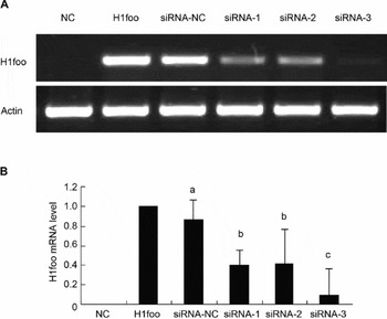
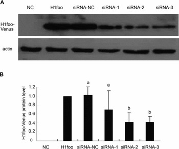
Before microinjection into oocytes, we examined the effects of individual siRNAs on H1foo mRNA and protein expression levels using RT-PCR (Fig. 1) and western blotting (Fig. 2), respectively. The results showed that H1foo mRNA and protein levels were down-regulated by all three siRNAs, and confirmed that siRNA-3 was the most effective in suppressing H1foo expression. Therefore, in the subsequent experiments, we used siRNA-3 to suppress H1foo expression and determine its biological effects on bovine oocyte maturation.
Efficiency of siRNAs on relative mRNA expression level in HeLa cells. (A) Gel electrophoresis analysis. (B) Semi-quantitative analysis of (A). NC: HeLa cells without transfection used for negative control. H1foo: positive control with pVenus–H1foo transfection. siRNA-NC, siRNA-1, siRNA-2, siRNA-3: cells cotransfected with plasmid pVenus-H1foo and siRNAs of negative control, siRNA-1, siRNA-2, siRNA-3, respectively. Note: H1foo expression is quantified using a value of H1foo/actin, and the value in the H1foo group is set to 1. Different letters (a, b, c) indicate significant difference (P < 0.05) among groups, and the same letter (b) indicates no significant difference (P > 0.05) between the groups.

Efficiency of siRNAs on relative protein expression level in HeLa cells. (A) Gel electrophoresis analysis. (B) Semi-quantitative analysis of (A) as above. NC, H1foo, siRNA-NC, siRNA-1, siRNA-2 and siRNA-3: as in Figure 1. Note: Efficiency of siRNAs on H1foo protein expression was analyzed indirectly by checking Venus expression using goat anti-GFP antibody.

Temporal expression pattern of H1foo transcripts during bovine oocyte maturation
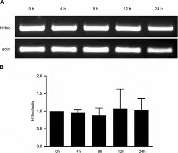
H1foo mRNA expression was measured throughout oocyte maturation, from 0 h (GV-stage oocyte) to 24 h (MII oocyte). Unlike the previous report that found the metaphase II (MII) oocytes conserved only 59% of the initial quantity of mRNA compared with the GV-stage oocytes (McGraw et al., Reference McGraw, Vigneault, Tremblay and Sirard2006); our results here showed that the H1foo transcripts maintained high values and were stable during oocyte maturation (Fig. 3). Therefore, we presumed that abundance of H1foo is required throughout meiotic maturation and subsequently designed the experiments for H1foo RNAi and overexpression at 8 h.
Bovine H1foo transcript expression profiles during oocyte meiotic maturation. The transcript expression levels were measured at differential time-points (0, 4, 8, 12 or 24 h) during bovine oocyte maturation in vitro by RT-PCR and its quantitative analysis.

Effect of microinjection of H1foo mRNA and siRNA on bovine H1foo expression and oocyte maturation
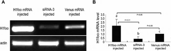
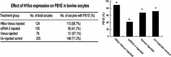
In order to assess the effect of mRNA and siRNA microinjection on H1foo expression, the transcript was tested among the different groups by semi-quantitative RT-PCR with the optimized parameters (Fig. S1). Compared with the Venus injection group (Control), H1foo transcript was increased by 200% in the H1foo-Venus group, and reduced by 70% in the siRNA-3 group (Fig. 4). Having established H1foo overexpression and knockdown in bovine oocytes, we next sought to determine whether H1foo expression level had any effect on meiotic maturation. Consistent with the previous observation in mouse oocytes (Furuya et al., Reference Furuya, Tanaka, Teranishi, Matsumoto, Hosoi, Saeki, Ishimoto, Minegishi, Iritani and Yoshimura2007), we found that knockdown of H1foo by siRNA-3 impaired bovine oocyte maturation (Fig. 5). Intriguingly, our data additionally showed that its overexpression significantly promoted meiotic progression (Fig. 5), a process in which maturation processing factor (MPF) activity regulation might be involved. Maturation processing factor is a heterodimer that consists of Cdk1 kinase and its regulatory partner cyclin B1; its activity is regulated by accumulation and degradation of cyclin B1, which then drives the events of meiosis including nuclear envelope breakdown and chromosome segregation at anaphase (Liu et al., Reference Yun, Zhao, Wu, Li and Lei2012). It has been shown that H1foo depletion arrested mouse oocytes at MI, and in these arrested oocytes MPF activity remained low (Furuya et al., Reference Furuya, Tanaka, Teranishi, Matsumoto, Hosoi, Saeki, Ishimoto, Minegishi, Iritani and Yoshimura2007). Therefore together with the previous data, our results suggested an association of H1foo expression and MPF activity via unknown pathways.
Detection and analysis of H1foo mRNA and siRNA-3 microinjection in oocytes. (A) Gel electrophoresis analysis. (B) Quantitative analysis of (A) as above (the value in Venus injected group is set to 1). Note: Different letters (a, b, c) indicate significant difference (P < 0.05) among groups.

Effect of H1foo expression on bovine oocyte maturation. PB1E (%) for different treatment groups. Note for bar chart: Different letters (a, b, c) indicate significant difference (P < 0.05) among groups, and the same letter (c) indicates no significant difference (P > 0.05) between the groups.

Effect of H1e overexpression on bovine oocyte maturation
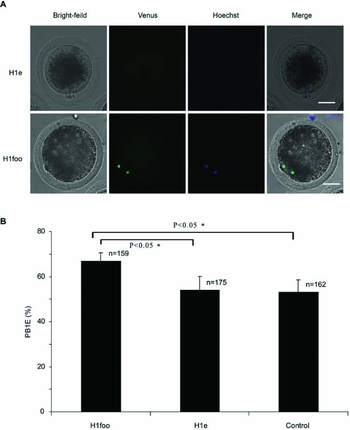
Having known that H1foo overexpression promoted bovine oocyte maturation, we wondered whether the somatic linker histones had the similar effect. To address this question, H1e was overexpressed in bovine oocytes, using the same procedure as for H1foo, to determine its effect on maturation. Our data showed that expressed exogenous H1e-Venus proteins cannot specifically bind to oocyte chromatin, while H1foo-Venus does bind (Fig. 6 A). Moreover, H1e overexpression could not improve bovine oocyte maturation as H1foo was shown to do (Fig. 6 B).
Effect of H1e expression on bovine oocyte maturation. (A) MII eggs microinjected with H1e-Venus or H1foo-Venus mRNA (green). Oocytes were incubated with Hoechst 33342 (blue) to stain chromatin. Note: Bar = 50 μm. (B) Effect of H1e or H1foo overexpression on bovine oocyte maturation given as PB1E(%).

H1foo RNAi-induced PB1E decrease can be rescued by co-injection of M-H1foo-Venus mRNA
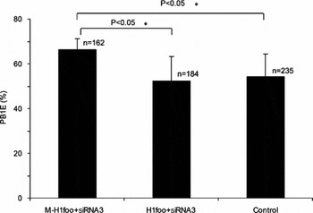
Consistent with the previous report in mouse oocytes (Furuya et al., Reference Furuya, Tanaka, Teranishi, Matsumoto, Hosoi, Saeki, Ishimoto, Minegishi, Iritani and Yoshimura2007), down-regulation of H1foo also significantly impaired oocyte maturation in the bovine; our study further demonstrated that only oocyte-specific types of linker histone H1foo overexpression rather than any somatic types could promote oocyte maturation (Fig. 6). To further confirm the function of H1foo, we applied a rescue experiment in H1foo-depleted oocytes, using a modified H1foo cDNA (M-H1foo) that encoded a functional protein but could not be targeted for degradation by siRNA-3 (sequence in Table 3). Similar to what was observed in the H1foo overexpression group, co-injection of M-H1foo mRNA with siRNA-3 significantly increased the PB1E rate when compared with the control groups (Fig. 7).
Modified Hlfoo tagged sequence for rescue from siRNA3 (letters in italics show the modified sequence)

PB1E reduction was rescued by co-injection of M-H1foo mRNA. Effect of different versions of H1foo mRNA expression with siRNA-3 on bovine oocyte maturation.

Discussion
Down-regulation of H1foo expression during bovine oocyte maturation impaired meiotic progression, a conclusion that is consistent with a previous report (Furuya et al., Reference Furuya, Tanaka, Teranishi, Matsumoto, Hosoi, Saeki, Ishimoto, Minegishi, Iritani and Yoshimura2007). Moreover, our results showed that only H1foo rather than somatic type of linker histones overexpression could stimulate extrusion of the first polar body, suggesting its indispensible role during bovine oocyte meiotic progression. Based on our data, it is possible that a threshold level of H1foo expression is required for oocyte maturation, such that only those oocytes that possess more than the threshold limit will be able to or have a higher chance to continue for maturation. This hypothesis would, in some way, explain why individual GV oocytes have different fates (arrested at GV, MI or matured to MII) after in vitro maturation. The in vitro collected GV-stage oocytes come from different follicles or different ovaries with diverse conditions, thus accumulating variable level of H1foo; this factor may finally decide their fate. Only the proportion of oocytes that had accumulated sufficient H1foo could mature to MII eggs ready for fertilization. It is also a reason to explain why in vitro matured oocytes are comparatively not as successful as in vivo oocytes, because the former might experience sub-optimal conditions during in vitro maturation, resulting in the loss of components globally, including H1foo. Our observations that H1foo overexpression stimulated meiotic maturation in vitro added evidence to the H1foo-threshold hypothesis, although further investigations are needed to reveal the unknown pathways.
Compared with somatic linker histones, which can compensate for each other (Fu et al., Reference Fu, Ghadam, Sirotkin, Khochbin, Skoultchi and Clarke2003), H1foo is indispensable in the process of oocyte maturation (Furuya et al., Reference Furuya, Tanaka, Teranishi, Matsumoto, Hosoi, Saeki, Ishimoto, Minegishi, Iritani and Yoshimura2007). One explanation is related to the abnormal ratio of H1 to nucleosomes that has been shown to be essential for mammalian development (Fan et al., Reference Fan, Nikitina, Morin-Kensicki, Zhao, Magnuson, Woodcock and Skoultchi2003). Oocytes are transcriptionally silent, and H1foo is the unique, or at least the predominant, linker-histone subunit required for oocyte maturation (Tanaka et al., Reference Tanaka, Hennebold, Macfarlane and Adashi2001, Reference Tanaka, Kihara, Meczekalski, King and Adashi2003b). Therefore, down-regulation of H1foo would directly result in a dramatic decrease in the total amount of H1. The other explanation for the importance of H1foo in oocyte maturation is demonstrated by the lack of functional H1foo studies. It has been reported that only H1foo, rather than somatic linker histones, is capable of correcting chromatin association in oocytes because of the presence of divergent N-terminal and globular domains (Becker et al., Reference Becker, Becker, Miyara, Han, Kihara, Brown, Hager, Latham, Adashi and Misteli2005). A study that used fluorescence recovery after photobleaching (FRAP) showed that H1foo binds chromatin more tightly, this finding provided molecular evidence for the replacement of somatic histones with H1foo during fertilization and nuclear transfer (Gao et al., Reference Gao, Chung, Parseghian, King, Adashi and Latham2004; Teranishi et al., Reference Teranishi, Tanaka, Kimoto, Ono, Miyakoshi, Kono and Yoshimura2004; Becker et al., Reference Becker, Becker, Miyara, Han, Kihara, Brown, Hager, Latham, Adashi and Misteli2005; Jullien et al., Reference Jullien, Astrand, Halley-Stott, Garrett and Gurdon2010; Mizusawa et al., Reference Mizusawa, Kuji, Tanaka, Tanaka, Ikeda, Komatsu, Kato and Yoshimura2010; Yun et al., Reference Yun, Zhao, Wu, Li and Lei2012). All these observations indicated that H1foo possesses both structure and function diversity compared with somatic types histones, and performs an indispensable role during oocyte maturation and early embryo development.
Due to the absence of a bovine H1foo antibody, we performed the co-transfection of plasmid pH1foo-Venus (GFP variant) and siRNA in HeLa cells to screen out an effective siRNA against H1foo in order that the GFP antibody could be used to detect the interference efficiency (Yun et al., Reference Yun, Zheng, Liu, Dou and Lei2008). The RNAi results in bovine oocytes showed that H1foo transcripts decreased by 70%, consistent with the data on HeLa cells. Here, we have developed a way of overcoming the dilemma of having no standard antibody.
In mice, antisense morpholino oligonucleotides (MO) against H1foo were injected into GV oocytes. After arrest for 20 h, dbcAMP was washed off from the culture medium to allow the spontaneous resumption of meiosis. Only 33.1% of H1foo-depleted oocytes extruded a first polar body, compared with 81.1% in the control (Furuya et al., Reference Furuya, Tanaka, Teranishi, Matsumoto, Hosoi, Saeki, Ishimoto, Minegishi, Iritani and Yoshimura2007). However, the GV was not visible in bovine oocytes. Recently we showed that the removal of cumulus cells at the GV stage did not impair bovine oocyte maturation. Here, we denuded the granulose cells and microinjected siRNA or mRNA at 8 h after in vitro maturation of oocytes, when the majority of the oocytes had completed GV breakdown (GVBD; Liu et al., Reference Liu, Yin, Zhao, Yun, Wu, Jones and Lei2012). In our H1foo knockdown group, the oocytes had a 41.2% first polar body extrusion rate, compared with 67.1% in the control. Our data indicate that H1foo expression after GVBD is still essential for bovine oocyte meiotic progression. This investigation demonstrates conclusively that H1foo is essential for the process of bovine oocyte maturation.
Supplementary material
To view supplementary material for this article, please visit http://dx.doi.org/10.1017/S0967199414000021
Acknowledgements
This work was supported by the National Natural Science Foundation of China (grant no. 31172280).