Introduction
Pigs are increasingly used as a dual-purpose model that benefits agricultural and biomedical research (Ireland et al., Reference Ireland, Roberts, Palmer, Bauman and Bazer2008; Perleberg et al., Reference Perleberg, Kind and Schnieke2018). The agricultural and biomedical applications of pigs in gene editing, human disease models and stem cell research rely on a large number of SCNT embryos obtained in vitro. However, early developmental efficiency of SCNT embryos in pigs is still suboptimal compared with naturally fertilized embryos and cloned embryos from other species (Fowler et al., Reference Fowler, Mandawala, Griffin, Walling and Harvey2018; Yang et al., Reference Yang, Wu and Li2019; Wang et al., Reference Wang, Qu, Li, He, Liu and Huan2020). This is mainly due to an inadequate understanding of unique molecular mechanisms underlying porcine early embryonic development (Ramos-Ibeas et al., Reference Ramos-Ibeas, Nichols and Alberio2018). Accumulating evidence has indicated that removal and re-establishment of epigenetic information is an inherent hallmark of early embryonic development in mammals (Marcho et al., Reference Marcho, Cui and Mager2015; Canovas and Ross, Reference Canovas and Ross2016; Xu and Xie, Reference Xu and Xie2018). Functional studies have indicated that writers and erasers for DNA and histone methylation play an essential role in preimplantation development of mammalian fertilized embryos (Lee et al., Reference Lee, Hamm, Whitworth, Spate, Park, Murphy and Prather2014; Huang et al., Reference Huang, Zhang, Wang, Dobbs, Yao, Qin, Whitworth, Walters, Prather and Zhao2015; Fraser and Lin, Reference Fraser and Lin2016; Cheng et al., Reference Cheng, Zhang, Zhang, Zhai, Jiang, An, Ma, Zhang, Li and Tang2019). Abnormal expression of these epigenetic modifiers even severely hindered both genomic reprogramming of somatic cells and early development of SCNT embryos (Matoba and Zhang, Reference Matoba and Zhang2018). Although epigenetic regulation of early embryonic development at the DNA and histone levels has been studied extensively, little information is known about the role of RNA modifications in early embryonic development.
N6-Methyladenosine (m6A) is the most prevalent RNA modification on eukaryotic message RNAs (mRNAs) (Nachtergaele and He, Reference Nachtergaele and He2018). Recently, m6A methylation was proved to be a reversible chemical modification that is deposited by methyltransferases (METTL3, METTL14 and WTAP) and removed by demethylases (FTO and ALKB5H), respectively (Shi et al., Reference Shi, Wei and He2019). The m6A modification has been shown to regulate many steps in RNA metabolism, such as stability, degradation, translation, splicing and localization (Huang et al., Reference Huang, Weng and Chen2020). Accumulating studies have shown that m6A modification in the animal reproduction field has been implicated in a variety of cellular and physiological events including spermatogenesis (Lin et al., Reference Lin, Hsu, Xing, Fang, Lu, Zou, Zhang, Zhang, Zhou, Zhang, Zhang, Song, Jia, Yang, He and Tong2017b), oogenesis (Hu et al., Reference Hu, Ouyang, Sui, Qi, Li, He, Cao, Cao, Lu, Zhou, Liu, Shen, Shu and Huo2020), germ cell maturation (Wang et al., Reference Wang, Yu, Liu, Li, Liu, Wang, Liu, Miao, Du and Yang2018; Xia et al., Reference Xia, Zhong, Wu, Chen, Tao, Xia, Shi, Zhu, Trudeau and Hu2018), maternal-to-zygotic transition (Zhao et al., Reference Zhao, Wang, Beadell, Lu, Shi, Kuuspalu, Ho and He2017; Hu et al., Reference Hu, Ouyang, Sui, Qi, Li, He, Cao, Cao, Lu, Zhou, Liu, Shen, Shu and Huo2020) and blastocyst formation (Kwon et al., Reference Kwon, Jo, Namgoong and Kim2019). In addition, m6A modification also plays a critical role in pluripotency maintenance, differentiation of embryonic stem cells (ESCs) (Wang et al., Reference Wang, Li, Toth, Petroski, Zhang and Zhao2014), and reprogramming of somatic cells into pluripotent stem cells (Chen et al., Reference Chen, Hao, Zhang, Li, Wang, Han, Wu, Lv, Hao, Wang, Li, Yang, Jin, Zhao, Li, Ping, Lai, Wu, Jiang, Wang, Sang, Wang, Yang and Zhou2015). It is worth noting that inconsistent results on the function of m6A modification in maintenance and differentiation of ESCs have been manifested in different studies (Park and Hong, Reference Park and Hong2017). Similarly, m6A modification also exhibited stimulatory and inhibitory effects on transcription factor-mediated somatic reprogramming (Guo et al., Reference Guo, Liu, Zheng, Huang and Chen2017). However, the role of m6A modification in porcine early embryonic development and its characteristics in nuclear transfer-mediated somatic reprogramming remain unclear.
In the present study, we identified a persistent occurrence of m6A methylation in porcine early embryos. Using a pharmacological approach, we demonstrated that m6A modification is indispensable for both early embryonic development and lineage allocation. Furthermore, m6A methylation exhibited an aberrant reprogramming in porcine early cloned embryos. Our findings provide new insights into epitranscriptomic regulation of early embryonic development and nuclear transfer-mediated somatic reprogramming.
Materials and methods
All chemicals in this study were purchased from Sigma (Sigma-Aldrich, St Louis, MO) unless otherwise stated.
Preparation of methylation inhibitor cycloleucine
Cycloleucine (Sigma, HY-19160) was dissolved in porcine zygote medium (PZM-3) to make 80 mM stock solutions. Then, the stock was further diluted using PZM-3 to obtain the desired working solution (10 mM, 20 mM and 40 mM).
Oocyte in vitro maturation
Ovaries were collected from gilts at a local slaughterhouse and follicular fluid was aspirated from antral follicles at 3–6 mm in diameter. Cumulus–oocyte complexes (COCs) were selected under a stereomicroscope. Subsequently, COCs were cultured in one well of 4-well plate containing 400 μl of in vitro maturation medium (TCM-199 supplemented with 5% FBS, 10% porcine follicular fluid, 10 IU/ml eCG, 5 IU/ml hCG, 100 ng/ml l-cysteine, 10 ng/ml EGF, 0.23 ng/ml melatonin, 2.03 × 10⁻⁵ ng/ml LIF, 2 × 10⁻⁵ ng/ml IGF-1, 4 × 10⁻⁵ ng/ml FGF2, 100 U/ml penicillin and 100 mg/ml streptomycin) for 44 h at 38.5ºC, 5% CO2 and saturated humidity. Cumulus cells surrounding oocytes were removed using 1 mg/ml hyaluronidase following maturation.
Parthenogenetic activation (PA)
Metaphase II (MII) oocytes were stimulated using two pulses of direct current (1.56 kV/cm for 80 ms) in activation medium (0.3 M mannitol supplemented with 0.1 mM CaCl2, 0.1 mM MgCl2 and 0.01% polyvinyl alcohol). Subsequently, activated oocytes were washed with PZM-3 and were incubated in chemically assisted activation medium (PZM-3 supplemented with 10 μg/ml cycloheximide and 10 μg/ml cytochalasin B) for 4 h. Then, embryos were cultured in PZM-3 droplets at 38.5°C, in 5% CO2 and 95% air with saturated humidity.
Somatic cell nuclear transfer
Donor cells and oocytes were placed in the operation medium TCB (TCM-199 with 7.5 mg/l cytochalasin B and 2% fetal bovine serum). The oocytes were enucleated by blind suction method, and a single donor cell was injected into the enucleated cytoplasm. The reconstructed oocytes were restored in PZM-3 droplets for 30 min. Then, the somatic cells and oocytes were fused using a direct current pulse (1.56 kV/cm for 100 ms) and further activated with chemical-assisted activation solution for 4 h. The fused embryos were cultured in PZM-3 droplets at 38.5°C, in 5% CO2 and 95% air with saturated humidity.
Real-time quantitative PCR
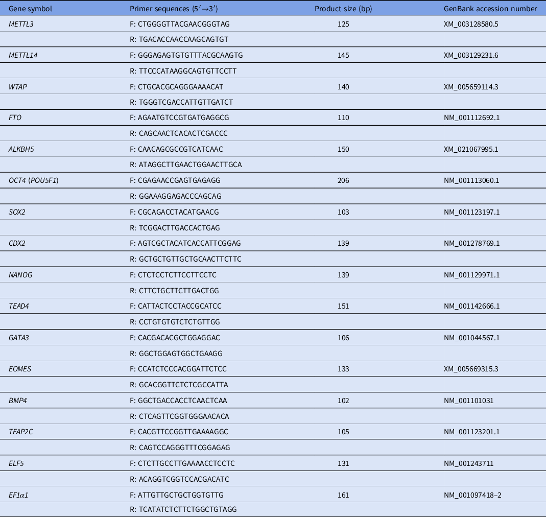
Total RNAs were extracted from oocytes and embryos using the RNeasy Mini Kit (Qiagen, 74104). The RNA was quantified by spectrophotometry at A260/A280 nm on a NanoDrop 2000 instrument (Thermo Scientific, Waltham, MA, USA). Reverse transcription was immediately performed using a QuantiTect Reverse Transcription Kit (Qiagen, 205311). The cDNA was aliquoted and stored at −80 ºC until ready for use. The primers used in the present study are listed in Table 1. The assembly for polymerase chain reaction (PCR) was prepared in the FastStart SYBR Green Master mix (Roche, 04673514001) and was run on a StepOne Plus (Applied Biosystems, Foster, USA). The following qPCR procedures were performed: pre-denaturation at 95°C for 10 min, followed by 45 amplification cycles of 95°C denaturation for 15 s, 60°C annealing for 10 s, and 72°C extension for 20 s. The quantification cycle (Cq) samples for each target gene were normalized to the value of the EF1α1 mRNA transcript. qPCR was performed in triplicate to minimize variations in operator techniques. The Cq values were obtained and transferred to a Microsoft Excel spreadsheet for further relative quantification analysis using the 2−ΔΔCt method. The samples were collected three times and three biological replicates were collected for each gene.
Porcine-specific primer sequences used in this study

F: forward; R: reverse.
Immunofluorescence staining
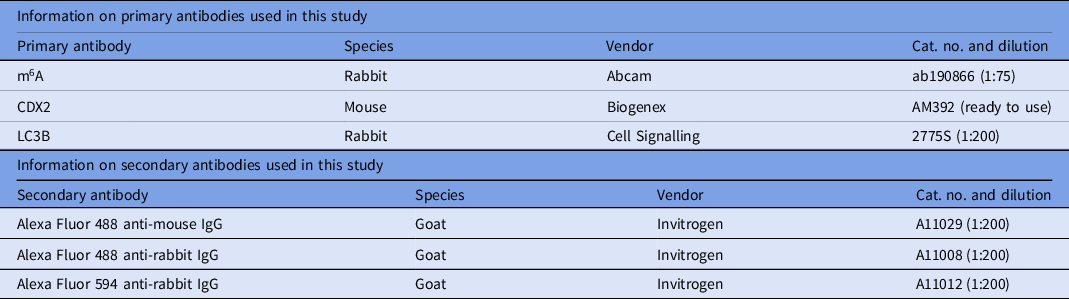
Oocytes or embryos were fixed in 4% paraformaldehyde (PFA) solution for 15 min, permeabilized with 1% Triton X-100 in Dulbecco’s phosphate-buffered saline (DPBS) for 30 min at room temperature (RT) and then blocked with 2% BSA in DPBS at RT for 1 h. Samples were incubated overnight at 4°C in blocking solution containing primary antibodies. The detailed information regarding primary antibodies used is listed in Table 2. Following several washes, samples were incubated in the dark at 37°C for 1 h in blocking solution containing secondary antibodies. After washing, samples were counterstained for 10 min in 4,6-diamidino-2-phenylindole dihydrochloride (DAPI) or propidium iodide (PI) solution and were then loaded onto glass slides and covered with a glass coverslip. Finally, samples were imaged under an inverted fluorescence microscope (Olympus, Japan). The average pixel intensity of embryos was determined using ImageJ software.
Antibodies used in this study

Terminal deoxynucleotidyl transferase (TdT)-mediated dUDP nick-end (TUNEL) staining
Blastocysts were fixed in 4% PFA for 15 min at RT. Following several washes, blastocysts were incubated in DNase I (TIANGEN, RT411, China) for 1 h at 38.5°C. After washing, blastocysts treated with label solution (In Situ Cell Death Detection Kit, 7791-13-1, Roche, Germany) served as the negative control group. Blastocysts treated with enzyme solution served as the positive control group. Blastocysts without DNase I treatment from other groups were incubated directly in enzyme solution for 1 h. All blastocysts were stained with DAPI for 15 min and were then imaged using laser scanning confocal microscopy (Olympus, Japan). Total numbers of cells and numbers of apoptotic dead cells were counted and the apoptotic ratio was calculated by dividing the number of dead cells by the total number of cells, which included dead cells.
Statistical analysis
All experiments were carried out at least three times. The data were analyzed using one-way analysis of variance (ANOVA) or Student’s t-test (SPSS 17.0) and were presented as mean ± standard error of the mean (mean ± SEM). A P-value < 0.05 was considered to be statistically significant.
Results
Cycloleucine treatment impaired developmental competence and quality of porcine early embryos
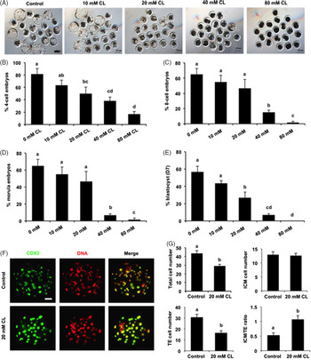
To explore the role of m6A methylation in porcine early embryonic development, embryos at the pronuclear stage were cultured for 7 days in medium containing different concentrations of cycloleucine. The results revealed that treatment with 10 mM cycloleucine did not affect the rates of 4-cell stage embryos (81.67 ± 8.81% vs. 63.33 ± 8.33%), 8-cell stage embryos (65.00 ± 7.64% vs. 55.00 ± 8.66%), morula (65.00 ± 7.64% vs. 55.00 ± 8.66%) and blastocysts (56.67 ± 6.67% vs. 43.33 ± 3.33%) (Fig. 1A–E). Treatment with cycloleucine (40 mM and 80 mM) significantly reduced the rates of 4-cell stage embryos (81.67 ± 8.81% vs. 38.33 ± 6.01%, 16.67 ± 4.41%), 8-cell stage embryos (65.00 ± 7.64% vs. 15.00 ± 2.88%, 1.67 ± 1.67%), morula (65.00 ± 7.64% vs. 6.67 ± 1.67%, 1.67 ± 1.67%) and blastocysts (56.67 ± 6.67% vs. 6.67 ± 1.67%, 0.00 ± 0.00%) (Fig. 1A–E) (P < 0.05). Interestingly, treatment with 20 mM cycloleucine only significantly decreased the rates of 4-cell stage embryos (81.67 ± 8.81% vs. 50.00 ± 10.40%) (Fig. 1B) and blastocysts (56.67 ± 6.67% vs. 26.67 ± 6.67%) (Fig. 1A, E) (P < 0.05), but did not affect development to the 8-cell stage embryos (65.00 ± 7.64% vs. 46.67 ± 11.67%) and morula (65.00 ± 7.64% vs. 46.67 ± 11.67%) (Fig. 1C, D). Furthermore, there were no blastocysts in the high dosage cycloleucine treatment groups (40 mM and 80 mM), while a small portion of blastocysts was still present in the 20 mM cycloleucine treatment group (Fig. 1A). Therefore, the 20 mM cycloleucine concentration was used in subsequent experiments. To analyze lineage allocation of trophectoderm (TE) and inner cell mass (ICM), blastocysts were stained with CDX2 to determine the TE cell number (Fig. 1F). The number of ICM cells was indirectly determined by subtracting the TE number from the total cell number. We found that the ICM cell number did not change between control and treatment groups (12.89 ± 1.15 vs. 12.63 ± 0.79) (Fig. 1G). However, cycloleucine treatment led to a significant reduction in the number of total cells (43.67 ± 3.09 vs. 29.15 ± 1.75) and TE cells (30.78 ± 2.71 vs. 16.52 ± 1.86) (Fig. 1G) (P < 0.05). Moreover, the ratio of ICM cells to TE cells in blastocysts in the treatment group significantly increased compared with the control group (0.53 ± 0.08 vs.1.08 ± 0.13) (Fig. 1G) (P < 0.05). Altogether, these data demonstrated that cycloleucine treatment was detrimental to early embryonic development and normal lineage allocation in pigs.
Effects of different concentrations of cycloleucine on porcine early embryonic development and quality. (A) Representative images of blastocysts in each group. Blastocysts from control and CL treatment group were imaged. CL, cycloleucine. Scale bars: 100 µm. Developmental rates of embryos at the 4-cell (B), 8-cell (C), morula (D) and blastocyst stage (E). The developmental rates of embryos in each group were recorded and statistically analyzed. (F) Immunofluorescence staining of blastocysts. Representative images obtained using confocal microscopy are shown. The experiment was independently repeated three times with at least 20 blastocysts per group. Scale bar: 50 µm. (G) Lineage allocation analysis of blastocysts from the control and CL treatment group. Total cell numbers, ICM cells, TE cells, and the ratio of ICM cells to TE cells were separately recorded and were subjected to statistical analysis. ICM: inner cell mass; TE: trophectoderm. All data are represented as mean ± SEM and different letters on the bars indicate significant differences (P < 0.05).

Cycloleucine treatment decreased m6A modification levels in porcine early embryos
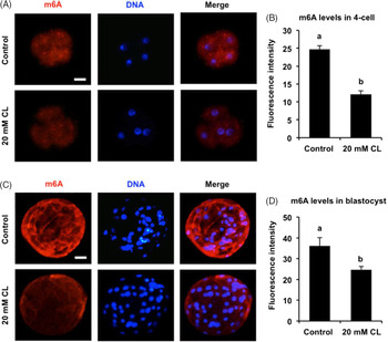
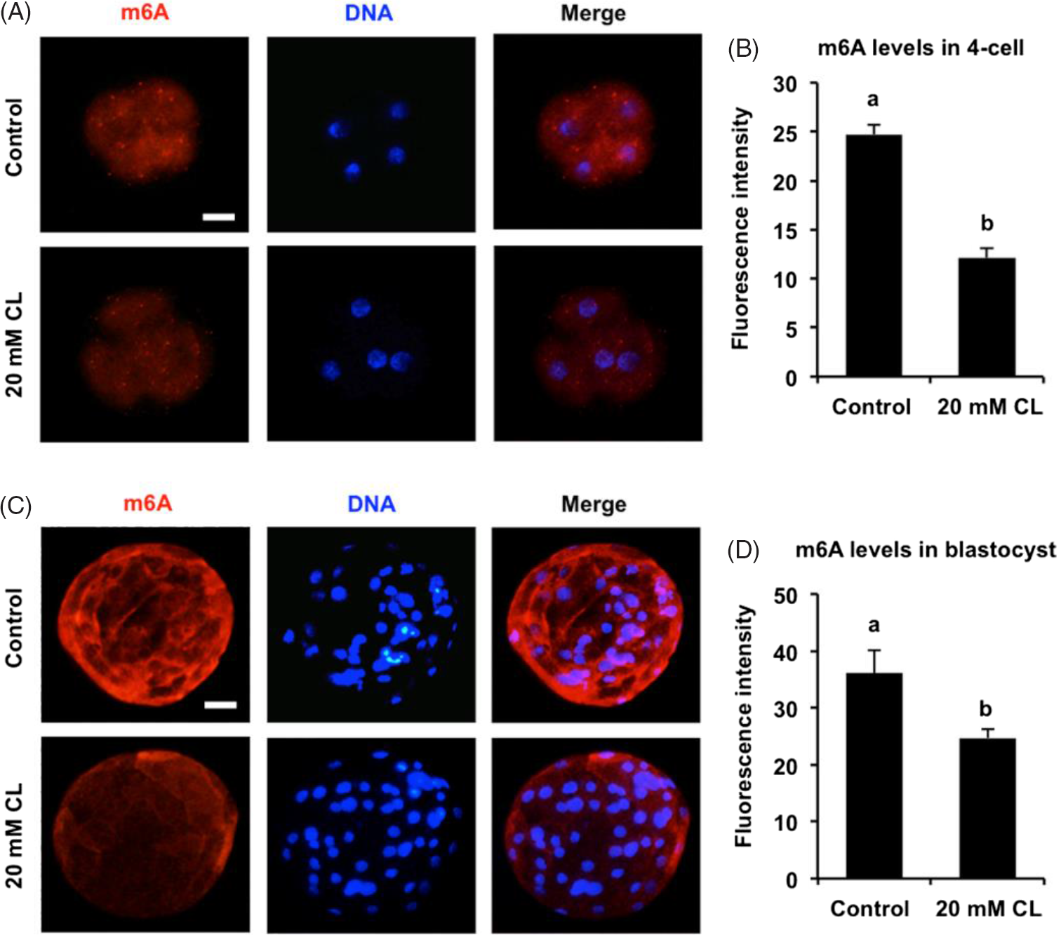
To investigate whether cycloleucine treatment affected m6A levels in embryos, immunofluorescence staining of 4-cell embryos (Fig. 2A) and blastocysts (Fig. 2C) was performed to analyze signal intensity of m6A. The results showed that cycloleucine treatment significantly reduced m6A levels in 4-cell embryos and blastocysts compared with the control group (Fig. 2B, D) (P < 0.05). To determine whether cycloleucine treatment affected the expression of genes associated with ICM and TE lineage specification, blastocysts were subject to qPCR analyses. The results showed that there was no change in expression levels of several genes between control and treatment groups, while cycloleucine treatment resulted in a significant reduction in TFAP2C expression levels (Fig. S1) (P < 0.05). Therefore, these results suggested that cycloleucine treatment effectively reduced m6A modification levels in porcine early embryos.
Effect of cycloleucine treatment on m6A modification levels in porcine early embryos. (A) Immunofluorescence staining of 4-cell embryos. Representative images obtained using confocal microscopy are shown. The experiment was independently repeated three times with at least 30 embryos per group. Scale bar: 50 µm. (B) Quantitative analysis of m6A levels in 4-cell embryos. (C) Immunofluorescence staining of blastocysts. Representative images are shown. The experiment was independently repeated three times with at least 24 embryos per group. Scale bar: 50 µm. (D) Quantitative analysis of m6A levels in blastocysts. All data are presented as mean ± SEM and different letters on the bars indicate significant differences (P < 0.05).
Reduced m6A methylation levels increased apoptosis and autophagy in porcine blastocysts
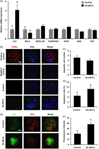
To study whether reduced m6A levels affected expression of genes associated with apoptosis and autophagy, blastocysts were subject to qPCR analyses. The results revealed that there were no changes in expression of genes including ATG5, BECL1N1, CASPASE3, BAX and P53 between control and treatment groups (Fig. 3A). However, cycloleucine treatment significantly decreased the expression of anti-apoptotic gene BCL2, and increased the expression of autophagy gene LC3 (Fig. 3A) (P < 0.05). Furthermore, TUNEL staining showed that cycloleucine treatment did not affect the number of TUNEL-positive cells (Fig. 3B, C), but resulted in a higher apoptosis ratio in blastocysts (Fig. 3B, D) (P < 0.05). Immunofluorescence analyses revealed that cycloleucine treatment significantly enhanced the expression levels of LC3 protein in blastocysts (Fig. 3E, F) (P < 0.05). Therefore, our data indicated that reduction of m6A levels increased apoptosis and autophagy in porcine blastocysts.
Effect of cycloleucine treatment on apoptosis and autophagy in porcine blastocysts. (A) Effect of cycloleucine treatment on expression of genes associated with apoptosis and autophagy. The relative abundance of mRNA of the indicated genes in blastocysts in each group was determined by qPCR. (B) TUNEL staining in blastocysts. Red spots indicate apoptotic cells, blue spots mark nuclei. Representative images obtained by confocal microscopy are shown. Scale bar: 50 µm. (C) Analyses of number cells TUNEL-positive in blastocysts. (D) Analyses of apoptosis ratio in blastocysts. (E) Immunofluorescence staining of LC3 protein. Blastocysts in each group were stained for LC3 and DNA. The experiment was independently repeated three times with at least 25 blastocyst per group. Scale bar: 50 µm. (F) Quantitative analysis of LC3 protein levels in blastocysts. All data are shown as mean ± SEM and different letters on the bars indicate significant differences (P < 0.05).

Aberrant reprogramming of m6A methylation during early development of porcine cloned embryos
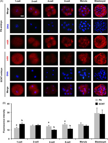
To elucidate the dynamic reprogramming of m6A modification in PA and SCNT embryos, immunofluorescence staining of embryos at different developmental stages was performed to detect signal intensity of m6A modification (Fig. 4A). Immunofluorescence analyses revealed that m6A methylation was present in the cytoplasm of PA embryos between 1-cell to blastocyst stages and m6A levels significantly increased during the morula-to-blastocyst transition (Fig. 4A, B). There were no significant differences in m6A levels in 2-cell, morula and blastocysts between PA and SCNT (Fig. 4A, B). However, m6A levels in SCNT 1-cell embryos were significantly higher than that in their PA counterparts (Fig. 4A, B) (P < 0.05). In contrast, m6A levels in SCNT 4-cell and 8-cell embryos were apparently lower than that in PA counterparts (Fig. 4A, B) (P < 0.05). Therefore, these results demonstrated that m6A methylation exhibited an aberrant reprogramming in porcine SCNT embryos.
Dynamic reprogramming of m6A methylation during early development of porcine cloned embryos. (A) Immunofluorescence staining of embryos derived from PA and SCNT. Embryos at the indicated developmental stages were stained for m6A (red) and DNA (blue). The experiment was independently repeated three times with at least 30 embryos per stage. Scale bar: 50 µm. (B) Quantitative analysis of m6A levels in embryos derived from PA and SCNT. All data are shown as mean ± SEM and different letters on the bars indicate significant differences (P < 0.05).

Abnormal expression of genes encoding m6A writers and erasers in porcine early cloned embryos
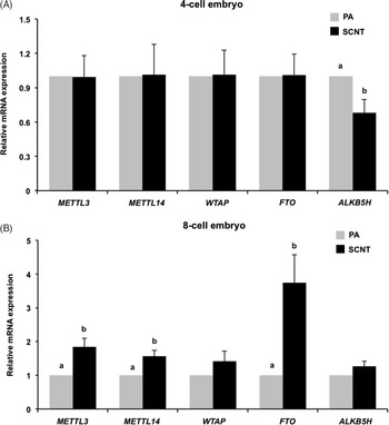
To further determine the relative expression levels of genes encoding m6A writers and erasers between PA and SCNT embryos, qPCR was performed to address the question. We found that expression levels of METTL3, METTL14, FTO and WTAP in SCNT 4-cell embryos were similar to that in their PA counterparts (Fig. 5A). However, ALKB5H mRNA levels in SCNT 4-cell embryos were significantly lower than that in PA 4-cell embryos (Fig. 5A) (P < 0.05). Additionally, at the 8-cell stage, expression levels of METTL3, METTL14 and FTO in SCNT embryos were significantly higher than that in PA embryos (Fig. 5B) (P < 0.05), while there was no significant difference in expression levels of WTAP and ALKB5H between PA and SCNT embryos (Fig. 5B). Therefore, these data indicated that genes encoding m6A writers and erasers displayed an abnormal expression in porcine SCNT embryos.
Expression of genes encoding m6A writers and erasers in porcine PA and SCNT embryos. (A) Expression of genes in 4-cell embryos derived from PA and SCNT. (B) Expression of genes in 8-cell embryos derived from PA and SCNT. The relative abundance of METTLE3, METTL14, WTAP, FTO and ALKB5H mRNA was determined by qPCR. All data are shown as mean ± SEM and different letters on the bars indicate significant differences (P < 0.05).

Discussion
Despite the essential role of m6A methylation in porcine oocyte maturation being demonstrated (Wang et al., Reference Wang, Yu, Liu, Li, Liu, Wang, Liu, Miao, Du and Yang2018), the role of m6A modification in early embryonic development and dynamics of m6A reprogramming in SCNT embryos remains unknown. Here our results showed that m6A modification is required for porcine early embryonic development, while it exhibited an aberrant reprogramming pattern in SCNT embryos. To our knowledge, the reprogramming pattern of m6A modification was characterized here for the first time in SCNT embryos.
Establishment of the functional TE lineage is an essential prerequisite for mammalian blastocyst formation (Choi et al., Reference Choi, Carey, Wilson and Knott2012; Posfai et al., Reference Posfai, Rovic and Jurisicova2019). Proliferation and differentiation of TE cells is accompanied by the establishment of a functional TE lineage. In this study, we found that reduced m6A levels induced by cycloleucine treatment significantly decreased TE cell numbers. It is therefore possible that m6A modification mediated TE proliferation and differentiation to allow blastocyst formation in pigs. Conversely, our data showed that expression of anti-apoptotic gene BCL2 and apoptosis ratio significantly increased in cycloleucine-treated blastocysts. Previous studies have indicated that increased apoptosis ratio frequently impaired the formation of both TE cells and blastocyst (Lin et al., Reference Lin, Lee, Oqani, Kim, Cho, Jeong, Baek and Jin2017a). In addition, reduced m6A levels also resulted in a higher level of autophagy in porcine blastocysts. One study indicated that induction of autophagy resulted in defects in TE cell differentiation in mice (Lee et al., Reference Lee, Hwang, Sun, Xu and Kim2011), and excessive levels of autophagy were detrimental to blastocyst development (Kim et al., Reference Kim, Kim, Lee, Lee, Jin and Lee2017; Song et al., Reference Song, Kim, Kim, Sim, Yoon, Cha, Choi, Yang, Mun, Park, Jeong, Huh, Lee, Kim, Kim and Chang2014). Therefore, we speculated that high levels of apoptosis and autophagy induced by cycloleucine might inhibit TE cell proliferation and differentiation, thereby impeding blastocyst formation.
Epigenetic reprogramming of chromatin after fertilization allows zygotic genome activation (ZGA) and development to full term (Fraser and Lin, Reference Fraser and Lin2016; Xu and Xie, Reference Xu and Xie2018). Aberrant epigenetic reprogramming is closely related to poor developmental competence in fertilized embryos and somatic cloned embryos (Shi and Haaf, Reference Shi and Haaf2002; Matoba and Zhang, Reference Matoba and Zhang2018). Recently, m6A modification was reported to regulate ZGA in zebrafish (Zhao et al., Reference Zhao, Wang, Beadell, Lu, Shi, Kuuspalu, Ho and He2017) and mice (Hu et al., Reference Hu, Ouyang, Sui, Qi, Li, He, Cao, Cao, Lu, Zhou, Liu, Shen, Shu and Huo2020). Our results in this study showed that reduced m6A levels induced by cycloleucine treatment caused developmental arrest at the 4-cell stage. In pigs, ZGA mainly occurs at the 4-cell stage (Zhou et al., Reference Zhou, Niu, Nie, Kim, Xu, Yan and Cui2020). Therefore, it is likely that m6A modification mediates ZGA to sustain normal development of 4-cell embryos.
Natural reprogramming of epitranscriptomic modifications during ZGA under the physiological context provides a reliable reference for artificial reprogramming in SCNT embryos (Yartseva and Giraldez, Reference Yartseva and Giraldez2015). Reprogramming patterns of m6A modification were characterized in detail during early development of porcine PA and SCNT embryos. Results indicated that m6A levels were significantly increased in 1-cell cloned embryos, whereas they dramatically decreased in 4-cell and 8-cell embryos compared with PA embryos, suggesting an aberrant reprogramming of m6A modification in porcine SCNT embryos. Correspondingly, genes encoding m6A writers and erasers also exhibited an abnormal expression in cloned embryos, which might be used to explain the aberrant reprogramming of m6A modification. In addition, previous studies have indicated that m6A methylation facilitated or inhibited transcription factor-mediated somatic reprogramming (Wu et al., Reference Wu, Liu, Zhao, Bi, Yao, Liu, Wang, Wang and Wang2019), suggesting that m6A methylation indeed functions in somatic reprogramming. Therefore, functional research on m6A methylation in nuclear transfer-mediated somatic reprogramming would be warranted in the future.
In conclusion, our results showed that m6A methylation is essential for early embryonic development in pigs and exhibits an aberrant reprogramming in SCNT embryos. These findings provide new insights into the function of m6A modification in mammalian preimplantation embryonic development.
Supporting information
Supporting information is available for this article at the publisher’s website.
Ethical standards
All experiments were conducted in accordance with the Institutional Animal Care and Use Committee (IACUC) guidelines under currently approved protocols at Anhui Agricultural University.
Financial support
This work was supported by grants from the Anhui Provincial Natural Science Foundation (1908085MC97), the Science and Technology Major Project of Anhui province (18030701185 and 17030701061), the National Natural Science Foundation of China (31802059 and 31902226), the Hefei Innovation and Entrepreneurship Support Plan for Returnee Scholar, and the Anhui Provincial Innovation and Entrepreneurship Support Plan for Returnee Scholar (2020LCX015).
Authors contribution
ZC, JGK, and YZ conceived the project and wrote the manuscript. TY, XQ, LZ, WN, DG and TX performed the experiments. JGK and AS analyzed the data. All authors read and approved the final manuscript.
Conflict of interest
The authors declare no conflicts of interest with regard to the study.
Supplementary material
To view supplementary material for this article, please visit https://doi.org/10.1017/S0967199420000799