Introduction
Infection by gastrointestinal nematodes is an important cause of production loss in cattle and sheep that results in poor general performance, particularly in young animals (Ploeger et al., Reference Ploeger, Kloosterman, Eysker, Borgsteede, Van Straalen and Verhoeff1990). The distribution and abundance of nematode parasites vary according to weather conditions, particularly rainfall and temperature, as well as within and between ruminant species. Haemonchus spp. and Cooperia spp. are the most important nematodes in subtropical/tropical environments (Waller, Reference Waller2006). Cooperia species are intestinal nematodes of ruminants. As a rule, males are 10–15 mm long and females are 15–20 mm long. The most distinguishing morphological features of the genus are the cephalic vesicle and cuticle transverse striations. Females present with a protuberant vulva, whereas males present with short spicules and the absence of gubernaculum. Additionally, the shape of the spicules may be used to identify different species (Ueno & Gonçalves, Reference Ueno and Gonçalves1998). Cooperia punctata is the most important species of the Cooperia genus found in cattle raised in Brazil (Lima, Reference Lima1998; Bricarello et al., Reference Bricarello, Zaros, Coutinho, Rocha, Kooyman, Vries, Goncalves, Lima, Pires and Amarante2007; Condi et al., Reference Condi, Soutello and Amarante2009), whereas C. curticei is the most important species found in sheep (Amarante et al., Reference Amarante, Bricarello, Rocha and Gennari2004). Studies on parasite host specificity with nematodes that parasitize sheep and cattle, raised either together or separately in Brazil, show that C. punctata is more adapted to cattle than sheep (Amarante et al., Reference Amarante, Bagnola, Amarante and Barbosa1997; Rocha et al., Reference Rocha, Bresciani, Barros, Fernandes, Silva and Amarante2008). Moreover, C. spatulata appears to be more adapted to cattle, whereas C. curticei is more adapted to sheep. The infection of sheep with C. punctata is relatively common when cattle and sheep share the same pasture (Amarante et al., Reference Amarante, Bagnola, Amarante and Barbosa1997; Rocha et al., Reference Rocha, Bresciani, Barros, Fernandes, Silva and Amarante2008). In naturally infected sheep, C. curticei is considered a mild pathogen (Ahluwalia & Charleston, Reference Ahluwalia and Charleston1975) and its effect is additive to that of other primary gastrointestinal nematodes, such as Ostertagia and Haemonchus contortus.
The intensity of gastrointestinal nematode infections can be estimated by the egg counts in faeces (eggs per gram, EPG), but eggs are difficult to identify at both species and genus levels. Therefore, it is necessary to produce infective third-larval stages from faecal cultures. Larval culture methods are used to determine the genera of worms and their proportions, but larval cultures may not match the exact proportion of worms (Amarante, Reference Amarante2000). In addition, this technique is time consuming, and identification of larvae requires microscopy expertise (Lichtenfels et al., Reference Lichtenfels, Hoberg and Zarlenga1997). Furthermore, necropsy studies are essential for determining the epidemiological pattern of infections and identifying the species present (Eysker & Ploeger, Reference Eysker and Ploeger2000) because, in some cases, species-specific identification is performed exclusively by morphological identification of the adult specimens.
DNA technology may help overcome these limitations and may lead to the development of improved diagnostic methods for nematodes of domestic animals (Bott et al., Reference Bott, Campbell, Beveridge, Chilton, Rees, Hunt and Gasser2009). Genetic markers in the first and second internal transcribed spacers (ITS-1 and ITS-2, respectively) or external transcribed spacer (ETS) of nuclear ribosomal DNA (rDNA) have been used successfully for nematode identification (Zarlenga et al., Reference Zarlenga, Chute, Gasbarre and Boyd2001; Von Samson-Himmelstjerna et al., Reference Von Samson-Himmelstjerna, Harder and Schnieder2002; Gasser et al., Reference Gasser, Bott, Chilton, Hunt and Beveridge2008). Sensitive, reliable and cost-effective laboratory test systems are desirable for conducting epidemiological surveys and are a prerequisite for routine, on-farm parasite monitoring (Demeler et al., Reference Demeler, Schein and Von Samson-Himmelstjerna2012). Species-specific diagnosis has important implications for the rapid identification of nematodes, approaches to anthelminthic treatment, geographical surveillance of anthelminthic resistance, and for understanding the epidemiology of nematodes in sheep flocks from different geographical regions (Sweeny et al., Reference Sweeny, Ryan and Robertson2012). The primary objective of this study was to obtain a species-specific primer pair to identify C. curticei.
Materials and methods
Collection and examination of nematodes
Male C. curticei specimens were obtained from lambs (Ovis aries) naturally infected with gastrointestinal nematodes. Lambs used in this study came from a Bergamacia sheep flock of approximately 200 animals raised on the university (UNESP) farm located in Botucatu, state of Sao Paulo. Lambs were sacrificed for other experimental purposes on 14 December 2011. Male C. punctata and C. spatulata specimens were obtained from a 16-month-old Simental bull naturally infected with gastrointestinal nematodes that died of undetermined causes on 20 June 2012. This animal had been raised with approximately 300 cattle (Bos taurus, Simental breed) on a farm located in the municipality of Avaré, state of Sao Paulo.
Molecular analysis
DNA samples were also obtained from H. contortus, Haemonchus placei, Haemonchus similis, Oesophagostomum columbianum, Oesophagostomum radiatum, Trichostrongylus axei, Trichostrongylus colubriformis, Trichuris discolor, Trichuris globulosa, Bunostomum phlebotomum, Bunostomum trigonocephalum, Moniezia spp., Paramphistomum sp. and Fasciola hepatica specimens found in cattle and sheep raised in farms located in the state of Sao Paulo. These animals were previously sacrificed for other experimental purposes. The species-level identification of adult male nematodes was based on the morphology of the spicules (Ueno & Gonçalves, Reference Ueno and Gonçalves1998). Genomic DNA was extracted from the cattle and sheep blood samples and from a single specimen of each of 17 helminthic species using the QIAamp® DNA Mini Kit (QIAGEN, Hilden, Germany) according to the manufacturer's instructions.
Cooperia curticei, C. punctata and C. spatulata DNA samples were polymerase chain reaction (PCR) amplified with the Hel primer pair described by Vrain et al. (Reference Vrain, Wakarchuk, Levesque and Hamilton1992) to amplify the rDNA (ITS-1, 5.8S ribosomal RNA gene, ITS-2 and 28S ribosomal RNA gene). The C. curticei PCR products were sequenced and used to design the following species-specific primer pair: CcurticeiF, 5′-GATGGATCATCGTCGAAACC-3′ and CcurticeiR, 5′-CAACCAATCCTCATAGAGTA-3′, using Primer3 (Rozen & Skaletsky, Reference Rozen, Skaletsky, Krawetz and Misener2000).
The GeneAmp® PCR System 9700 thermocycler (Applied Biosystems, Foster City, California, USA) was used to perform the PCR reactions. Amplification was performed in a 10 μl reaction mix containing 10 mm Tris-HCl, 1.5 mm MgCl2, 50 mm KCl (pH 8.0), 100 μ m of each deoxynucleoside triphosphate (dNTP), 20–50 ng genomic DNA and 0.5 U Taq polymerase (Invitrogen, Carlsbad, California, USA). The cycling conditions were as follows: 95°C for 5 min followed by 35 cycles of 95°C for 30 s, 55°C for 30 s and 72°C for 30 s followed by 5 min at 72°C and 4°C to finalize. The PCR products were electrophoresed on a 2% agarose gel in 1% Tris–acetate–EDTA (TAE) buffer containing ethidium bromide and photographed under UV light using a Sony Cyber-shot DSC-HX1 camera (Sony Electronics, San Diego, California, USA). All PCR products were purified using ExoSAP-It (GE Healthcare, Piscataway, New Jersey, USA) and fully sequenced using the Big Dye Terminator V3.1 Cycle sequencing Kit (Applied Biosystems). Sequencing reactions were analysed on a 3730xl DNA Analyzer (Applied Biosystems) and assembled with BioEdit (Hall, Reference Hall1999). The C. curticei sequences were compared to known sequences by BLAST analysis against the National Center for Biotechnology Information (NCBI) database.
Results
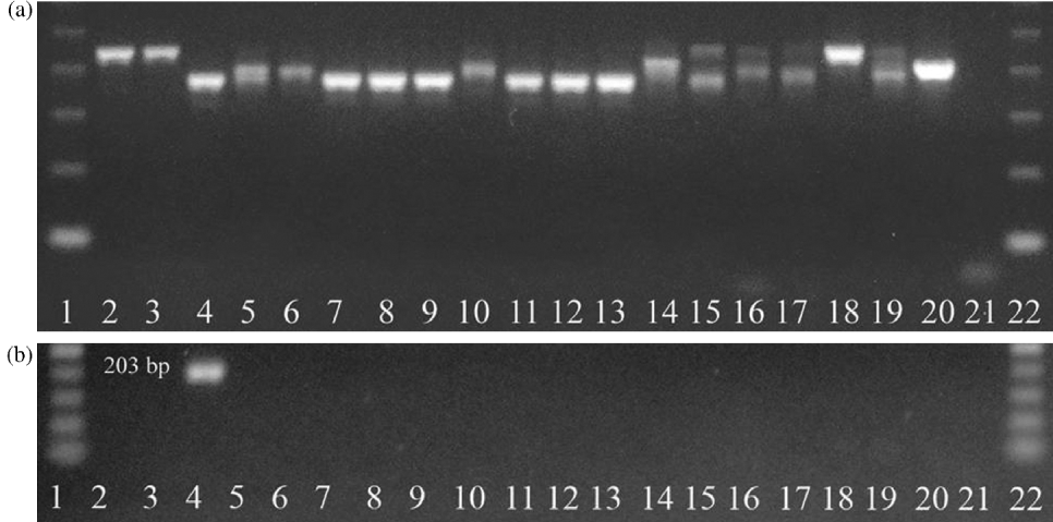
In this study, DNA samples were extracted from single, male adult specimens of C. curticei, C. punctata and C. spatulata identified based on the spicule morphology (fig. 1). Cooperia curticei, C. punctata and C. spatulata DNA samples were amplified using the Hel primer pair. A 946 bp amplification product (data not shown) was then sequenced from six C. curticei specimens, and the species-specific primer pair CcurticeiF/R was designed, resulting in the amplification of a 203 bp PCR product. Additionally, PCR reactions using the primers SSUAF and SSU22R (Dorris et al., Reference Dorris, Viney and Blaxter2002) were performed to amplify the small subunit (SSU) rDNA gene segment in all DNA samples, including the two hosts (bovine and ovine) and 17 species of helminths, such as C. curticei, C. punctata and C. spatulata (fig. 2a). To verify the CcurticeiF/R primer pair specificity, PCR reactions with the same DNA samples were performed and a 203 bp band was observed only in lane 4 (fig. 2b), which corresponds to the C. curticei DNA sample.
Spicules of (a) Cooperia curticei, (b) C. punctata and (c) C. spatulata used for DNA extraction.

PCR reactions with (a) the primer pair described by Dorris et al. (2002) and (b) the species-specific primer pair Ccurticei using DNA samples from two hosts and 17 helminths. Lanes: 1 and 22 show 100 and 50 bp molecular markers, respectively (GE Healthcare); 2, ovine and 3, bovine DNA (hosts); 4, Cooperia curticei; 5, C. punctata; 6, C. spatulata; 7, Haemonchus contortus; 8, H. placei; 9, H. similes; 10, Oesophagostomum columbianum; 11, O. radiatum; 12, Trichostrongylus axei; 13, T. colubriformis; 14, Trichuris discolour; 15, T. globulosa; 16, Bunostomum phlebotomum; 17, B. trigonocephalum; 18, Moniezia spp.; 19, Paramphistomum spp.; 20, Fasciola hepatica; and 21, reagents without DNA.
It was possible to verify the identification of the Cooperia genus among the different species. The 203 bp band was amplified in all six C. curticei DNA samples (isolate numbers 1, 11, 15, 29.1, 34.1 and 34.2) (fig. 3, lanes 4–9), and there was no amplified band in the C. punctata and C. spatulata DNA samples. The sequences of isolates 1, 11, 15, 29.1, 34.1 and 34.2 are deposited in GenBank (accession numbers: KC167179.1 KC167180.1, KC167181.1, KC167182.1, KC167183.1, KC167184.1, respectively). The multiple sequence alignment of the six isolates indicates no genetic diversity. These sequences were assembled and compared to known sequences by BLAST analysis against the NCBI database, resulting in 97% similarity with 49 C. oncophora partial ITS-1 clones (AJ544290.1–AJ544377.1). Additionally, BLAST analysis showed 99% similarity with the C. curticei sequence JF680982, which is 682 bp long and corresponds to ITS-1, the 5.8S ribosomal RNA gene, ITS-2 and the 28S ribosomal RNA gene. No similarity was found by BLAST analysis with sequence AJ000033, which corresponds to ITS-2. These data suggest that the C. curticei sequence in this study corresponds to the ITS-1 rDNA.
PCR reactions with the species-specific primer pair Ccurticei and DNA samples. Lanes 1 and 11, 100 bp molecular marker (GE Healthcare); 2, ovine and 3, bovine DNA (hosts); 4–9 isolates of Cooperia curticei (isolates 1–34.2); and 10, reagents without DNA.
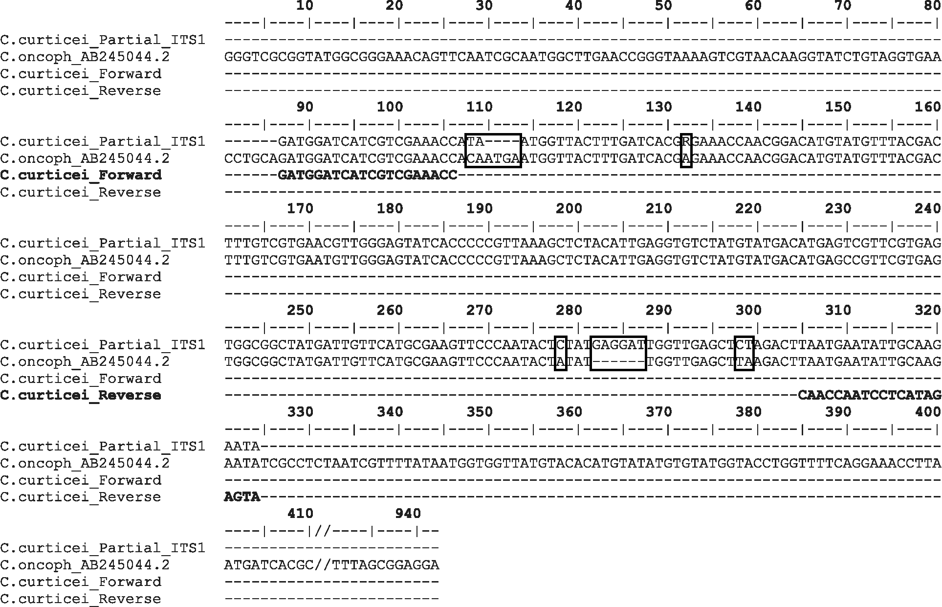
Cooperia oncophora, which does not occur in tropical areas of Brazil, is an economically important nematode in temperate regions of the world and has been the subject of several studies, with significant data available in the NCBI database. A BLAST 2 sequence analysis of 936 bp of C. oncophora (AB245044.2) and 234 bp of C. curticei (part of the 946 bp sequence) that corresponds to the partial ITS-1 was performed and showed 94% identity between these sequences (189/201 bp). The AB245044.2 sequence includes genes for the small subunit rRNA, ITS-1, 5.8S rRNA, ITS-2 and large subunit rRNA. The alignment of the species-specific primer pair CcurticeiF/R (forward and reverse) and the nucleotide sequences of C. curticei (234 bp) and C. oncophora (AB245044.2) is presented in fig. 4. This analysis was performed with the DAMBE program, version 5.3.10 (Xia & Xie, Reference Xia and Xie2001). To emphasize the alignment among these sequences, and because C. curticei is a small sequence (234 bp), the C. oncophora nucleotides from 411 to 931 bp were suppressed. The beginning of this region is marked by two slashes. Additionally, base changes and gaps are indicated in boxes. Hyphens represent the absence of nucleotides, and Ccurticei forward and reverse primers are represented in bold letters. The Ccurticei forward primer shows a perfect alignment to the C. curticei and C. oncophora sequences, while the Ccurticei reverse primer presents with an incomplete alignment to both sequences (fig. 4). As mentioned above, this primer set results in the amplification of a 203 bp band in C. curticei, and according to the sequence similarities, the same PCR amplification product is expected from C. oncophora.
Alignment of the species-specific primer pair Ccurticei forward and reverse (in bold) and the nucleotide sequences for the ITS-1 region of Cooperia curticei (234 bp) and C. oncophora (AB245044.2); base changes and gaps (boxed), absence of nucleotides (- -|- -), and nucleotide suppression from 411 to 931 bp of the AB245044.2 sequence (//).
Discussion
Because agricultural ruminants usually harbour mixed infections, a specific diagnosis is important to analyse the contribution of individual species to the infection. Different species can differ significantly in their fecundity, pathogenicity and susceptibility to classes of anthelmintics. Additionally, the accurate diagnosis of infections is necessary for their treatment and control. The present study shows that it is possible to identify C. curticei from a wide range of parasitic helminths using PCR with a species-specific oligonucleotide primer pair. Because DNA samples can be obtained from either eggs or larvae, this procedure will facilitate studies on the biology, ecology and epidemiology of this species. Although coproscopic methods have been traditionally used to diagnose the infections by gastrointestinal nematodes, eggs from different species of strongylids are morphologically indistinguishable by conventional microscopic examination. Larval culture methods are time consuming and usually do not allow for a specific identification, which occurs when different Cooperia species simultaneously infect the same animal. The results obtained with C. curticei are promising, and with the development of specific primer pairs to identify other Cooperia species, there will be no need for the morphological identification of adult specimens. Therefore, animal necropsies for obtaining nematode specimens will no longer be necessary. According to Roeber et al. (Reference Roeber, Jex, Campbell, Campbell, Anderson and Gasser2011), DNA-based methods have provided accurate, reproducible and rapid diagnostic results. PCR methods are useful for specific diagnoses because they rely on the specific enzymatic amplification of genomic DNA.
Molecular methods using PCR primer pairs have been able to identify nematode genera successfully (Schnieder et al., Reference Schnieder, Heise and Epe1999; Von Samson-Himmelstjerna et al., Reference Von Samson-Himmelstjerna, Harder and Schnieder2002). However, the present study is the first to present a species-specific primer pair to identify C. curticei. The present results also indicate that the same primer pair may be useful for the diagnosis of C. oncophora in cattle raised in temperate areas of the world. Nevertheless, this information must be confirmed with additional studies. Although all the C. curticei isolates in this study did not show genetic diversity, it is important to note that they were all obtained from the same region (the state of Sao Paulo). Additional studies are necessary to verify the existence of intraspecific sequence variation of the primer sites of C. curticei with specimens obtained from different regions.
Acknowledgements
This study was supported by the Fundação de Amparo à Pesquisa do Estado de São Paulo (FAPESP, grant number 07/07182-6). C.C.B., J.H.d.N. and A.F.T.A. received financial support from FAPESP (grant number 2010/18678-5), Coordenação de Aperfeiçoamento de Pessoal de Nível Superior (CAPES) and Conselho Nacional de Desenvolvimento Científico e Tecnológico (CNPq, grant number 302248/2009-9), respectively.