Introduction
Embolisation of vascular tumours of the head, neck and central nervous system has become an important option prior to surgical treatment, as it facilitates tumour removal.1 The introduction of this procedure has resulted in reduced morbidity and mortality of patients with such tumours. However, despite the good results obtained with embolisation of head, neck and central nervous system tumours, there is still only an extremely limited number of publications on the application of selective embolisation of thyroid arteries as a pre-resective treatment for primary thyroid tumours.Reference Ramos, Braga-Basaria, Haquin, Mesa, de Noronha and Sandrini2, Reference Dedecjus, Tazbir, Kaurzel, Lewinski, Strozyk and Brzezinski3
We describe the case of a patient with a large papillary thyroid cancer who was treated with pre-operative selective embolisation of the thyroid arteries followed by total thyroidectomy. We also review previous papers to discuss the efficacy of embolisation of thyroid cancer.
Case report
A 29-year-old man with a history of a large, painless, slowly growing cervical mass was referred to the otolaryngology department of the University of Toyama.
On physical examination, he had a large, firm, immobile, binodal neck tumour that occupied the entire anterior cervical area. The diameter of each of the nodes was approximately 6 cm, making the major axis of the tumour approximately 12 cm wide, as detected by palpation. Examination of the cervical lymph nodes was hindered by the presence of the large tumour.
Serum blood tests revealed normal thyroid function test results, with a thyrotropin (thyroid-stimulating hormone) level of 1.77 µIU/ml (normal range, 0.35–3.73 µIU/ml), free tri-iodothyronine level of 3.9 pg/ml (normal range, 2.2–4.1 pg/ml) and free thyroxine level of 1.5 ng/dl (normal range, 0.9–1.8 ng/dl). However, the patient's serum thyroglobulin level was extremely high, at 3396 ng/ml (normal range, 0–32.7 ng/ml).
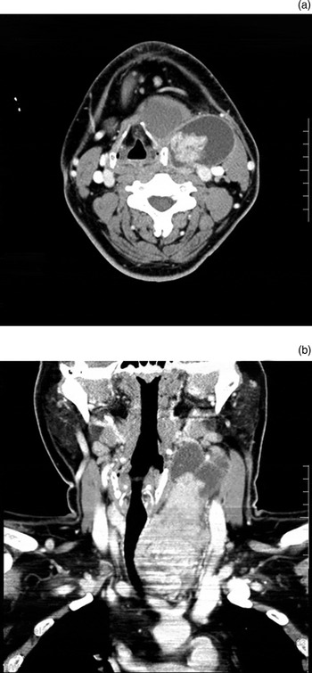
A computed tomography scan of the neck and thorax revealed the presence of a large tumour that possibly originated from the left lobe of the thyroid gland (Figure 1). The tumour extended to the mediastinum and submandibular region, and compressed the larynx and trachea to the right. The upper quarter of the tumour was cystic while the lower three-quarters was solid. The solid part was enhanced after contrast medium application, indicating a substantial vascular supply.
Enhanced computed tomography scans of the neck and thorax: (a) axial view and (b) coronal view. Note that the lower three-quarters of the large tumour is solid and enhancing, indicating a substantial vascular supply.

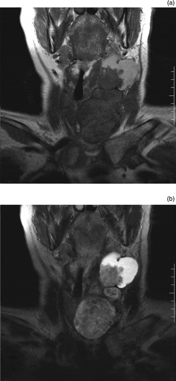
In a magnetic resonance imaging scan of the neck and chest, T1- and T2-weighted images of the upper, cystic part of the tumour showed an area of high signal intensity, indicating storage of blood (Figure 2).
Coronal magnetic resonance imaging scans of the neck and thorax: (a) T1-weighted image and (b) T2-weighted image. Note that the upper, cystic part of the tumour shows an area of high signal intensity indicating the presence of stored blood.

In order to reduce thyroid vascularisation and hence diminish intra-operative blood loss, we planned pre-operative selective embolisation of the thyroid arteries. After being informed of the risks and possible complications related to this radiological intervention procedure and to surgery, the patient gave his consent.
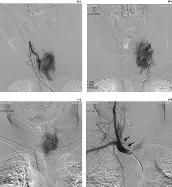
Selective angiography of both superior thyroid arteries and the left inferior thyroid artery was performed by a qualified radiologist using Seldinger's technique. Briefly, a 4-F size diagnostic angiographic catheter (angiocatheter; Create Medic, Yokohama, Japan) was advanced via the aorta to the bilateral external carotid and subclavian arteries. Then, a 2-F size microcatheter (Sirabe Pure Selective; Piolax Medical Devices, Yokohama, Japan) was passed through the 4-F size catheter and advanced to the superior and inferior thyroid arteries. Before the embolisation, contrast medium (Proscope; Mitsubishi Tanabe Pharma, Osaka, Japan) was injected into the vessels, thus allowing visualisation of the arteries and the regions of the thyroid tumour to which they supplied blood. After contrast medium injection, the intense enhancement of both superior thyroid arteries and the left inferior thyroid artery showed that these arteries provided substantial blood supply to the tumour (Figure 3a to 3c).
(a – c) Angiographic studies recorded during selective thyroid artery embolisation. Pre-embolisation studies show intense enhancement after contrast medium is injected into (a) the right superior thyroid artery, (b) the left superior thyroid artery and (c) the left inferior thyroid artery. (d) Post-embolisation angiographic study of the brachiocephalic artery; note that the pre-embolisation intense enhancement of the right superior thyroid artery had disappeared, but that the right inferior thyroid artery (arrows) is revealed.

To prepare the gelatin sponge particles used for embolisation, thick gelatin sponge sheets (Spongel; Astellas Pharma, Tokyo, Japan) were cut with scissors into cubes of approximately 1 × 1 mm and immersed in a small amount of contrast medium. These embolic agents were then slowly injected via a syringe into the three identified blood vessels, until the radiologist confirmed stasis of the bloodstream. After selective embolisation of the thyroid arteries, an angiogram of the brachiocephalic artery showed that the right inferior thyroid artery was patent (Figure 3d). Selective angiography of the three embolised vessels was not performed again, to avoid the perfused contrast medium dislodging the embolised granules and creating the risk of an iatrogenic embolic cerebral infarction. No immediate complications developed after the radiological procedure.
During the 3 days after interventional embolisation, the patient developed a mild fever of up to 37.6°C and complained of mild anterior neck pain, but he did not require nonsteroidal anti-inflammatory drugs. There was no reduction in tumour size as detected by palpation.
Four days after embolisation, the patient underwent total thyroidectomy. Dissection of the tumour surface from the surrounding tissue was time-consuming because of tight, vascularised adhesion. The thyroid tumour was completely resected from the pre-thyroid muscles, without any need for blood transfusion. Bilateral central (level VI) and left lateral (level III–IV) neck dissections were subsequently performed. During surgery, the total volume of blood loss was 775 ml. No blood transfusion was required during or after surgery. The mass removed weighed 324 g and measured 12 × 10 × 8 cm. The final diagnosis was a papillary thyroid carcinoma with regional lymph node metastasis.
Discussion
Pre-operative selective embolisation of the thyroid arteries involved in thyroid cancers has rarely been described as a strategy for decreasing surgical blood loss, facilitating tumour removal or reducing surgical complications. We found only two previously published papers reporting similar experiences.
Ramos et al. reported a case of pre-operative selective embolisation of the thyroid arteries involving a patient with a large, multinodular, cervico-mediastinal goitre with a papillary carcinoma.Reference Ramos, Braga-Basaria, Haquin, Mesa, de Noronha and Sandrini2 Seven days before surgery, selective embolisation of three of the four thyroid vessels was performed (i.e. both superior thyroid arteries and one inferior thyroid artery) using polyvinyl alcohol particles ranging in size from 150 to 300 µm, suspended in an emulsion with Histoacryl® particles. This resulted in a significant reduction in tumour size. During surgery, the manipulation and removal of the tumour, which measured 9 × 6 × 5 cm and weighed 250 g, were achieved easily.
Dedecjus and colleagues' Polish study presented 13 patients with medium-sized, differentiated carcinomas (with tumour (T) staging of T3 in six patients and T4 in seven).Reference Dedecjus, Tazbir, Kaurzel, Lewinski, Strozyk and Brzezinski3 These authors performed pre-resective selective embolisation of three thyroid vessels with polyvinyl alcohol, with particle sizes ranging from 150 to 750 µm. In addition, the superior thyroid arteries were embolised using a non-magnetic wire coil with synthetic fibres of appropriate size, depending on the diameter of the arterial lumen in question. Patients underwent thyroidectomy up to 6 days after selective embolisation of the thyroid arteries. These patients' operating times, intra-operative blood loss and drainage required in the first 24 post-operative hours were significantly decreased, compared with selected patients with similar thyroid volumes who had not received pre-operative selective thyroid artery embolisation.
• This patient with a large, vascular thyroid cancer underwent pre-operative selective thyroid artery embolisation
• Both superior thyroid arteries and only one inferior thyroid artery were embolised, to prevent hypoparathyroidism
• Results suggest that this minimally invasive procedure is effective for such tumours
In our study, we performed pre-operative selective embolisation of three thyroid arteries using gelatin sponge particles. Although dissection of the tumour from the surrounding tissues was time-consuming because of tight, vascularised adhesion, the large, vascular thyroid tumour was completely resected without any blood transfusion. This suggests that pre-operative selective embolisation of the thyroid arteries resulted in a significant reduction in tumour blood perfusion.
The thyroid gland has two superior arteries and two inferior arteries. More than 70 per cent of the blood supply comes from the two superior arteries.Reference Ramos, Braga-Basaria, Haquin, Mesa, de Noronha and Sandrini2 Because there is communication between both the superior and inferior arteries, embolisation of only the superior arteries has been suggested as sufficient to destroy most of the gland. However, in previous work and in our own case, pre-operative selective embolisation was performed on both superior thyroid arteries and one inferior thyroid artery, leaving a single inferior thyroid artery.
It has been previously recommended that both superior thyroid arteries and one of the inferior thyroid arteries should be embolised.Reference Xiao, Zhuang, Wang, Yu, Chen and Zhou4, Reference Tartaglia, Salvatori, Pichelli, Sgueglia, Blasi and Custureri5 The reason for this is that the parathyroid glands are mainly supplied by the inferior thyroid arteries, and occlusion of both inferior arteries might lead to hypoparathyroidism.Reference Miller6, Reference Nobori, Saiki, Tanaka, Harihara, Shindo and Fujimoto7 Indeed, Zhao et al. have reported the case of a female patient with Graves' disease who underwent embolisation of one superior thyroid artery and both inferior thyroid arteries, and who developed serious temporary hypoparathyroidism but recovered one week later.Reference Zhao, Gao, Yang, Li, Dong and Xiang8 Approximately one week is necessary to establish collateral circulation to the parathyroid glands and thereby recover normal secretion. Beers et al. reported the case of a male patient with medullary thyroid cancer who underwent embolisation of the left inferior thyroid artery one week after embolisation of the right inferior thyroid artery, and who did not develop any hypoparathyroidism.Reference Beers, Svendsen, Carter and Bell9 In the present study, as in the previous two studies performing selective embolisation of the same three arteries, there were a few minor complications, including fever and neck pain.
Conclusion
The reported case suggests that pre-operative selective embolisation of the thyroid arteries may be an effective, safe procedure for patients with large, vascular thyroid tumours.
Acknowledgement
We express our sincere appreciation to Dr Gakuto Tomizawa, Dr Hideto Kawabe and Hiroko Shojaku at the Department of Radiology, University of Toyama, and to Dr Hirohiko Tachino at the Department of Otolaryngology, University of Toyama, for their assistance and cooperation.