Since Kawasaki disease was first described 40 years ago, many studies have focused on coronary arterial lesions.Reference Iemura, Ishii, Sugimura, Akagi and Kato1–Reference Suzuki, Kamiya, Arakaki, Kinoshita and Kimura3 However, few pathological studies have examined the after-effects of Kawasaki disease on the myocardium using endomyocardial biopsy.Reference Yonesaka, Takahashi and Matubara4, Reference Yutani, Okano and Kamiya5
The long-term natural history of patients with Kawasaki disease with or without coronary artery lesions remains unknown. Conventional investigations such as coronary arteriography, two-dimensional echocardiography, and myocardial scintigraphy might not completely identify all Kawasaki patients at risk of future myocardial lesions or those who previously had lesions. Most patients with aneurysms related to Kawasaki disease, even those with giant aneurysms, without significant stenotic lesions, generally remain asymptomatic.
Our previous studies revealed residual myocardial damage in Kawasaki patients with coronary artery lesions even at the remote stage.
Considering these findings, we investigated long-term silent myocardial lesions in light and electron microscopy of endomyocardial biopsies from Kawasaki patients with giant coronary aneurysms.
Patients and methods
Patients
The study group comprised 16 consecutive Kawasaki patients with giant coronary aneurysms who were referred to Hirosaki University Hospital before April, 2006. They ranged from 2 to 12 years of age at the initial study and 4.9 to 16 years in the subsequent study, and the male-to-female ratio was 12:4. The interval between the onset of disease and the first cardiac catheterisation ranged from 2 months to 2.3 years with a median of 1.1 years, and that between the initial and the subsequent studies ranged from 1.2 and 10.8 years with a median of 3.3 years.
In the initial and subsequent studies, none of the patients with giant coronary aneurysms had total obstruction, whereas four developed obstruction during follow-up. However, the endomyocardial biopsies in this study were assessed before the obstructions appeared.
Kawasaki disease was diagnosed according to the guidelines of the Japan Kawasaki Disease Research Committee. We defined giant coronary aneurysms when the maximal diameter of coronary artery was larger than 8 millimetres either by two-dimensional echocardiography or by selective coronary arteriography.Reference Kawasaki6
Methods
Written informed consent for cardiac catheterisation, including selective coronary arteriography and endomyocardial biopsy, was obtained from the parents of each child in accordance with hospital policies.
All patients with Kawasaki disease underwent routine cardiac catheterisation and selective coronary arteriography when endomyocardial biopsy specimens were obtained from the apex of the right ventricle.
Specimens were histomorphometrically evaluated. The fraction of myocardial volume occupied by fibrillar collagen (%fibrosis) and myocyte diameter was determined by quantitative morphometry, using the automated Mac Scope® image analysis system (Mitani Corporation, Tokyo, Japan) in sections stained with Azan. The sections were assessed histopathologically according to the diagnostic guidelines of the Pathological Branch of the Idiopathic Cardiomyopathy Research Committee of the Ministry of Health and Welfare of the Japanese GovernmentReference Sekiguchi, Hiroe and Moritomo7 as 0, normal; 1 plus, mildly; 2 plus, moderately; and 3 plus, severely abnormal. We regarded more than 1 plus as significant. Inflammatory cell infiltrates were scored positive only in the presence of more than five inflammatory cells, namely lymphocytes, plasma cells or polymorphonuclear leucocytes per high-power field (×400).Reference Yonesaka and Becker8 Microangiopathy, mitochondrial abnormalities, and myofibril disarray were also compared by electron microscopy. We defined microangiopathy as the presence of microvascular dilation, red thrombus formation, narrow or occlusive lumens, and thickened arteriolar walls in intramural capillaries, small arteries, and arterioles. Histopathological findings were evaluated with special reference to baseline characteristics, such as patient age, duration of fever, left ventricular systolic function, severity of coronary involvement, and time since Kawasaki disease to biopsy.
During the acute phase of the disease, 7 of the 16 patients were administered with intravenous immunoglobulin for 5 days – 200 milligrams per kilogram of body weight per day in six patients; 400 milligrams per kilogram of body weight per day in one patient – at the acute phase of the disease, that is, 5th–10th day of illness. All patients received aspirin –30–80 milligrams per kilogram of body weight – or flurbiprofen – 2–5 milligrams per kilogram of body weight – daily during the acute phase with or without intravenous immunoglobulin.
All patients, except no. 6, received aspirin – 5–10 milligrams per kilogram of body weight – and dipyridamole – 3–5 milligrams per kilogram of body weight – daily as anticoagulants in the subsequent course. One patient, no. 6, was given warfarin.
Continuous variables are reported as means plus or minus standard deviation and comparisons between groups are based on Student’s t-test. Categorical variables are summarised as percentages and group comparisons are based on Fisher’s exact test using StatView 5.0 (SAS Institute Inc., Cary, North Carolina, United States of America) and considered significant when the p-value was less than 0.05.
Results
Baseline characteristics of Kawasaki disease with giant coronary aneurysms
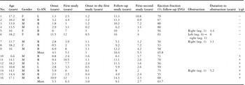
Baseline characteristics of the enrolled patients are shown in Table 1. They ranged from 2 to 12 years of age at the initial study and 4.9 to 16 years in the subsequent study, and the male-to-female ratio was 12:4. The interval between the onset of disease and the first cardiac catheterisation ranged from 2 months to 2.3 years with a median of 1.1 years, and that between the initial and the subsequent studies ranged from 1.2 and 10.8 years with a median of 3.3 years. In the study, the mean age of the patients treated without intravenous immunoglobulin was 15 years with a range from 10.2 to 18.2 years, and it was 13.6 years with a range from 6.6 to 18.2 years in those treated with intravenous immunoglobulin. The mean interval between the onset and the first examination was 1.0 with a range from 0.2 to 1.5 years and 1.0 with a range from 0.1 to 2.4 years in the patients without intravenous immunoglobulin and those treated with intravenous immunoglobulin, respectively. The size of giant coronary aneurysm ranged from 8 millimetres to 24 millimetres, and the site of giant coronary aneurysm was located on the left in four Kawasaki patients, right in six and bilateral in six patients. The left ventricular ejection fraction was 53–80% (with mean of 64%) in all Kawasaki patients and 63% in patients with bilateral giant coronary aneurysm, 64% in those with a right aneurysm, and 69% in those with a left aneurysm. There was one patient with low left ventricular ejection fraction, that is, less than 55%. There were no statistically significant differences in age at onset, age at the first and second examination, male gender, site of giant coronary arteries, and duration of follow-up, left ventricular ejection fraction between the patients with and without intravenous immunoglobulin. The time interval between the two biopsies was shorter in the patients treated with intravenous immunoglobulin than those without intravenous immunoglobulin, p-value was less than 0.05.
Clinical data of Kawasaki disease with giant coronary aneurysms.

γ-gl, intravenous gamma globulin administration (No. 10–15, 200 milligrams per kilogram of body weight per day × 5 days; No. 16, 400 milligrams per kilogram of body weight per day × 5 days); B, bilateral; G-AN, giant coronary aneurysm; L, left; LAD, left anterior descending artery; LVG, left ventriculography; obstruction, site of coronary artery obstruction; R, right; Seg., segment number according to the American Heart Association classification
Changes in the giant coronary aneurysms during the follow-up period
Giant coronary aneurysms progressed in the subsequent study to total obstruction in four patients, and remained unchanged in 12 of the 16 patients with Kawasaki disease. Four patients with total coronary artery obstruction were asymptomatic when the obstruction was discovered. Patient no. 6 developed bilateral coronary obstruction, and was given warfarin, aspirin, and dipyridamole after the onset of the disease. The histopathological study was undertaken before the obstruction of the giant coronary aneurysms was determined.
Myocardial changes in Kawasaki patients with giant coronary aneurysms at initial and follow-up studies
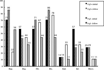
Histopathological findings in endomyocardial biopsy specimens were varied both at initial and follow-up studies (Fig 1).
Myocardial changes of Kawasaki patients with giant coronary aneurysms at initial and follow-up study. Hypertrophy, degeneration, and inflammatory cell infiltration are frequent in patients with intravenous immunoglobulin at initial study. Higher incidence of inflammatory cell infiltration, interstitial fibrosis, and disarray persist at follow-up endomyocardial biopsies, although statistical significance was not reached.

Hypertrophy, degeneration, and inflammatory cell infiltration were frequent in the patients with intravenous immunoglobulin therapy during the initial study. A higher incidence of inflammatory cell infiltration, interstitial fibrosis, and disarray tended to persist on follow-up endomyocardial biopsy, although the values did not reach statistical significance. The incidence of microangiopathy was 20% in initial and follow-up biopsies.
Comparison of the percentage of fibrosis in Kawasaki patients with giant coronary aneurysms at initial and follow-up endomyocardial biopsy
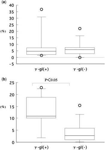
The percentage of fibrosis was similar in all patients with Kawasaki disease treated with or without intravenous immunoglobulin at initial endomyocardial biopsy (Fig 2a), but was significantly increased in those treated with intravenous immunoglobulin at follow-up endomyocardial biopsy, p-value was less than 0.05 (Fig 2b). The value of the percentage of fibrosis was 5.9% in the patients with bilateral giant coronary aneurysm, 12.7% in those with right aneurysms, and 8% in those with left aneurysms. There was no statistical correlation between the percentage of fibrosis and left ventricular ejection fraction. In addition, we could not find any correlation between the percentage of fibrosis and the site of the giant coronary aneurysm.
Comparison of %fibrosis in Kawasaki patients with giant coronary aneurysms at initial (a) and follow-up (b) studies. Area of fibrous tissue (%fibrosis) is significantly larger in patients with intravenous immunoglobulin on follow-up endomyocardial biopsies (p less than 0.05).

Histopathological changes in patients with Kawasaki disease with giant coronary aneurysms at follow-up endomyocardial biopsy
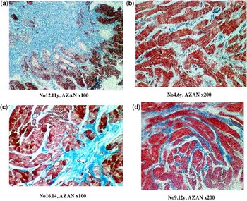
The various histopathological abnormalities in the follow-up endomyocardial biopsy specimens from patients with giant coronary aneurysms were evident, which comprised severe interstitial fibroses, vacuolar degeneration of myocytes, disarray, and branching abnormalities of the myocytes (Fig 3a–d).
Histopathological abnormalities in endomyocardial biopsy specimens from patients with giant coronary aneurysms at follow-up study. (a) Severe fibrosis and vacuolar degeneration of myocytes. (b) Disarray and interstitial inflammatory cell infiltrations with fine fibrosis. (c) Perivascular fibrosis and vacuolar degeneration. (d) Fine interstitial fibrosis, hypertrophy, and branching abnormalities of myocytes.

Ultrastructural changes in some patients with Kawasaki disease with giant coronary aneurysms at follow-up endomyocardial biopsy
Ultrastructural changes, such as myofibril lysis and mitochondrial alterations, indicated myocyte degeneration, interstitial fibrosis, and myofibril disarray.
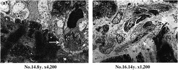
Microangiopathies included microvascular dilation, relatively old thrombus comprising degenerated platelets with a few fibrotic areas attached to damaged endothelial cells in microvessels (Fig 4a), and capillaries with swollen endothelial cells (Fig 4b).
Electron micrographs of endomyocardial biopsies from patients with giant coronary aneurysms at follow-up study. (a) Thrombus comprises degenerated platelets and a few fibrotic areas (white arrow) attached to injured endothelial cells of microvessels, 7-year-old boy with bilateral giant coronary aneurysms (no. 14). (b) Fibro-oedema in interstitium and capillaries with swollen endothelial cells (white arrow), 14-year-old boy with bilateral giant coronary aneurysms (no. 16).

Discussion
Kawasaki disease is an acute generalised self-limiting vasculitis of small- and medium-size arteries of unknown aetiology that mainly affects infants and children. Coronary arteries can be involved with aneurysms, thrombotic occlusion, acute coronary syndrome, and sudden death. The myocardium is damaged in many patients with Kawasaki disease, regardless of the presence or absence of coronary artery lesions during the acute stage.Reference Yutani, Okano and Kamiya5, Reference Yonesaka, Takahashi and Furukawa9 However, long-term pathological changes in the myocardium have not been precisely described, and there is little understanding of the long-term after-effects on the myocardium following Kawasaki disease with giant coronary aneurysms.
Here, the endomyocardial biopsy revealed persistent histopathological abnormalities such as fibrosis and disarray in patients with long-term Kawasaki disease with giant coronary aneurysms treated with intravenous immunoglobulin. Intravenous high-dose immunoglobulin administered during the acute phase of Kawasaki disease reduces not only the incidence of coronary artery aneurysms but also the severity of myocardial damage from moderate or severe to mild or slight,Reference Yutani, Go and Kamiya10, Reference Haneda and Mori11 whereas fulminant Kawasaki disease sometimes rapidly and extensively progresses during the early phase of the disease despite high-dose gamma globulin administration.Reference Mori, Tomono and Yokota12 We found noticeable myocardial damage among patients treated with or without intravenous immunoglobulin. The sites of coronary aneurysms did not seem to impact the endomyocardial biopsy findings, suggesting that the underlying pathophysiology is related to the original disease process.
As most of our patients with giant coronary aneurysms with a high incidence of myocardial damage did not have significant coronary stenotic lesions, the higher percentage of fibrosis reflected myocardial damage due to advanced vasculitis, resulting in diffuse myocarditis or microvascular disease. Ultrastructural evaluation of Kawasaki disease using endomyocardial biopsy can reveal various types of myocardial damage, such as a post-myocarditis state, hypertrophy, degeneration, mitochondrial abnormalities, fibrosis, and a cardiomyopathy-like state.Reference Liu, Ghazizadeh, Onouchi and Asano13 That study also found a higher incidence of microangiopathy in Kawasaki disease with coronary artery aneurysms than without. The incidence of microangiopathy was 20% in our patients with Kawasaki disease. The mechanism of microangiopathy formation remains unclear, but its presence could indicate more severe vasculitis. The persistence of large coronary artery aneurysms, despite high-dose intravenous immunoglobulin, should also be considered a complication of intramural microangiopathy.
Patients with Kawasaki disease-related aneurysms, even those with giant coronary aneurysms, who have no significant stenotic lesions, generally remain asymptomatic. Regardless of treatment, giant coronary aneurysms that develop over time do not seem to be resolved. The potential risks of developing coronary occlusions and of forming microthrombi within dilated intramural microvessels are high for patients with Kawasaki disease accompanied by giant coronary aneurysms. Onouchi et alReference Onouchi, Hamaoka and Sakata14 have described that the regimen of aspirin and warfarin was effective for the prevention of coronary artery occlusion/recanalisation in patients with giant coronary aneurysms, but the effects on intramural small vessels are less well understood. In our study, one patient, who was treated with warfarin, aspirin, and dipyridamole, developed complete occlusion of bilateral coronary artery. The efficacy of anticoagulation therapy using warfarin for intramural small coronary artery thrombosis has not been determined. In addition, the mechanism of microangiopathies has not yet been clarified. Further investigations should be mandatory to clarify the precise mechanisms and prognosis of these abnormalities and whether the more powerful anticoagulants may be of benefit to prevent occlusion of intramural small coronary artery thrombosis.
Recently, a single-averaged, non-invasive electrocardiogram has been used to assess the myocardial sequelae at the late stage of Kawasaki disease.Reference Takeuchi, Matsushita, Kurotobi, Sano, Kogaki and Ozono15, Reference Genma, Ogawa, Zhang and Yamamoto16 However, it is not easy to assess myocardial ischaemia or old myocardial infarction in Kawasaki disease only by non-invasive, objective, and convenient methods. In our study, although we could not evaluate the non-invasive cardiac function, left ventriculography was performed from the right anterior oblique view at follow-up study, and therefore, mild regional abnormalities might have been detected. However, we could not detect significant left ventricular wall motion abnormalities but only found mild global function impairment of the left ventricle in one case. There was not significant correlation between histopathology and left ventricular ejection function. Although whether abnormalities are due to direct myocardial injury, chronic ischaemia, repeated small-vessel thrombosis, or other problems associated only with endomyocardial biopsies is difficult to determine, endomyocardial biopsies could be useful for clarifying myocardial sequelae. In fact, endomyocardial biopsies revealed residual abnormal myocardial lesions in our subgroup with giant coronary aneurysms. In this study, we could not find evidence that myocardial abnormalities resulted in a remarkable left ventricular dysfunction. However, as a consequence of the myocardial changes, it is suggested that some cases of Kawasaki disease may end up as cardiomyopathy.Reference Yutani, Go and Kamiya10 Follow-up of these patients should be more aggressive to identify myocardial lesions that could exist even in asymptomatic patients.
Study limitations
This was not a case–control study and the cohort was very small. Therefore, more Kawasaki disease patients with giant coronary aneurysms treated with intravenous immunoglobulin should be investigated. In addition, cardiac function and changes in coronary artery lesions should be further evaluated by continuous long-term anticoagulation therapy. Endomyocardial biopsy was not read in a blinded manner for histopathological testing and there was no controlled histopathological study. Another limitation of this study is the lack of histological evaluation for autopsied cases.
Nevertheless, we believe that our study provides a realistic sample of patients referred for long-term myocardial histology in the Kawasaki patients with giant coronary aneurysms, because the long-term sequelae of the myocardium are uncertain and remain obscure.
Conclusion
We described the myocardial damages in Kawasaki disease with giant coronary aneurysms proven by biopsy and treated with and without intravenous immunoglobulin. Our findings seem to reflect the severity of the inflammatory processes in Kawasaki disease with giant coronary aneurysms regardless of intravenous immunoglobulin therapy.
As most cases of Kawasaki disease with coronary aneurysms are free of symptoms, clinicians need to be aware of the potentially serious myocardial association in some Kawasaki cases with giant coronary aneurysms.
Further investigations are necessary to determine whether the myocardial damages will persist or progress to a cardiomyopathy-like state.