

Introduction
Two-fifths of the world population lives in areas at-risk of acquiring dengue virus infection. These single-stranded RNA viruses (of the family Flaviviridae) are the most common cause of arboviral diseases that are transmitted by the bite of the Aedes mosquito.1,2 Dengue fever has a long association with suffering in humans, but only in recent years has it come to be recognized as a major public health concern. Dengue fever has emerged as a global epidemic—it is present in >100 tropical and subtropical nations. The first epidemic of dengue hemorrhagic fever (DHF) in recent times occurred in Manila, Philippines in 1953. In India, while the virus was first isolated in Kolkata in 1945, it was recognized as a major health problem following the first outbreak of DHF in 1963 in the same city. Rapid industrial and economic development over the previous few decades has brought about a massive infrastructural shift, and has an ideal environment for breeding of mosquitoes. In a recent epidemic, 182 districts across India reported cases of dengue fever, several resulting in a fatal outcome.3
Despite rapid advancement of knowledge in the last decade, there is no clinical or laboratory test that identifies the illness with a reasonable degree of reliability and accuracy. While the diagnosis most often is based on experiential assessment of the clinical presentation, the diagnosis often must be supported by serological evidence. In addition, the disease may be affected by the season of the year at time.4
Much has been said on the role of sonography in dengue and dengue hemorrhagic fever.4–16 Ultrasonographic examination has been reported to reveal ascites, hepatomegaly, splenomegaly, gall bladder wall edema, perihepatic fluid collection, pericholecystic fluid collection, perinephric fluid collection, acalculous cholecystitis, sludge in gall bladder, pleura-pericardial fluid collections, and pancreatic enlargement. However, several issues regarding the radiographic findings of the disease have not yet been clearly understood with relation to clinical and serological findings.
The current study was undertaken to analyze retrospectively the ultrasonographic (USG) features of patients presenting with probable dengue fever during the outbreak of dengue fever of 2006 in North India. Ultrasound has been used to determine the sonolographic profile of patients and assess if USG could be used as an adjunct to clinical and laboratory evaluation as a diagnostic marker for sero-positive dengue fever or a prognostic marker to determine a poor outcome defined by renal dysfunction. Further, an attempt was made to determine the correlation between clinico-radio-serological findings in subjects presenting with probable dengue fever.
Methods
Patient Selection and Classification
The current study was conducted in a 200-bed hospital in the north Indian city of Ghaziabad, India during the outbreak of dengue infection in 2006. Case records of a total of 169 patients who had presented with short febrile illness and thrombocytopenia during the time as other confirmed cases of DF and had undergone a sonographic examination were retrospectively included in the study as cases of probable dengue fever.(17) The patients with known hematological disorders or systemic illnesses causing thrombocytopenia were excluded from the study. Data were collected pertaining to history, clinical features, immunological status, and laboratory parameters of the patients.
All ultrasound examinations had been performed in fasting state by a trained radiologist (MM), using a Toshiba Nemio 30 machine using 3.5 MHz and 7.5 MHz probes. A total of 10 individual sonographic parameters were reviewed: (1) ascites; (2) hepatomegaly; (3) splenomegaly; (4) gallbladder wall edema (GBWE); (5) right pleural effusion; (6) left pleural effusion; (7) pleural effusion on both sides; (8) pericardial effusion; (9) pericholecystic collection; and (10) perinephric collection. Subjects who had gallbladder wall thickness >3 mm as measured by ultrasound were identified as positive for GBWE. Gallbladder wall thickness had been measured by placing the calipers between the two layers of anterior wall, while thoracic scanning was done in supine posture and both pleural spaces were evaluated using an intercostal approach. The pericardial space was evaluated using a subcostal approach. The radiologists were unaware of the serological status of the patient at the time of sonographic examination.
The demographic data including age and sex was abstracted along with the clinical presence of petechiae as documented in the case-records. The platelet count of the patient at the time of radiological assessment and the presence of renal dysfunction also were abstracted.
The serological status of each of the the patients was reviewed from the case records. Serological tests had been performed using paired sera in all of the patients. The patients were classified into five serological groups based on the serology reports such that there was overlap between the groups. Group I included subjects who had symptoms and signs consistent with DF and were IgM positive—this group was labeled Primary Dengue. Group II included patients who had symptoms and signs of DF and were both IgG and IgM positive and this group was labeled Secondary Dengue Serologically Confirmed. Group III included subjects who had signs and symptoms consistent with dengue fever, but tested IgG positive, some also were IgM positive—this group was called Secondary Dengue including Late Converters. Group IV included subjects who had signs and symptoms consistent with DF and tested positive either for IgG or IgM or both—this group was called All Serologically Positive Dengue irrespective of nature of positivity. Group V included subjects who presented with signs and symptoms consistent with dengue fever, but tested negative on serology—this group was called Sero-Negative Probable Dengue Fever.
Statistical Processing
After computation of frequency distributions, means, and standard deviations (SD) for various parameters, the ultrasound features were assessed for association with clinical and laboratory parameters. Independent samples t-test, and chi-square tests were used to compare means and test for statistical significance using SPSS version 11.0 (SPSS Inc, Chicago, IL, USA).
Results
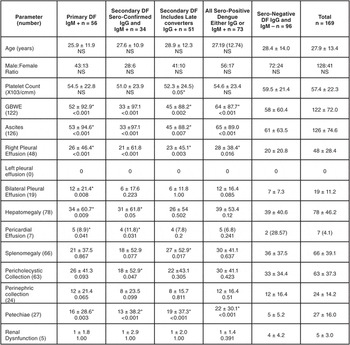
A total of 169 subjects were retrospectively included in the study of whom 56 were Primary Dengue Fever positive for IgM antibodies against dengue virus, 34 were Sero-positive Secondary Dengue Fever positive for both IgG and IgM. The distribution of subjects to different groups along with their clinical and radiological findings is in Table 1. The mean of the ages of the subjects was 27.9 + 13.4 years; minimum 3, maximum 75 years). The mean value of the platelet counts was 57.4 ± 22.3 x 10³/cmm (minimum 11.0, maximum 99.0 x 10³/cmm)
Distribution of study subjects. Data are presented as number (percentage) or mean +1 Standard Deviation and p-value for significance. Platelet count has been expressed as (x 103/cmm)

(GBWE = gallbladder wall edema; NS = not statistically significant)
The most common ultrasonographic feature found among subjects presenting with DF was ascites (126, 74.6%) followed by GBWE (122, 72%), hepatomegaly (78, 46.2%), splenomegaly (66, 39.1%) and pericholecystic collection (63, 37.3%); 48 (28.4%) demonstrated evidence of pleural effusion on the right side, and 19 (11.2%) had bilateral effusions. None of the subjects had an isolated left pleural effusion. Twenty-seven (16%) reported bleeding manifestations in the form of petechiae, and five (3%) developed acute renal dysfunction. Subjects who developed renal dysfunction had significantly lower platelet count (36.0 ± 13.25 x 10³/cmm) than those who did not (58.0 ± 22.3 x 10³/cmm). Those who had a perinephric collection seen on USG had a greater chance of developing renal dysfunction (Likelihood Ratio, 11.48; sensivity, 0.16; specificity, 0.99; p <0.01).
Significant association was noted between the presence of sonographic findings of ascites, GBWE, pleur-pericardial effusion and hepatomegaly as well as clinical presence of petechiae and serological positivity for IgM antibodies. The platelet count was found to be statistically significantly lower among subjects who tested positive for IgG (52.3 +/− 24.5 x 10³/cmm) as compared to those who tested negative (59.6 +/− 21.1 x 10³/cmm).
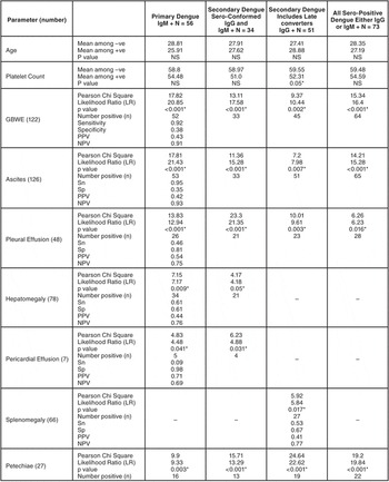
The sonographic findings of ascites (r = 0.325; p <0.001), GBWE >3 mm (r = 0.325; p <0.001), pleural effusion (r = 0.281; p <0.001), hepatomegaly (r = 0.206; p = 0.007) and pericardial effusion (r = 0.169; p = 0.028) had a significant correlation with cases of primary dengue fever cases. However, secondary dengue cases, were significantly associated with the presence of ascites (r = 0.29; p <0.001), GBWE (r = 0.301; p <0.001), and pleural effusion (r = 0.307; p = 0.012). The distribution of the clinical and laboratory markers found significant with relation to serological groups is in Table 2. Presence of pleural (Sensitivity (Sn) = 0.46; Specificity (SP) = 0.81; Positive Predictive Value (PPV) = 0.54; Negative Predictive Value (NPV) = 0.75) and pericardial (Sn = 0.09; Sp = 0.98; PPV = 0.71; NPV = 0.69) effusions were found to be specific while ascites (Sn = 0.95; Sp = 0.35; PPV = 0.42; NPV = 0.93) and GBWE (Sn = 0.92; Sp = 0.38; PPV = 0.43; NPV = 0.91) were identified as highly sensitive markers for seropositive positive dengue fever.
Association of serological positivity with radiological and clinical fi ndings

Discussion
In this retrospective analysis of the case records of 169 subjects who presented with probable dengue fever during the 2006 outbreak in North India, ascites, GBWE >3 mm as detected by non-invasive sonological examination are highly sensitive for dengue fever, and pleural and pericardial effusions are specific markers of DF. Further, subjects who developed renal dysfunction had lower platelet counts and evidence of perinephric collection on sonography.
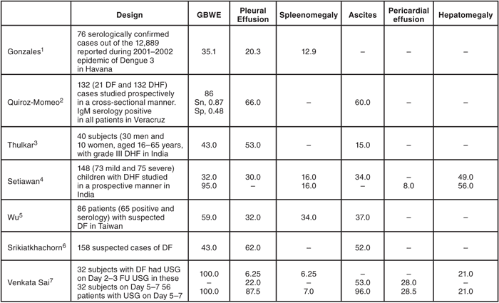
Several authors have reported sonological markers such as the presence of GBWE, pleur-pericardial effusion and hepato-splenomegaly in varying proportions in different parts of the world.4–16Table 3 contains a comparison of the salient findings of some authors who have reported sonographic findings in subjects with dengue fever.
Comparison of studies reporting ultrasound features in dengue fever

Abbreviations: Sn = sensitivity’ Sp = specificity
Data has been presented as percentage. Blanks indicate that the data has not been reported by authors.
1 Gonzalez D, Castro OE, Kouri G, et al.: Classical dengue hemorrhagic fever resulting from two dengue infections spaced 20 years or more apart: Havana, Dengue 3 epidemic, 2001–2002. Int J Infect Dis 2005;9(5):280–5.
2 Quiroz—Moreno R, Mendez GF, Ovando—Rivera KM.: [Clinical utility of ultrasound in the identification of dengue hemorrhagic fever]. Rev Med Inst Mex Seguro Soc 2006;44(3):243–8.
3 Thulkar S, Sharma S, Srivastava DN, et al.: Sonographic findings in grade III dengue hemorrhagic fever in adults. J Clin Ultrasound 2000;28(1):34–7.
4 Setiawan MW, Samsi TK, Wulur H, et al.: Dengue haemorrhagic fever: ultrasound as an aid to predict the severity of the disease. Pediatr Radiol 1998;28(1):1–4.
5 Wu KL, Changchien CS, Kuo CH, et al.: Early abdominal sonographic findings in patients with dengue fever. J Clin Ultrasound 2004:32(8):386–8.
6 Srikiatkhachorn A, Krautrachue A, Ratanaprakarn W, et al.: Natural history of plasma leakage in dengue hemorrhagic fever: A serial ultrasonographic study. Pediatr Infect Dis J 2007;26(4):283–90; discussion 91–2.
7 Venkata Sai PM, Dev B, Krishnan R.: Role of ultrasound in dengue fever. Br J. Radiol 2005;78)929):416–8.
Gallbladder Wall Edema
Changes in gallbladder wall thickness were described by Wang et al in Southern Taiwan in 1990. The thickening resolved completely with clinical recovery.18 Later, Pramuljo et al, who studied 29 children with DHF in Djakarta in 1991, further described these changes.15 In 1995, Setiawan et al proposed that GBWE of >3 mm in DHF indicated a poor prognosis and that that these patients require hospitalization. Furthermore, GBWE ≥≥5.14 mm predicted development of a hypovolemic shock state with sensitivity of 0.94 and specificity of 0.92 in subjects with DHF.13,14 They further noted that patients with ascites had significantly thicker gallbladder walls than did those without. While studying 57 seropositive subjects with dengue fever in Jakarta in 2004, Zulkarnain et al noted that GBWE was associated with the type of dengue virus causing infection, more commonly found in subjects with secondary dengue infection (55%).11 Lee et al noted GBWE in 64.7% of the 644 DHF cases they studied in Southern Taiwan in 2006.5 However, their patients were older with a mean age of 47.5 years as compared to the subjects in the current study (27.8 years of age). In the same year, Quirez-Moreno et al reported that GBWE >3 mm had a sensitivity of 0.87 and specificity of 0.48 in detecting seropositive dengue fever in Veracruz.8 They also reported mild correlation between GBWE >5 mm and presence of acalculous cholecystitis, and free fluid in the abdominal cavity. In the current study, GBWE was present in 122 (72%) of the 169 subjects with DF and is a sensitive marker of the disease (Sn = 0.92) as reported by other authors. Subjects with sero-confirmed secondary dengue fever (33; 97.1%) had GBWE as compared to 52 (92.9%) subjects with primary dengue fever. Further, there was a statistically significant correlation between presence of ascites and GBWE (r = 0.82; p < 0.001).
Ascites
Pramuljo et al noted a common occurrence of ascites, abnormal liver parenchyma, and bilateral pleural effusions in subjects with DF.15 They postulated that the abnormal liver parenchyma could be due to intraparenchymal and subcapsular hemorrhages. Balasubramanian et al proposed that sonography with a sensitivity of 0.91 and a negative predictive value of 0.84 is a good modality to detect plasma leakage in patients with dengue fever in Chennai, India.19 In the current study ascites was present in 126 (74.6%) subjects, and it had a statistically significant positive correlation with the presence of GBWE.
Pleuro-Pericardial Effusions
Several authors have noted that patients commonly present with right pleural or bilateral pleural effusions, but never with isolated left pleural effusions.9,15 However, Venkata Sai et al conducted serial ultrasounds on 32 patients with dengue in Chennai reported left pleural effusions, and noted that the time of performance of USG may influence the findings.4 It is unclear from their report whether their subjects had isolated left or this finding was a part of a bilateral pleural effusion. More recently, Srikiatkhachorn et al reported that pleural effusions occurring in 62% of 158 subjects with suspected dengue fever were the most common ultrasonographic evidence of plasma leakage starting two days before defervescence.10 Pelupessy et al reported the presence of minimal pericardial fluid on echocardiography in two children with DHF.20 Pericardial effusions also have been reported by Setiawan in 1998 (8%) and Venkata Sai in 2005 (28%) and in larger studies. In the current study, right and bilateral pleural effusions were found, but there were no isolated left sided pleural effusions recorded. Pericardial fluid was present in seven (4.1%) patients, five of whom were seropositive. It is suggested that pleuro-pericardial effusions are a specific marker for dengue fever during times of an outbreak of this disease.
Severity of Disease
In a prospective analysis, Setiawan et al reported that sonography of could be useful for early prediction of the severity of DHF while studying.13 Ultrasonography in children with mild disease revealed pleural effusions in 30%, ascites in 34%, gallbladder wall thickening in 32%, hepatomegaly in 49%, splenomegaly in 16%, and pancreatic enlargement in 14%. In the severe group, pleural effusions, ascites and gallbladder wall thickening were found in 95%, pararenal and perirenal fluid collections in 77%, hepatic and splenic subcapsular fluid collections in 9%, pericardial effusion in 8%, hepatomegaly in 56%, splenomegaly in 16%, and pancreatic gland enlargement in 44%. Recently, Lee et al proposed a decision algorithm to predict severity and assist management of DF though it must be validated externally.21 While the current study was a retrospective analysis, there was no association between the sonographic or clinical (petechiae, age) markers of the disease with a poor prognosis as defined by a renal dysfunction and development of an acute renal failure. However, those subjects who developed renal dysfunction had significantly lower platelet count than those who did not. It is suggested that lower platelet counts could indicate a poor prognosis and, in some cases, the development of renal dysfunction may lead to the need for renal replacement therapy.
Advantages, Limitations, and Areas of Uncertainty
The current study is the first report of a clinic-radio-serological correlation of findings in subjects with dengue fever. Since this is a retrospective analysis of the data, it was not possible to definitively establish the timing of the ultrasound examination with relation to the onset of symptoms, which would have helped to better characterize the changes. The changes in pancreatic echotexture and size were not recorded during the sonographic examinations, and hence, could not be retrospectively analyzed. The level of platelet count that portends a poor prognosis and could indicate a need for transfusion of platelet rich concentrates must be established in larger, prospective studies. While several mechanisms have been proposed including higher permeability of the right diaphragm and increased secretion from the surface of the liver, yet the etiological causes and pathophysiological significance of the absence of isolated left pleural effusions in dengue fever patients (barring an isolated report)4 still eludes researchers.
Conclusions
In this era of technological revolution, an ultrasonographic evidence of ascites, pleuro-pericardial effusion, and gallbladder wall edema may be used as rapid, non-invasive markers during outbreaks of dengue before serological investigations become available. These findings may not only indicate severity of the disease, but also may predict onset of bleeding in form of petechiae or the development of renal dysfunction. With focus on evidence-based emergency management, as handheld-bedside sonography machines become available, these markers would become increasingly important in high volume, resource-restricted settings.
The authors have no conflicts of interest in the subject of the paper.
Abbreviations:
DF: dengue fever
DHF: dengue hemorrhagic fever
GBWE: gall bladder wall edema
USG: ultrasonographic