Introduction
The flexible crinoid genus Zenocrinus Moore and Strimple, Reference Moore and Strimple1973, and its type species, Z. zeus Moore and Strimple, Reference Moore and Strimple1973, was described based on two specimens from the lower part of the Morrowan (Lower Pennsylvanian, Bashkirian) Wapanucka Formation of Canyon Creek, Pontotoc County, Oklahoma (Moore and Strimple, Reference Moore and Strimple1973). Moore and Strimple noted significant discrepancies between the holotype (SUI 35487) and the paratype (SUI 35488) in the number and arrangement of plates in the posterior interray. They also noted differences in the number of secundibrachials, both within each specimen and between the two specimens, with some half-rays containing two secundibrachials, and others containing three. Moore and Strimple were convinced that the two specimens were conspecific, but concluded, pending the availability of additional data, that the species is somewhat unstable. Their diagnosis for the genus and type species included two distinct morphologies for the posterior interray, with one based on the holotype and the other based on the paratype (Moore and Strimple, Reference Moore and Strimple1973). The diagnosis including both morphologies was followed in the Treatise on Invertebrate Paleontology (Moore, Reference Moore1978).
Modification of the posterior plating has provided one of the most important bases for taxonomic classification of flexible crinoids (Springer, Reference Springer1920, p. 52; Moore and Laudon, Reference Moore and Laudon1943), and has been the most important character for distinguishing higher-level taxa (Springer, Reference Springer1920, p. 76). The number of anal plates, and their arrangement and location, is central to the diagnosis of flexible crinoid genera.
A re-examination of the type specimens reconciled the discrepancies in the features of the posterior interray of Zenocrinus zeus as described by Moore and Strimple (Reference Moore and Strimple1973). This reconciliation was achieved by a reinterpretation of the location of the posterior interray in the paratype. This reinterpretation of the Carpenter ray designations of the paratype makes the posterior plating of the paratype consistent with the holotype, and simplifies the diagnosis for the genus and type species.
Additional morphological description not included in the original account of the species is also provided herein. Newly described features include the presence of a generating columnal between the proxistele and mesistele portions of the column and a ratcheting profile for the exterior of the calyx ray plates.
The reconciliation of the morphological discrepancies of Zenocrinus zeus was only possible through direct examination of the type specimens, because the existing literature did not adequately illustrate the posterior of the paratype. A corrected morphology for the genus is required for proper taxonomic classification and also for proper phylogenetic analyses of the Flexibilia.
Methods
Terminology follows Ubaghs (Reference Ubaghs1978) and Ausich et al. (Reference Ausich, Brett, Hess and Simms1999); classification follows Ausich (Reference Ausich1998). Repository abbreviation: SUI=State University of Iowa, Iowa City, Iowa.
Systematic paleontology
Class Crinoidea Miller, Reference Miller1821
Subclass Flexibilia von Zittel, Reference Zittel1895
Order Sagenocrinida Springer, Reference Springer1913
Superfamily Sagenocrinitoidea Roemer, 1854
Family Dactylocrinidae Bather, Reference Bather1899
Genus Zenocrinus Moore and Strimple, Reference Moore and Strimple1973
Type species
Zenocrinus zeus Moore and Strimple, Reference Moore and Strimple1973, by monotypy.
Zenocrinus zeus Moore and Strimple, Reference Moore and Strimple1973
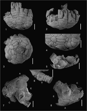
Zenocrinus zeus Moore and Strimple, Reference Moore and Strimple1973, specimens coated with ammonium chloride. (1–4) Holotype, SUI 35487; (1) D ray view; (2) anterior view, centered on right branch of B ray; (3) dorsal view, with posterior (CD) interray centered above proximal column; (4) close-up D ray view, showing small steps down from the distal end of one calyx ray plate to the proximal end of the next plate. (5–8) Paratype, SUI 35488; (5) view with anal X centered above column; (6) close-up view, showing wedge-shaped generating columnal (indicated by arrow); (7) dorsal view, with A ray centered above column; (8) dorsal view, with CD interray centered above column. A, C, and E=Carpenter rays. Scale bars=5 mm.

Type specimens
Holotype (SUI 35487), paratype (SUI 35488).
Diagnosis
Medium-sized, low crown with wide, shallow, bowl-shaped calyx. Fixed brachials in proximal arms branching isotomously on primibrachials two and secundibrachials two (or exceptionally three); arms abutting, but free above axillary secundibrachials, with heterotomous branching of biendotomous type in upper part of crown; each complete ray with 16 slender arm tips bent inward (indicating 80 or more arm tips in perfect crown). Infrabasals and most of basal circlet concealed by large proximal columnals; radials exposed all around, unequal, with C and D radials asymmetric and lower on sides adjoining CD basal, laterally abutting except where separated by CD basal; CD interray with enlarged CD basal followed by anal X, followed by two plates, followed by single series of two plates; other interrays with single large plate touching radials and separating fixed brachials, with one or two small plates above them; single intrabrachial at level of secundibrachials in each ray. Proxistele circular in cross section, with low homeomorphic columnals and peripherally finely crenulate facets, tapering away from calyx, separated from mesistele by a wedge-shaped generating columnal; mesistele with circular, heteromorphic columnals, with nodals bearing coarse tubercles.
Occurrence
Morrowan (Lower Pennsylvanian, Bashkirian); Pontotoc County, Oklahoma, USA.
Remarks
The diagnosis is emended for the genus and species by Moore and Strimple (Reference Moore and Strimple1973), and for the genus by Moore (Reference Moore1978). Because Zenocrinus is monospecific (Webster, Reference Webster2014), the diagnosis for the species is also that for the genus.
Discussion: reconciliation of the holotype and paratype morphology
A careful examination of the holotype and paratype has allowed reconciliation of perceived differences in the posterior plating as described by Moore and Strimple (Reference Moore and Strimple1973). Differences in the number of secundibrachials between the holotype and paratype as noted by those authors are herein confirmed, but deemed insufficient to merit a species-level distinction.
Posterior Interray
The description for the genus and type species provided by Moore and Strimple (Reference Moore and Strimple1973, p. 36) reads in pertinent part: “…radials exposed all around, laterally abutting except where separated by CD basal or (?abnormally) arching over this plate to allow C and D radials to meet; CD interray variable, in holotype of type species with primanal followed by 6 additional anals but in paratype with only 2 plates, lower one very large; other interrays with single large plate touching radials and separating fixed brachials, with or without 1 to 3 small plates above them…” As Moore and Strimple (Reference Moore and Strimple1973, p. 37) admitted, “Such great dissimilarities are hard to explain.” This discrepancy in the description of the anal area motivated the current author to re-examine the type specimens of Zenocrinus zeus. A careful examination of both the holotype and paratype has led to the conclusion that Moore and Strimple (Reference Moore and Strimple1973) misidentified the AB interray of the paratype specimen as the posterior (CD) interray.
A combination of factors might have led to their misidentification of the posterior interray in the paratype specimen. First, the paratype preserves only the anal X and parts of two additional plates of the posterior interray, making positive identification of the posterior interray more difficult. This difficulty is compounded by the curvature of the proximal column and a small amount of supporting matrix, which obscures the basals and radials on that side of the paratype.
Second, the first interradial plate of the AB interray of the paratype appears upon casual examination to be slightly narrower than those of the other regular interrays, although it is similar in shape, and this perceived difference in width is exacerbated by a longitudinal crack along the left side of the plate (Fig. 1.7). It is actually not the narrowest of the first interradial plates; that of the DE interray in the paratype is narrower. The actual height of the first AB interradial plate is ~5.4 mm, and the width is ~3.8 mm. This is within the range of variation in height (4.8–5.6 mm) and width (3.5–4.4 mm) for the first interradial plates in the other regular interareas of the paratype specimen. Measurements for the same plate in the holotype are consistent with the paratype. The AB first interradial plate height is on the high end of the range, and its width is on the narrow end of the range. The H:W ratio is the second highest of the eight regular interrays measured from the two specimens, with only the DE interray of the paratype having a slightly higher ratio. The high H:W ratio of the first AB interradial in the paratype might have added to the perception of narrower width, and this could have been another factor in the original authors’ choice of the AB interray as the posterior for the paratype.
Third, Moore and Strimple (Reference Moore and Strimple1973) might have assumed that the direction of curvature of the proximal column would be consistent for both specimens. This assumption is evidenced by their comparisons to other flexible crinoids, namely ‘Paramphicrinus multiramosus’ and P. magnus Moore and Strimple, Reference Moore and Strimple1973. They noted different orientations for proxistele curvature in these two species. Note: Moore and Strimple’s (Reference Moore and Strimple1973, p. 37) reference to ‘P. multiramosus’ was a lapsus calumni (error); they also referred to it in their discussion of P. magnus, in which they indicated P. multiramosus as the type for the genus, but this is clearly an erroneous reference to P. oklahomaensis Strimple, Reference Strimple1939 (Moore and Strimple, Reference Moore and Strimple1973, p. 39).
For Zenocrinus zeus, Moore and Strimple (Reference Moore and Strimple1973) described the curvature of the proximal column as toward the A ray, apparently having interpreted the holotype and paratype to agree in this character. This direction is correct for the holotype specimen, but as interpreted herein, the curvature of the proximal column in the paratype is initially in the direction of the posterior (CD) interray and more distally, in the mesistele, toward the D ray. It would be speculative to say which, if any, of these three factors influenced the original authors’ interpretation of the Carpenter ray designations, but it is likely that one or more in combination could have contributed.
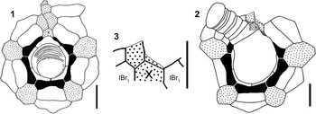
The interpretation provided herein reconciles the paratype with the holotype. The AB interray of the paratype that Moore and Strimple (Reference Moore and Strimple1973) identified as the posterior (CD) interray resembles the regular interrays, with a large interradial plate on the shoulders of abutting radials and not in contact with the basal circlet (Fig. 2.2). This large interradial plate is bounded laterally by the two primibrachials of each ray plus the first secundibrachials and, above, by another smaller interradial plate. The basal plate aligned with this interray is distally similar to the adjoining basals and is not enlarged distally as is the CD basal of the holotype (Figs. 1.3, 2.1). The paratype interray herein interpreted to be the posterior is proximally obscured by matrix surrounding the column and partly missing distally, but the anal X plate is preserved and exposed except for its most proximal portion (Figs. 1.5, 2.2, 2.3). Parts of the two plates that follow are also preserved. The plate interpreted herein as the anal X is pointed on its distal margin, for the reception of two plates above, as is the corresponding plate in the holotype. Laterally, the anal X of the paratype is bounded by the first primibrachials of the C and D rays, which is consistent with the anal X of the holotype.
Camera lucida drawings of Zenocrinus zeus Moore and Strimple, Reference Moore and Strimple1973. (1) Dorsal view of holotype, SUI 35487. (2) Dorsal view of paratype, SUI 35488; note that two anal plates (shaded area) partially obscured from view by column are shown and proxistele column divisions are omitted. (3) Dorsal view of close-up of posterior interray of paratype. Black=radial plate; stippled=interradial plate; gray=matrix; dashed line=plate suture obscured by matrix; thin line=broken plate margin; unbounded plate margin=plate boundary obscured by matrix; X=anal X plate; IBr1=first primibrachial plate. Scale bars=5 mm.

The shapes of the exposed radials and basals in the paratype are consistent with the A, B, and E rays of the holotype. In the holotype, the C and D radials are highly asymmetric, with both radials reduced in height on the side of the CD interray, in the portion in contact with the CD basal. The CD basal of the holotype is enlarged relative to the other basals and is essentially truncate, although slightly convex distally where it supports the heptagonal anal X. In the paratype, the three radials that are fully exposed are all symmetrical. Unfortunately, the C and D radials are covered by matrix beneath the overarching proximal column, but if they could be seen, the author predicts that they would be asymmetric as in those of the holotype. The basals visible in the paratype are also similar to the regular basals of the holotype and not enlarged or truncate as in the CD basal of the holotype. The enlarged, truncate basal is not visible in the paratype, but is presumed to be present beneath the obscuring matrix and proximal column.
Moore and Strimple (Reference Moore and Strimple1973, p. 36) described the posterior plating of the holotype specimen as having a “primanal (anal X as interpreted herein) followed by 6 additional anals.” However, in their discussion following (Moore and Strimple, Reference Moore and Strimple1973, p. 37), they referred to the holotype as having “what is evidently a primanal followed by 5 other anals above the cup rim.” A re-examination of the holotype shows that it in fact has a total of five anal plates following the CD basal, in a 1-2-1-1 configuration.
The plate diagram of Zenocrinus zeus is revised (Fig 2.1) to show the CD basal followed by an anal X, followed by four additional anal plates. This new plate diagram is also a revision of the Treatise plate diagram (Moore, Reference Moore1978, p. T808), which shows the CD basal followed by an unlabeled plate (presumably the primanal of Moore and Strimple, Reference Moore and Strimple1973), followed by an anal X on its left shoulder and a radianal on its right shoulder.
The relabeling of the plates in Figure 2.1 is an attempt to achieve consistency within the family Dactylocrinidae. In other genera within the family, the first plate following a truncate CD basal is considered to be the anal X plate.
Secundibrachials
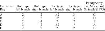
In addition to the perceived morphological differences of the posterior interray between the holotype and paratype of Zenocrinus zeus, Moore and Strimple (Reference Moore and Strimple1973) noted differences in the number and distribution of secundibrachials of the various rays. Some half-rays of each specimen have two secundibrachials, whereas other half-rays have three. The number of secundibrachials for each Carpenter ray for the holotype and paratype is given in Table 1.
Number of secundibrachials in left and right branches of each Carpenter ray in Zenocrinus zeus. Asterisk (*) denotes a branch reattached with glue; ‘greater than’ symbol (>) indicates that the distal-most brachial preserved is non-axillary, implying additional secundibrachial(s) in the branch.
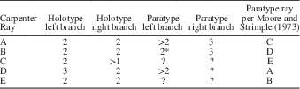
The number of secundibrachials present in each half-ray of each specimen agrees with those of Moore and Strimple, once the Carpenter rays for the paratype specimen are rotated to account for their misidentification of the posterior side. This rotation does not reconcile the holotype and paratype for this character, however. In the ten secunditaxis branches of the holotype, only one ray, the left branch of the D ray, has three secundibrachials; all of the rest have two (except for the right branch of the C ray, where only the first secundibrachial is preserved). In the five preserved secundibrachitaxis branches of the paratype, only the left branch of the B ray has two secundibrachials; the other four have at least two preserved, or three.
The discrepancy in the number of secundibrachials within the various rays is considerably less troubling than the perceived discrepancies in the posterior interray. Whereas the number of primibrachials is considered to be relatively stable and is frequently a basis for generic distinctions within the Flexibilia, the number of secundibrachials, although sometimes used for species-level distinctions, is not always consistent between individuals within a species (Springer, Reference Springer1920). An argument for a species-level distinction based on the number of secundibrachials might be warranted if a sufficient number of specimens existed and there were consistent differences, i.e., two populations separated geographically or temporally or distinct in other morphological features. With only two available specimens, both of which are in agreement in other important characters, the difference in the number of secundibrachials is insufficient to warrant a species-level distinction.
The combination of the perceived differences between the holotype and paratype in the posterior interray, and the differing number and arrangement of secundibrachials, prompted Moore and Strimple (Reference Moore and Strimple1973, p. 37) to “conclude that the species is somewhat unstable.” The same authors also suggested that the “possibility of sexual dimorphism is highly conjectural but not ruled out.” In light of the discussions above, the species need not be considered particularly unstable and there is insufficient evidence of sexual dimorphism based on the two known specimens.
Discussion: additional morphological features
Two morphological features of Zenocrinus zeus not previously noted are described herein. The first is the presence of a generating columnal separating the proxistele and mesistele portions of the column. The second is a ‘ratcheting’ profile for the exterior surfaces of calyx ray plates.
Generating columnal
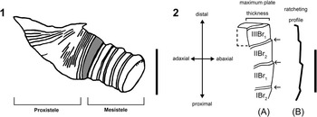
The paratype specimen (SUI 35488; Figs. 1.5–1.8, 2.2, 3.1) has a differentiated column, with a proxistele composed of homeomorphic, wide, low columnals that initially taper distally below the base of the calyx but that reach a constant width before joining the mesistele. The mesistele has higher, discoidal, heteromorphic columnals. Between the proxistele and the mesistele, there is a significantly higher columnal that tapers from the width of the proxistele columnals to those of the mesistele. This columnal (Figs. 1.6, 3.1) has a morphology that is distinct from other columnals in either the proxistele or the mesistele and resembles the ‘generating columnal’ of Strimple and Frest (Reference Strimple and Frest1979). According to the hypothesis advanced by Wulff and Ausich (Reference Wulff and Ausich1989), new columnals for the proxistele were generated on the proximal side of this specialized columnal, and new columnals for the mesistele were generated on its distal end. At the time Moore and Strimple (Reference Moore and Strimple1973) described Zenocrinus zeus, the phrase ‘generating columnal’ had not yet been coined and its significance was not yet appreciated, so it is hardly surprising that the authors did not include this character of the column in their description.
Zenocrinus zeus Moore and Strimple, Reference Moore and Strimple1973, additional morphological features. (1) Line trace of column of paratype, SUI 35488, from photograph in Figure 1.6, showing wedge-shaped generating columnal (gray shading), separating proxistele from mesistele columnals. (2) Holotype, SUI 35487, left side of E ray; (A) diagrammatic cross section, with steps down between successive brachial plates indicated by arrows; (B) profile of adaxial-abaxial cross section at exterior of calyx, resembling the teeth of a ratchet. Scale bars=5 mm.

The generating columnal of Zenocrinus zeus resembles that of Euonychocrinus simplex Strimple and Moore, Reference Strimple and Moore1971 (Strimple and Frest, Reference Strimple and Frest1979, text-figs. 1, 2c), in that it tapers distally in width from ~5.0 mm to ~4.3 mm to bridge the wider proxistele and narrower, immature mesistele columnals. The generating columnal of Z. zeus is wedge-shaped, with the tallest side (~1.6 mm) facing the D ray and the lowest side (~0.8 mm) facing the B ray.
This columnal in Zenocrinus zeus does not create a sharp bend in the column, unlike the strongly wedge-shaped generating columnals of the Permian (Wolfcampian) flexibles Nevadacrinus geniculatus Lane and Webster, Reference Lane and Webster1966, and Trampidocrinus phiala Lane and Webster, Reference Lane and Webster1966, the facets of which set the proxistele and mesistele columnals at an ~45° angle to one another. In the paratype of Z. zeus, the highest part of the generating columnal is located on the inside bend of the column, so that the wedge-shaped profile of this columnal has the net effect of straightening the column. In both the holotype and the paratype, a sharp bend in the proxistele is present just below the base of the cup.
Because these bends occurred in different directions relative to the Carpenter rays, it seems likely that the direction of the bend in the proxistele column was not genetically determined, as assumed by Moore and Strimple (Reference Moore and Strimple1973), but rather might have been either taphonomically or ecologically determined. Whereas dorsal cup morphology is generally constant in form, implying genetic control, a combination of genetic control and environmental factors might explain some of the intraspecific variation found in crinoid attachment structures (Brett, Reference Brett1981). Similarly, Donovan (Reference Donovan1992) suggested that observed individual variations in the dorsal cup of extant Holopus sp. crinoids, in which the dorsal cup itself forms the attachment structure, could be attributable to environmental factors.
For the bend to have occurred as a result of early taphonomic processes without fracturing, a certain inherent flexibility in the proxistele would be required. In some modern isocrinid crinoids, the proxistele portion of the column, with its thinner columnals, is more flexible than dististele portions that have thicker columnals (Donovan, Reference Donovan1984, p. 831). In some other modern crinoids, however, the proxistele is not the most flexible part of the column (Donovan and Pawson, Reference Donovan and Pawson1994). Based on the actual preserved state of Mississippian flexible crinoids from Crawfordsville, Indiana, Baumiller and Ausich (Reference Baumiller and Ausich1996) concluded that, contrary to some model predictions, the proxistele portion of the column was a very rigid portion of the stalk for at least some Paleozoic flexible crinoids. In their sample, only three of 27 specimens (25 of which were flexibles) with differentiated proxisteles had even a slight flexure within the proxistele (Baumiller and Ausich, Reference Baumiller and Ausich1996, p. 57–58). Even if the proxistele portion of the column in Zenocrinus zeus was rigid and immutable during life, it is still possible that the direction of curvature was ecologically as opposed to genetically determined.
Ratcheting profile
The exterior surfaces of calyx plates comprising the rays of Zenocrinus zeus have an unusual ratcheting profile. This profile, or cross section, results from a hypothetical planar section made perpendicular to the exterior of the calyx ray plates, along the adaxial-abaxial axis (Fig. 3.2). The proximal facet of each ray plate meets the distal facet of the plate below at a point beneath the exterior surface of the plate below. Proceeding distally from the base of the calyx along each ray, there is a small ‘step down’ from one ray plate to the next. Viewed from above the exterior of the calyx, the calyx ray plates form a series of descending terraces or long steps, with the face of each step on the distal facet of each plate (Fig. 1.4). When viewed in cross section, the exterior surfaces of the plates resemble the profile of teeth on the gear or rack of a ratchet (Fig. 3.2).
Although the ray plates have the appearance of being imbricated when viewed from above, there is little, if any, actual overlap of plates as seen from the exposed distal ends of brachial plates, and the thickness of the step down to the next plate distally is only 0.3–0.4 mm. The exterior of each plate of the holotype has a brownish coloration that extends to this depth, but not deeper, so that although the calyx plates are ~2.5 mm in total thickness, only the outer 0.3–0.4 mm is raised above the proximal end of the next distal plate and the brownish color extends adaxially only for this depth. The paratype is similar to the holotype in the thickness of these features, although the brownish color of the outer portion of each plate is not as dark. This difference in the coloration of the calcite could be attributable to differential diagenesis and/or a preservational phenomenon. The exterior of the paratype also appears to have been abraded to a greater degree than that of the holotype. Note that the coloration in the preceding discussion is not apparent in the photographs in Figure 1, which are printed in gray tones, and for which the specimens were whitened to improve contrast.
This arrangement of the ray plates does not appear to be an artifact of preservation or a condition of partial disarticulation. The ratcheting profile occurs only within the ray plates and does not occur within the interrays. It is present in all rays, and occurs in both the holotype and the paratype. The profile is most obvious in the primibrachials and secundibrachials of the calyx, but also extends to more distal brachials. Because the ratcheting profile is a subtle feature, it is not remarkable that it was not described by Moore and Strimple (Reference Moore and Strimple1973).
This profile might be an adaptation to accommodate a rapidly expanding calyx and reorientation of the arms from a mostly outward orientation in the primibrachials to a mostly upward orientation in the secundibrachials. Another possibility is that the ratcheting profile helped to passively orient the crown in a strong, directional current so that the oral side of the crown, including the mouth and ambulacral grooves, would be on the lee side. Water flowing against the base of the crown would encounter a hydrodynamic surface if the crown were oriented with the oral surface in the lee of the current, which is the typical feeding position of some modern stalked crinoids that form a parabolic filtration fan (Macurda and Meyer, Reference Macurda and Meyer1974). In any other orientation of the crown, the water would encounter the exposed distal faces of the ray plates, and the resistance to a strong current would tend to rotate the crown. Although intuitively plausible, this proposed function of the ratcheting profile is speculative at this point and testing this hypothesis would require simulation in an experimental flume study.
Summary
This study was instigated by a need to resolve the conflicting descriptions of the posterior interray of the holotype and paratype of Zenocrinus zeus. A simple explanation for the prior discrepancy became apparent after closer examination of the two available specimens. Moore and Strimple (Reference Moore and Strimple1973) had misidentified the posterior interray in the paratype specimen. By reinterpreting the orientation of the calyx of the paratype, it became possible to reconcile the morphology of the paratype with the holotype, thereby simplifying the diagnosis for the type species and genus. Previously undescribed morphological features are documented, i.e., the presence of a generating columnal and the ratcheting profile of the calyx ray plates.
Acknowledgments
This study was part of the NSF-funded Assembling the Echinoderm Tree of Life (AEToL) project (DEB 1036416). The author thanks W.I. Ausich, S.K. Donovan, and D.L. Meyer for reviewing and improving this manuscript, and T. Adrain from State University of Iowa for loaning the type specimens.