Introduction
Angiostrongyliasis is caused by the rat lung worm Angiostrongylus cantonensis, a nematode first indentified and classified by Chen (Reference Chen1935). Humans are accidental hosts, occasionally infected by eating raw or improperly cooked freshwater snails or paratenic hosts such as shrimps (Wallace & Rosen, Reference Wallace and Rosen1966), frogs (Lai et al., Reference Lai, Yen, Chin, Chung, Kuo and Lin2007) and slugs, etc. (Eamsobhana et al., Reference Eamsobhana, Yoolek, Punthuprapasa and Yong2009). Eosinophilic meningitis is caused by larval invasion of the human central nervous system (CNS). In China, hundreds of suspected cases of eosinophilic meningitis or meningo-encephalitis due to A. cantonensis infection, and several angiostrongyliasis outbreaks, have been reported in recent years (Chen et al., Reference Chen, Li and Lun2005; Wang et al., Reference Wang, Lai, Zhu, Chen and Lun2008). Several endemic foci of A. cantonensis have been identified in Hainan, Yunnan, Guangdong, Guangxi, Fujian and Zhejiang provinces (Chen et al., Reference Chen, Li and Lun2005; Lv et al., Reference Lv, Zhang, Chen, Wang, Fang, Chen, Jiang, Li, Du and Zhou2009). Consequently, angiostrongyliasis is considered to be a serious foodborne disease in China.
Eosinophilic meningitis is associated with cerebrospinal fluid (CSF) eosinophilia, which is an important criterion for A. cantonensis infection (Diao et al., Reference Diao, Chen, Yin, Wang, Qi and Ji2009). Matrix metalloproteinase-9 (MMP-9) plays an important role in the neuro-immunological response and the blood–brain barrier dysfunction (Rosenberg et al., Reference Rosenberg, Kornfeld, Estrada, Kelley, Liotta and Stetler-Stevenson1992; Leppert et al., Reference Leppert, Leib, Grygar, Miller, Schaad and Holländer2000). It is associated with the development of eosinophilic meningitis and can be a useful marker for this disease (Lai et al., Reference Lai, Twu, Jiang, Hsu, Chen, Chiaing, Wang, Tseng, Shyu and Lee2004; Lee et al., Reference Lee, Chou, Chen and Lai2004). The level of eotaxin in the CSF is worm-load dependent (Chang et al., Reference Chang, Chung and Yen2004) and correlates with CSF eosinophilia (Intapan et al., Reference Intapan, Niwattayakul, Sawanyawisuth, Chotmongkol and Maleewong2007). In mice, body weight loss and mortality are associated with this infection (He et al., Reference He, Lv, Li, Yu and Wu2009). Evaluations of motor activities and sensation are relatively objective measures of mouse neurological function (Parra et al., Reference Parra, McGirt, Sheng, Laskowitz, Pearlstein and Warner2002).
Albendazole has been found to be highly effective against angiostrongyliasis when administered early (before day 5 post-inoculation). This drug exerts its effects through larvicidal activity and reduction of MMP-9 activity (Lan et al., Reference Lan, Wang, Lai, Chen, Lee, Hsu and Lee2004; Tu & Lai, Reference Tu and Lai2006; Chen & Lai, Reference Chen and Lai2007). However, if treatment is begun on or after day 14 post-inoculation (PI), dead worms in the brain can evoke a severe immune response resulting in brain damage (Wang et al., Reference Wang, Jung, Chen, Wong, Wan and Wan2006). Clinically, patients usually become symptomatic after day 14 PI (Wang et al., Reference Wang, Lai, Zhu, Chen and Lun2008). Recently, albendazole has been used in combination with corticosteroids to prevent the inflammatory reaction (Tu & Lai, Reference Tu and Lai2006). However, the use of corticosteroid therapy is still controversial (Chen & Lai, Reference Chen and Lai2007). Baicalein is a flavonoid from the roots of Scutellaria baicalensis Georgi, a medicinal plant documented in the Chinese Pharmacopoeia (Pharmacopoeia Commission of the People's Republic of China, 2000) and Japanese Pharmacopoeia (Society of Japanese Pharmacopoeia, 2001). Scutellaria baicalensis Georgi has been widely employed as a popular antibacterial, antiviral and anti-inflammatory agent for many centuries. Recent investigations have shown that baicalein has broad anti-inflammatory and anti-oxidation activities (Lin & Shieh, Reference Lin and Shieh1996; Li & Chen, Reference Li and Chen2005; Chen et al., Reference Chen, Chow and Lin2006; Huang et al., Reference Huang, Lee and Yang2006). It can inhibit eotaxin production through the prevention of eotaxin mRNA expression (Nakajima et al., Reference Nakajima, Imanishi and Yamamoto2001). At the usual dosage, baicalein is not cytotoxic (Ong et al., Reference Ong, Len, Lee, Chui and Chooi2004).
The objective of the present study was to assess the efficacy of albendazole combined with baicalein for the treatment of eosinophilic meningitis in a mouse model. Administration of the drugs was initiated on day 14 PI. Efficacy parameters included survival time, neurological function, body weight, MMP-9 activity, eotaxin level, worm recovery, histopathological examination, leucocytes and eosinophil counts.
Materials and methods
Parasitological procedures
The infective larvae (L3) of A. cantonensis, originally obtained from wild giant African snails (Achatina fulica), were propagated for several months in our laboratory by cycling through rat and the intermediate host Ampullarum crossean. The shells were crushed and the tissues homogenized and digested in a pepsin–HCl solution (pH 2, 500 IU pepsin/g tissue) with agitation at 37°C for 2 h. The larvae in the sediment were collected by serial washing in double-distilled water and counted under the microscope (Chen & Lai, Reference Chen and Lai2007).
Animal treatment
Sixty-eight five-week-old male BALB/c mice (weight 20–25 g, Grade III, Certificate 2009B012) were supplied by the Laboratory Animal Center of Sun Yat-sen University. The animals were purchased a week before the experiment, raised at a 12 h light/dark cycle photoperiod, and provided with solid chow and water ad libitum.
Eight mice were randomly selected as the uninfected control, orally dosed with distilled water for 8 days starting from day 14 of the experiment. The remaining mice were randomly divided into four groups (15 mice in each group), each orally infected with 25 larvae. Daily treatment orally for 8 days with (a) distilled water; (b) albendazole (Sigma, St. Louis, Missouri, USA) at 20 mg/kg/day; (c) baicalein (Toppharman Co, Shanghai, China) at 100 mg/kg/day; or (d) a combination of albendazole (20 mg/kg/day) and baicalein (100 mg/kg/day) started on day 14 PI. All procedures were conducted according to the ethical guidelines of the Animal Care and Use Committee of Sun Yat-sen University.
Survival, body weight and assessment of neurological function
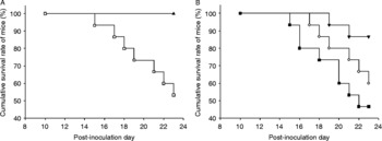
Mortality was documented daily, and analysed through a cumulative survival curve. The surviving animals from all five groups (see fig. 1) were sacrificed by cervical dislocation on day 23 PI for pre-planned analyses.
Cumulative survival of mice. The surviving mice were expressed as a percentage. (A) Uninfected control (▲), untreated infected group (□). (B) Treatment with albendazole alone (■), baicalein alone (⋄) and baicalein plus albendazole (▾).

Assessment of body weight and neurological examination were performed on days 0 and 23 PI. The mean body weight of each treatment group was compared with the untreated infected group.
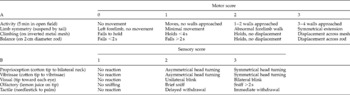
Neurological examination was performed as previously described (Parra et al., Reference Parra, McGirt, Sheng, Laskowitz, Pearlstein and Warner2002). A motor score (0–12) was derived from spontaneous activity, symmetry of limb movements, climbing, balance and coordination (table 1A). A sensory score (5–15) was derived from body proprioception, vibrissae, visual, olfactory and tactile responses (table 1B).
Evaluation of neurological function in mice by (A) motor and (B) sensory scores.

0, 1, 2 and 3 represent the respective scores a mouse acquired in an inspected item.
Cell count, eotaxin detection and gelatin zymography in CSF
On day 23 PI, the surviving mice were sacrificed. The brains were placed in 35-mm dishes. The cranial cavity and cerebral ventricles (lateral, third and fourth ventricles) were rinsed with 1 ml of 0.15 m phosphate-buffered saline (PBS) (pH 7.0). CSF was harvested and identified as ‘CSF-like fluid’ (K.M. Chen et al., 2004; Chen & Lai, Reference Chen and Lai2007). The CSF-like fluids were centrifuged at 400 g for 5 min, and the sediments were resuspended with 30 μl PBS for a total leucocyte count. The cells were calculated with Wright–Giemsa staining in 5 μl for each smear. The supernatants from the CSF-like fluids were centrifuged again at 12,000 g at 4°C for 10 min, the resulting supernatants were used for biochemical analysis.
The concentrations of eotaxin were assayed by commercial enzyme-linked immunosorbent assay (ELISA) kit (Biosource, Camarillo, California, USA) according to the manufacturer's instructions.
The activity of MMP-9 was detected by gelatin zymography as described previously (K.M. Chen et al., 2004). In brief, the samples were loaded on to 7.5% (mass/volume) SDS-polyacrylamide gels that had been co-polymerized with 0.1% gelatin. Stacking gels were 4% (mass/volume) polyacrylamide and did not contain gelatin substrate. Electrophoresis was performed in the running buffer (25 mm Tris, 250 mm glycine, 1% SDS) at room temperature at 120 V for 1 h. After electrophoresis, the gel was washed twice at room temperature for 30 min each in 2.5% Triton X-100, and then washed twice with double-distilled water for 10 min each. Subsequently, the gel was incubated in the reaction buffer (pH 7.5, 50 mm Tris–HCl, 200 mm NaCl, 10 mm CaCl2, 0.01% NaN3) at 37°C for 18 h. The gel was stained with 0.25% Coomassie brilliant blue R-250 for 1 h, destained in 15% methanol and 7.5% acetic acid for about 10 min. Gelatinase activity was detected as unstained bands on a blue background. Quantitative analysis of the gelatinolytic enzyme was performed with a computer-assisted imaging densitometer system.
Larval recovery and histopathological examination
Each brain was shredded in 10 ml of 0.15 m PBS and examined for viable larvae under 20 × magnification using a dissecting microscope. The mean number of larvae was compared with the untreated infected group to evaluate the treatment efficacy.
The mouse brains were fixed separately in 10% neutral buffered formalin for 24 h, then dehydrated in a graded ethanol series (50, 75 and 100%) and xylene, and embedded in paraffin at 55°C for 24 h. Sections were cut at 4 μm thickness for each embedded specimen. Paraffin was removed by heating the sections at 65°C for 5 min. The sections were dewaxed by washing three times for 5 min each in xylene, then rehydrated through 100, 95 and 75% ethanol for 5 min each, and finally rinsed with double-distilled water. After staining with haematoxylin and eosin, pathological changes were examined under 200 × magnification using a light microscope.
Data analysis
Comparisons of the survival rate were analysed by chi-square test for homogeneity. Other data from different groups of mice were compared using a non-parametric Kruskal–Wallis test followed by post-testing using Dunn's multiple comparisons of means. These results were presented as mean ± standard deviation (SD). P values of < 0.05 were considered as statistically significant.
Results
Survival, mean body weight and neurological function
On day 23 PI all uninfected control mice were surviving but only eight mice (53.33%) from the untreated infected group (fig. 1). The combination of albendazole and baicalein treatment (86.67% or 13 mice survived) significantly prolonged the survival of the infected mice, compared to the untreated infected mice (P < 0.05) (fig. 1). The cumulative survival rate of infected mice treated with albendazole-alone (46.67% or 7 mice survived on day 23 PI) or baicalein-alone (60% or 9 mice survived) was not significantly different from the cumulative survival rate of untreated infected mice (P>0.05).
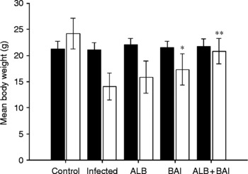
The changes in the body weight of treated and control mice during the 23 days of infection are shown in fig. 2. The animals in the uninfected control group gained weight. The animals in all the four infected groups lost weight. In infected mice treated with baicalein plus albendazole or baicalein-alone, the weight loss was less than that of the infected control group.
Mean body weight changes in surviving mice. The black solid columns and the white columns represent the mean body weights of the mice on days 0 and 23 PI, respectively. ALB, BAI and ALB+BAI represent mice treated with albendazole-alone, baicalein-alone and baicalein plus albendazole, respectively. *P < 0.05, **P < 0.01 compared to the untreated infected mice.

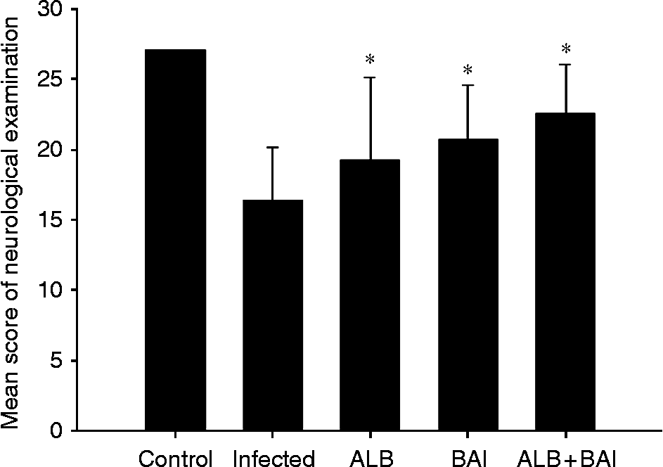
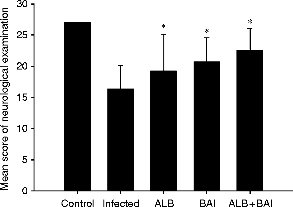
On day 23 PI, the mice in the uninfected group maintained normal neurological function (27 points) (fig. 3). The neurological functions in the other mice had deteriorated, as evidenced by reduced neurological scores. However, the neurological scores of the three drug-treated groups were significantly higher than that of the untreated infected mice (fig. 3).
Neurological examination score changes in surviving mice. Values and bars represent the mean and SD. ALB, BAI and ALB+BAI represent mice treated with albendazole-alone, baicalein-alone and baicalein plus albendazole, respectively. *P < 0.05 compared to the untreated infected mice.
Leucocyte counts, eotaxin levels and MMP-9 activities in CSF
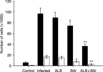
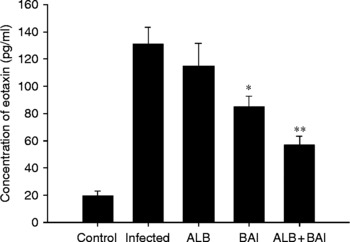
The numbers of eosinophils and leucocytes (fig. 4) and the concentrations of eotaxin (fig. 5) in the CSF-like fluid of the mice treated with albendazole-alone were close to that of the infected control group (P>0.05). The albendazole plus baicalein and baicalein-alone treated groups had fewer eosinophils and leucocytes and lower levels of eotaxin in their CSF-like fluid than the infected control group.
Leucocyte and eosinophil counts of surviving mice on day 23 PI. The black and white columns indicated number of leucocytes and eosinophils, respectively. ALB, BAI and ALB+BAI represent mice treated with albendazole-alone, baicalein-alone and baicalein plus albendazole, respectively. *P < 0.05, **P < 0.01 compared to the untreated infected mice.

Concentration of eotaxin in CSF of surviving mice on day 23 PI. Values and bars represent the mean and SD. ALB, BAI and ALB+BAI represent mice treated with albendazole-alone, baicalein-alone and baicalein plus albendazole, respectively. *P < 0.05, **P < 0.01 compared to the untreated infected mice.

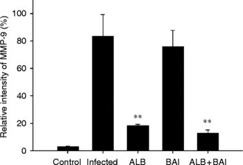
The relative activities of MMP-9 in the infected mice treated with baicalein-alone were not significantly different from that of the untreated infected mice (P>0.05). However, albendazole plus baicalein and albendazole-alone groups showed a reduction in MMP-9 activity (fig. 6).
Relative MMP-9 activity in CSF of surviving mice. Values and bars represent the mean and SD of relative intensity. ALB, BAI and ALB+BAI represent mice treated with albendazole-alone, baicalein-alone and baicalein plus albendazole, respectively. **P < 0.01 compared to the untreated infected mice.

Larval recovery and histological observations
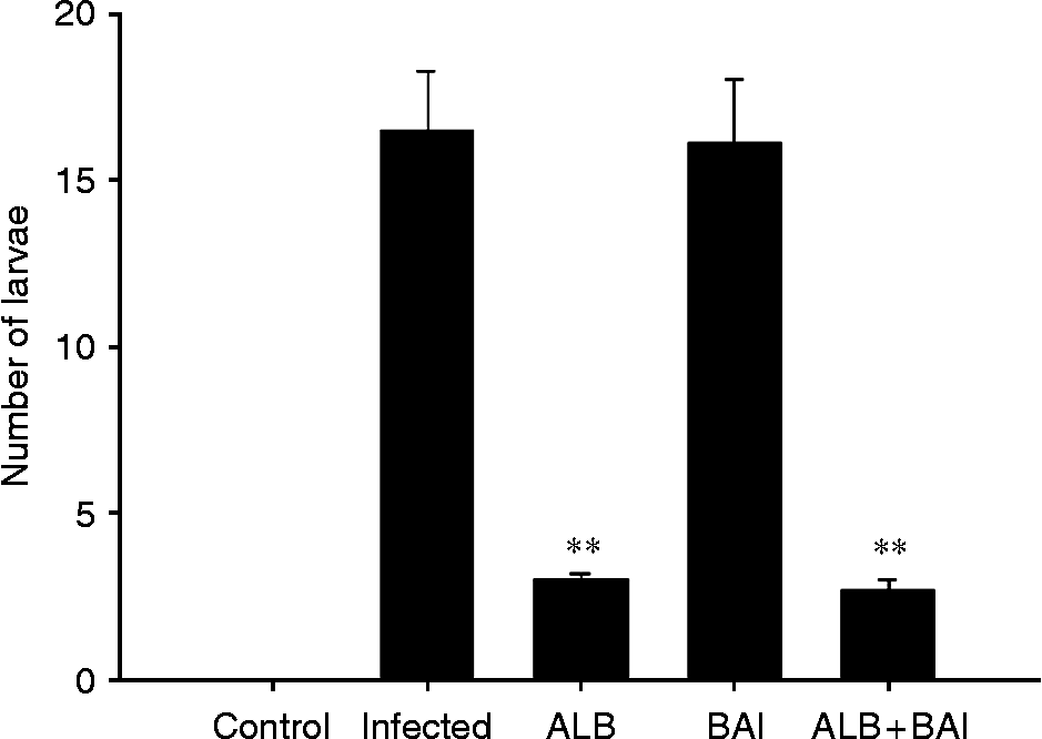
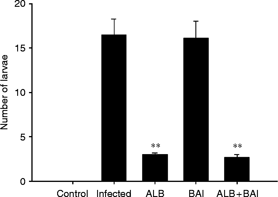
The numbers of recovered larvae were significantly decreased by albendazole-alone and albendazole plus bacailein therapy compared to the untreated infected group (fig. 7). No significant difference in worm recovery was observed between the untreated infected group and baicalein-alone treated group.
Larval recovery in surviving mice. The solid columns and the range bars represent the numbers of larvae and standard error of the mean. ALB, BAI and ALB+BAI represent mice treated with albendazole-alone, baicalein-alone and baicalein plus albendazole, respectively. **P < 0.01 compared to the untreated infected mice.
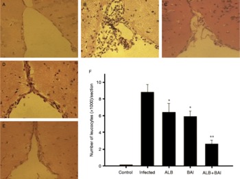
Optical microscopic examination of tissues stained with haematoxylin and eosin revealed that eosinophilic meningitis was induced in mice by A. cantonensis infection. The large-scale infiltration of the subarachnoid space by leucocytes was evident on day 23 PI (fig. 8). Leucocytes were reduced by treatment with baicalein and albendazole singly, and by albendazole plus baicalein in combination, but the reduction was more significant in the combination therapy group (fig. 8).
Histological observations of surviving mice, haematoxylin and eosin staining (magnification × 200): (A) normal meninx in uninfected control; (B) leucocyte accumulation in the subarachnoid space of the untreated infected mice; (C) infected mice treated with albendazole; (D) infected mice treated with baicalein; (E) infected mice treated with albendazole plus baicalein. (F) Mean number of leucocytes in mice. ALB, BAI and ALB+BAI represent mice treated with albendazole-alone, baicalein-alone and baicalein plus albendazole, respectively. *P < 0.05, **P < 0.01 compared to the untreated infected mice. (See online for a colour version of this figure.)

Discussion
Baicalein, a component from the Chinese herbal medicine S. rivularis, can be absorbed from the gastrointestinal tract and passes through the blood–brain barrier (Tarragó et al., Reference Tarragó, Kichik, Claasen, Prades, Teixidó and Giralt2008). The drug is generally regarded as safe and effective for the treatment of inflammation (Lin & Shieh, Reference Lin and Shieh1996; Hong et al., Reference Hong, Jin, Cho and Cyong2002; Li & Chen, Reference Li and Chen2005; Huang et al., Reference Huang, Lee and Yang2006). The present study supports an anti-inflammatory role of baicalein in eosinophilic meningitis. Eotaxin, leucocytes and eosinophils in the infected mice treated with baicalein-alone were reduced compared to the infected control group, similar to the effect of curcumin (Shih et al., Reference Shih, Lee, Lai, Chen, Jiang and Chen2007), Herba Artemisiae Capillaris (Lai, Reference Lai2006) and Longdan Xiegan Tang (Lai et al., Reference Lai, Chen, Chang and Lee2008) in the treatment of eosinophilic meningitis. Body weight loss and neurological function deterioration in infected mice were also lessened by baicalein treatment. Baicalein might possess pharmacological activities beneficial to the inhibition of parasite-induced inflammation, oxidation stress, neurological dysfunction (C.J. Chen et al., 2004; Cheng et al., Reference Cheng, He, Mu, Zhang, Li, Hu, Xu and Du2008) and production of eotaxin (Nakajima et al., Reference Nakajima, Imanishi and Yamamoto2001). However, our study suggests that anti-inflammatory treatment with baicalein alone shows little benefit in cumulative survival rate, larval elimination and MMP-9 expression. A possible reason might be that baicalein could not kill the larvae, so that the live larvae persisted to stimulate proteolytic enzymes or secrete MMP-9 (we have found that the live larvae could secrete MMP-9-like protein, unpublished data). We postulate that baicalein, when combined with an anthelminthic drug may be a more effective treatment for eosinophilic meningitis.
Albendazole is a broad-spectrum anthelmintic drug, which acts by binding to parasite β-tubulin, inhibiting its polymerization and impairing glucose uptake, causing death of the parasite (Venkatesan, Reference Venkatesan1998). The drug can penetrate into the subarachnoid space (John & Martinez, Reference John and Martinez1975). The ability of albendazole to kill the larvae of A. cantonensis has been demonstrated (John & Martinez, Reference John and Martinez1975; Lan et al., Reference Lan, Wang, Lai, Chen, Lee, Hsu and Lee2004; Tu & Lai, Reference Tu and Lai2006; Chen & Lai, Reference Chen and Lai2007). The effect of albendazole in angiostrongyliasis may be through larvicidal activity and reduction of MMP-9 activity (Lan et al., Reference Lan, Wang, Lai, Chen, Lee, Hsu and Lee2004). However, the timing of medication administration is important and closely related to anthelmintic efficacy of the drug (Tu & Lai, Reference Tu and Lai2006; Chen & Lai, Reference Chen and Lai2007). At the same dosage, earlier administration (before day 5 PI) has shown better results (Chen & Lai, Reference Chen and Lai2007). In our present study, the result of mice treated with albendazole alone was disappointing. Although the activity of MMP-9 and the number of larvae were reduced, the drug did not improve survival or lessen body weight loss in infected mice. At this time point of treatment, the dead worms could cause more severe inflammation, through activating eosinophils in the host brain (Wang et al., Reference Wang, Jung, Chen, Wong, Wan and Wan2006). Our results are quite comparable, suggesting that albendazole alone is not sufficient for the treatment of cerebral angiostrongyliasis at this time point (on day 14 PI).
The present study showed that combination of albendazole and baicalein had enhanced efficacy compared to monotherapy. This effect might be attributed to reduction of worm burden and inflammation. The survival rate was significantly different between the untreated infected mice and the mice treated with a combination of albendazole and baicalein. Combination therapy reduced the number of leucocytes, the activity of MMP-9, the body weight loss, as well as improving neurological function of the infected mice. We hypothesize that in combination treatment albendazole kills the worms, while baicalein reduces the hypersensitivity reaction (Lin & Shieh, Reference Lin and Shieh1996; Huang et al., Reference Huang, Lee and Yang2006) evoked by the dead worms. In severe infection, combination therapy with an anthelmintic drug (to kill the larvae) and an anti-inflammatory agent (to provide symptomatic relief) may well be a better treatment approach (Lai et al., Reference Lai, Chen, Chang and Lee2008). In a similar study, John & Martinez (Reference John and Martinez1975) found that worms could remain in the central nervous system and developed into young adults on day 14 PI, indicating that larvicidal treatment at this time point was more difficult than early treatment. As with albendazole alone, there were still a few larvae alive in combination therapy. It is possible that the eight consecutive day regimen was insufficient to kill all the larvae; a longer duration of treatment might kill all the larvae and lead to better recovery. However, further investigation is needed to confirm this hypothesis.
In conclusion, the combination treatment of albendazole and baicalein prolonged the survival time, reduced worm burden and hypersensitivity reaction in infected mice. The present study suggests that the combination treatment with albendazole and baicalein probably exploits the potential synergistic effects in eosinophilic meningitis induced by A. cantonensis.
Acknowledgements
We are grateful to Professor Yong Hoi Sen from the University of Malaya and Professor Barry Koehler from University of British Columbia for their helpful discussion and comments on the manuscript. We also thank Miss Li Yan for excellent technical assistance. Financial support was provided by: the National ‘973’ Program of China (No. 2010CB530004), the National ‘863’ Program of China (No. 2006AA02Z422), the National Natural Science Foundation of China (No. 30640028) and the Natural Science Foundation of Guangdong Province Key Project (No. 8251008901000007).