Introduction
Intrauterine growth restriction (IUGR) is generally regarded to increase susceptibility toward cardiovascular disease in adulthood,Reference Barker, Winter, Osmond, Margetts and Simmonds 1 a phenomenon referred to as fetal or developmental programming.Reference Alexander, Dasinger and Intapad 2 Various animal models of IUGR have been established to study the underlying mechanisms, including fetal hypoxia, placental insufficiency or maternal undernutrition during pregnancy.Reference Blackmore and Ozanne 3 In sheep, chronic hypoxia resulting from placental insufficiency reduces the number of cardiomyocytes in the heart.Reference Botting, McMillen, Forbes, Nyengaard and Morrison 4 Studies in rats have shown that IUGR induced by maternal low-protein diet (LPD) during pregnancy or placental insufficiency reduces heart weight and cardiomyocyte number in neonatal offspring.Reference Corstius, Zimanyi and Maka 5 , Reference Black, Siebel, Gezmish, Moritz and Wlodek 6 Such cardiac hypoplasia is likely due to impaired cardiomyocyte proliferation in the fetal or neonatal heart upon IUGR.Reference Aroutiounova, Fandrich, Kardami and Tappia 7 – Reference Paradis, Gay, Wilson and Zhang 9 Although heart weight and cardiomyocyte number appear to normalize during early postnatal cardiac growth in rodents,Reference Black, Siebel, Gezmish, Moritz and Wlodek 6 , Reference Lim, Zimanyi and Black 10 the reduced complement of cells at birth is considered an important determinant of cardiovascular health in adulthood.Reference Botting, Wang and Padhee 11 Indeed, rat hearts exposed to prenatal protein restriction show various alterations during postnatal life and in adulthood under baseline conditions, including reduced left ventricular (LV) function and altered myocardial tissue composition,Reference Menendez-Castro, Toka and Fahlbusch 12 increased apoptosis, increased LV wall thickness and reduced output,Reference Cheema, Dent, Saini, Aroutiounova and Tappia 13 and increased LV stiffening.Reference Xu, Williams, O’Brien and Davidge 14 Importantly, isolated adult rat hearts after intrauterine LPD are more susceptible to ischemia and reperfusion (I/R) injury compared with controls on a standard diet, resulting in impaired recovery of LV contractility and increased infarct size.Reference Xu, Williams, O’Brien and Davidge 14 , Reference Elmes, Gardner and Langley-Evans 15 Similarly, increased susceptibility to I/R injury was observed in offspring after fetal hypoxia.Reference Rueda-Clausen, Morton, Lopaschuk and Davidge 16 , Reference Xue and Zhang 17 Although such studies generally confirm the concept of developmental programming in the heart, the available data mainly relies on isolated and ex vivo perfused hearts, whereas in vivo data were sparse. In addition, most studies concentrate on I/R injury to investigate disease susceptibility, whereas other pathological conditions such as pressure overload or pharmacological challenges have rarely been applied.
We have previously characterized a mouse model of embryonic heart regeneration based on the heart conditional knockout (KO) of the X-linked Holocytochrome c synthase (Hccs) gene.Reference Drenckhahn, Schwarz and Gray 18 HCCS is required for normal function of the electron transport chain in mitochondria,Reference Babbitt, Sutherland, San Francisco, Mendez and Kranz 19 such that loss of HCCS activity in the developing heart results in respiratory chain dysfunction, disturbed cardiomyocyte differentiation and reduced cell cycle activity.Reference Drenckhahn, Schwarz and Gray 18 The Hccs gene is subject to random X chromosome inactivation, a process that permanently and irreversibly silences one of the two X chromosomes in mammalian female cells in order to normalize gene dosage compared with males.Reference Payer 20 Consequently, in female mice heterozygous for the cardiac Hccs KO (hereafter referred to as cHccs +/− ), approximately 50% of cardiomyocytes keep the defective X chromosome active and develop mitochondrial dysfunction while the other 50% inactivate the mutated X and remain unaffected. This leads to a tissue mosaic of 50% healthy and 50% HCCS deficient cardiomyocytes in the mid-gestational cHccs +/− myocardium, but the contribution of HCCS deficient cells decreases to 10% at birth. This regeneration of the prenatal heart is mediated by increased proliferation of the healthy cardiac cell population, which compensate for the defective cells and build up a fully functional organ.Reference Drenckhahn, Schwarz and Gray 18 It is important to note that the vast majority (⩾90%) of cardiomyocytes in the postnatal cHccs +/− heart is uncompromised. Inactivation of one X chromosome is the physiological state in female cells, such that silencing of the defective X does not affect Hccs gene dosage. We have subsequently shown that prenatal compensatory proliferation of healthy cells is insufficient to completely build up a heart. Neonatal cHccs +/− females exhibit cardiac hypoplasia characterized by reduced heart size and a 25% reduction in cardiomyocyte numbers compared with controls.Reference Drenckhahn, Strasen and Heinecke 21 Although heart size is normalized until early adulthood, we did not find evidence for a postnatal normalization of cardiomyocyte number. The latter would require increased cardiomyocyte proliferation, which was not detected in cHccs +/− hearts at any postnatal stage.Reference Drenckhahn, Strasen and Heinecke 21 Furthermore, cardiomyocyte size is increased in adult cHccs +/− hearts in the absence of increased heart weight or LV mass, and this compensatory cardiomyocyte hypertrophy cannot be maintained in ageing cHccs +/− hearts resulting in reduced heart weight with age, likely due to a persistently reduced cardiomyocyte number throughout life.Reference Drenckhahn, Strasen and Heinecke 21 We therefore concluded that postnatal normalization of heart weight in cHccs +/− hearts is mediated by accelerated hypertrophic growth of cardiomyocytes but not normalization of cell number.
Given that a reduced cardiomyocyte complement after IUGR is proposed to contribute to developmental programming in the heart, we speculated that the cHccs +/− mouse model might help to better understand the latter. Importantly, however, there are considerable differences between conditional genetic models, like cHccs +/− mice, and animal models of IUGR. Maternal LPD during pregnancy, fetal hypoxia or placental insufficiency all have systemic effects on the fetal organism, such that alterations in other organs impact on the heart (as e.g. by inducing hypertension in adulthood).Reference Alexander, Dasinger and Intapad 2 , Reference Blackmore and Ozanne 3 Instead, the cHccs +/− model provides an organ-specific impairment of heart development. Birth weight is normal in cHccs +/− mice,Reference Drenckhahn, Strasen and Heinecke 21 such that growth and stress response in the adult heart can be investigated without confounding secondary effects induced by other organ systems. We therefore believe that the heart conditional Hccs KO model could complement classical IUGR animal studies to better understand fetal programming specifically in the heart. We have previously shown that adult cHccs +/− females show significant cellular and molecular alterations when challenged by chronic angiotensin II (Ang II) infusion.Reference Drenckhahn, Strasen and Heinecke 21 To further clarify how hypoplastic cHccs +/− hearts cope with different challenges in adulthood, in the current study we applied LV pressure overload by abdominal aortic constriction (AAC).
Materials and methods
Heart conditional Hccs KO mice
The generation and characterization of heart conditional Hccs KO mice has been described previously.Reference Drenckhahn, Schwarz and Gray 18 In brief, ‘floxed’ (fl) Hccs mice were bred to mice expressing Cre recombinase under the control of the Nkx2.5 promoter. All mice were maintained on a mixed 129Sv/C57Bl6 genetic background and all experiments were performed on heterozygous Hccs KO females (Hccs fl/+ /Nkx2.5Cre, referred to as cHccs +/− ) and Cre positive female controls (Hccs +/+ /Nkx2.5Cre, referred to as Hccs +/+ ).
Surgery and echocardiography
AAC was performed on 10-week-old mice for a period of 4 weeks with echocardiographic measurements immediately before the intervention (baseline) as well as after 2 and 4 weeks [Hccs +/+ n=6 (three pairs of littermates from three different litters), cHccs +/− n=6 (each mouse from a different litter)]. For surgery, animals were anaesthetized by 2% isoflurane inhalation. The abdominal cavity was opened by a midline incision, the abdominal organs placed to the right and the aorta dissected from the retroperitoneum. The constriction was placed at the suprarenal level by a 6.0 silk suture tied around a blunt 27-gauge needle. The needle was then removed immediately to restore blood flow. Ultrasound measurements of the aortic diameter 2 weeks after surgery confirmed a ~50% narrowing at the site of ligation in all animals. Sham operated animals underwent all surgical and echocardiography procedures but did not receive a ligature [Hccs +/+ n=3 (each mouse from a different litter), cHccs +/− n=3 (one individual mouse and one pair of littermates from two different litters)]. The same three sham operated mice per genotype were used for all experiments throughout the study. For analgesia, animals received Carprofen [5 mg/kg body weight (BW)] before surgery as well as for the following 3 days. Mice were euthanized by cervical dislocation and hearts prepared 1 day after the final echocardiography.
Echocardiography was performed using a VisualSonics Vevo 2100 high frequency ultrasound system. Measurements were performed on anesthetized mice (2% isoflurane inhalation) and body temperature was kept constant at 37°C using a heat lamp and a rectal temperature probe.
Investigators performing echocardiography and surgery were blinded for mouse genotypes and treatment groups during data acquisition and analysis. All animal procedures were performed following institutional guidelines and had previously been approved by the responsible authorities [Landesamt für Gesundheit und Soziales (LaGeSo), Berlin, approval number G 0191/06].
Histology and assessment of myocardial fibrosis
Hearts were excised, rinsed in cold phosphate buffered saline (PBS) and fixed in 4% paraformaldehyde/PBS for 48 h. The tissue was subsequently dehydrated through an increasing ethanol series, cleared in toluol and embedded in paraffin; 5 µm paraffin sections were stained with hematoxylin & eosin (Roth) to assess overall cardiac morphology and tissue composition or with Sirius Red (Sigma) to visualize myocardial fibrosis. For quantification of interstitial fibrosis the entire LV myocardium of two non-adjacent sections was imaged at 5× magnification (using a Keyence BZ-8100E microscope) and the fibrotic (i.e. pink) area was measured using ImageJ analyses software (http://rsb.info.nih.gov/ij/). Perivascular fibrosis was excluded. Fibrosis was calculated as the percentage of the fibrotic area in relation to the total area of myocardial tissue.
Evaluation of cardiomyocyte size
To determine cardiomyocyte size, paraffin sections were stained with fluorescence conjugated wheat germ agglutinin (WGA Alexa Fluor 555; Invitrogen) to visualize cell membranes while nuclei were stained with 4',6-Diamidino-2-Phenylindole (DAPI) (Invitrogen). Subendocardial areas of the left ventricle from two non-adjacent sections per heart were imaged at 20× magnification using fluorescence microscopy (Keyence BZ-8100E). For each heart, 200 circular or symmetrically shaped cardiomyocytes with visible nuclei were selected to measure their cross-sectional area (CSA) (using Keyence BZ image analysis software) and the mean cell size was calculated for each heart.
Evaluation of cell death
Apoptotic cells were detected on paraffin sections using Terminal deoxynucleotidyl transferase dUTP nick end labeling (TUNEL) assay (ApopTag Fluorescein Apoptosis Detection Kit; Merck Millipore) according to the manufacturer’s instructions. Nuclei were stained with DAPI. The LV myocardium of two non-adjacent sections per heart was imaged at 2.5× magnification using an Axio Scope.A1 fluorescence microscope (Zeiss) and the entire LV tissue area was measured using ZEN blue software (Zeiss). The average nuclear density per mm2 tissue was calculated for each section by manually counting the number of nuclei on three randomly selected high-powered fields (40× magnification) within a defined LV tissue area (using ImageJ). Based on nuclear density per mm2 and the overall LV tissue area, the total number of LV nuclei could be estimated for each section. TUNEL positive nuclei were counted manually and related to the total number of nuclei.
Evaluation of cell proliferation
Paraffin sections were deparaffinized, rehydrated and heat mediated antigen retrieval was performed in sodium citrate buffer (10 mM, pH 6.0) for 20 min. After blocking in antibody solution containing 5% normal goat serum for 1 h, Ki67 (Thermo Scientific RM-9106) and troponin T (Developmental Studies Hybridoma Bank, CT3) primary antibodies were applied at 4°C over night. Secondary antibody detection was performed at room temperature for 1 h using Alexa 555 goat anti-rabbit and Alexa 488 goat anti-mouse antibodies (Invitrogen). Nuclei were stained with DAPI and sections were mounted in Prolong Gold antifade reagent (Invitrogen). The LV and interventricular septum (IVS) myocardium of two non-adjacent cross-sections was imaged at 20× magnification (resulting in ~40 random fields per heart). Ki67 positive nuclei were manually counted using ImageJ software and related to the total number of DAPI stained nuclei.
Western blot analyses and quantitative real-time polymerase chain reaction (qRT-PCR)
Western blot and qRT-PCR procedures have been described previouslyReference Drenckhahn, Strasen and Heinecke 21 and are furthermore available in the Supplementary Material (including details about antibodies and primer sequences).
Statistical analyses
Differences between two groups were analyzed using unpaired, two-tailed Student’s t-test. Differences between multiple groups were determined using one-way ANOVA followed by Bonferroni’s post-hoc test. Echocardiography data from the same groups of animals at different time points were analyzed using two-way ANOVA for repeated measures. For animal studies of developmental programming it is recommended to use only one representative male and/or female per litter in order to compensate for differences in intrauterine or postnatal conditions between mice from different litters or within the same litter.Reference Dickinson, Moss and Gatford 22 If more than one mouse per litter is used, this should be accounted for in the statistical analyses.Reference Dickinson, Moss and Gatford 22 Thus, for all echocardiography and histological data additional analyses using mean values per litter were performed (see Supplementary Tables S4 and S5). All data are presented as mean±standard error of the mean (s.e.m.). A probability value of P<0.05 was considered to indicate statistical significance (*P<0.05, **P<0.01, ***P<0.001).
Results
Pressure overload results in increased LV mass and wall thickness in cHccs +/− hearts compared with controls
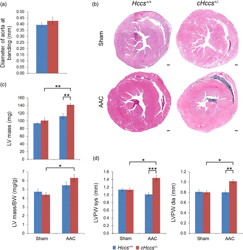
We applied pressure overload by suprarenal constriction of the abdominal aorta in 10-week-old cHccs +/− and control female mice for the duration of 4 weeks. Sham operated animals of both genotypes were included in the study to account for changes induced by the operation procedure. Echocardiography was recorded before as well as 2 and 4 weeks after the intervention. At the 2 week time point we furthermore assessed the degree of aortic constriction by ultrasound measurements, which confirmed that the aortic diameter at the site of banding was identical in both groups (Fig. 1a and Supplementary Fig. S1). After 4 weeks AAC cHccs +/− females showed significantly increased LV mass (calculated from echocardiography data) as well as LV mass normalized to BW when compared with their respective sham group (Fig. 1b and 1c and Supplementary Table S2). Interestingly, LV mass and LV mass/BW ratio were mainly unchanged in AAC v. sham controls, resulting in increased LV mass in cHccs +/− compared with control females after AAC (Fig. 1c). These differences in heart size were also reflected by LV wall thickness, which was significantly increased in cHccs +/− hearts 4 weeks after AAC compared with controls as well as compared with the cHccs +/− sham group (Fig. 1b and 1d and Supplementary Table S1). In contrast, wall thickness was not different in AAC compared with sham controls after 4 weeks. Thus, in response to pressure overload cHccs +/− females exhibit a more pronounced or sustained hypertrophic response of the LV myocardium compared with control animals.
Left ventricular (LV) mass and wall thickness are increased in heterozygous cardiac Holocytochrome c synthase knockout (cHccs +/− ) hearts compared with controls 4 weeks after abdominal aortic constriction (AAC). (a) Ultrasound measurements 2 weeks after AAC revealed a similar diameter of the abdominal aorta at the site of banding in both genotypes (n=6). (b) Cardiac cross-sections revealed increased LV wall thickness in cHccs +/− hearts compared with controls 4 weeks after AAC. Total cardiac dimensions are mainly preserved in all groups (hematoxylin & eosin staining, scale bar=300 μm). (c) LV mass calculated from echocardiography data as well as LV mass normalized to body weight is increased in cHccs +/− females after 4 weeks AAC when compared with the respective sham group. In controls both parameters are only slightly increased after AAC compared with sham operated mice. (d) Echocardiography revealed that LV wall thickness [LV posterior wall (LVPW)] in systole (sys) as well as diastole (dia) is increased in cHccs +/− females 4 weeks after AAC compared with controls as well as the respective sham group (n=3 for sham groups and n=6 for AAC groups in (c) and (d), *P<0.05, **P<0.01, ***P<0.001).

Pressure overload reduces LV contractility in controls but not cHccs +/− hearts
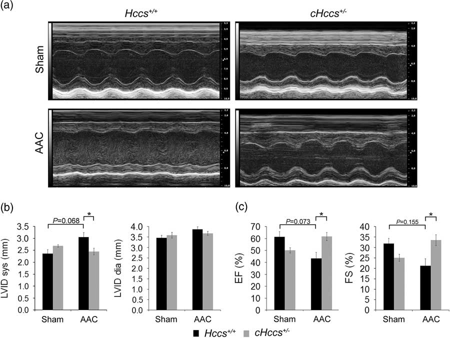
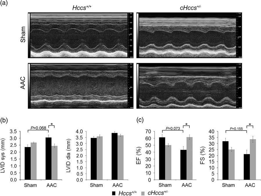
Given the marked differences in LV mass and wall thickness between control and cHccs +/− females 4 weeks after AAC, we investigated LV internal diameter (LVID) and contractility in echocardiography recordings. Compared with sham operated animals, control hearts developed a slight increase in systolic but not diastolic LVID after 4 weeks AAC, which, however, did not reach statistical significance. In contrast, LVID was not altered in cHccs +/− hearts after AAC when compared with the respective sham group (Fig. 2a and 2b), resulting in a significantly smaller systolic but not diastolic LVID in cHccs +/− females compared with controls after AAC. These data suggest that neither control nor cHccs +/− animals exhibit overall cardiac dilation associated with heart failure, which normally coincides with increased diastolic LVID. In contrast, control but not cHccs +/− mice appear to develop systolic contractile dysfunction as a result of pressure overload, as inferred from increased systolic LVID when comparing AAC groups (Fig. 2b and Supplementary Table S3). Indeed, whereas control animals show a clear trend toward reduced ejection fraction (EF) and fractional shortening (FS) 4 weeks after AAC compared with their respective sham group, both parameters where not altered (or even slightly increased) in cHccs +/− females (Fig. 2a and 2c and Supplementary Table S3). Consequently, the latter show a significantly better LV function after AAC compared with controls (Fig. 2c). Taken together, whereas control hearts develop systolic dysfunction after 4 weeks of pressure overload, cHccs +/− females fully maintain LV dimensions resulting in uncompromised contractility.
Preserved left ventricular (LV) contractility and diameter in heterozyogous cardiac Holocytochrome c synthase knockout (cHccs +/− ) hearts compared with controls 4 weeks after abdominal aortic constriction (AAC). (a) M-mode echocardiography recordings 4 weeks after AAC or sham operation, respectively, indicate reduced LV contractility in controls but not cHccs +/− females when exposed to pressure overload. (b) LV internal diameter (LVID) in systole (sys) but not diastole (dia) is increased in controls compared with cHccs +/− females after AAC. (c) Reduced LV function assessed by ejection fraction (EF) and fractional shortening (FS) in control animals compared with cHccs +/− females 4 weeks after AAC (n=3 for sham groups and n=6 for AAC groups in (b) and (c), *P<0.05).
LV hypertrophy, internal diameter and contractility are well maintained in cHccs +/− females during pressure overload
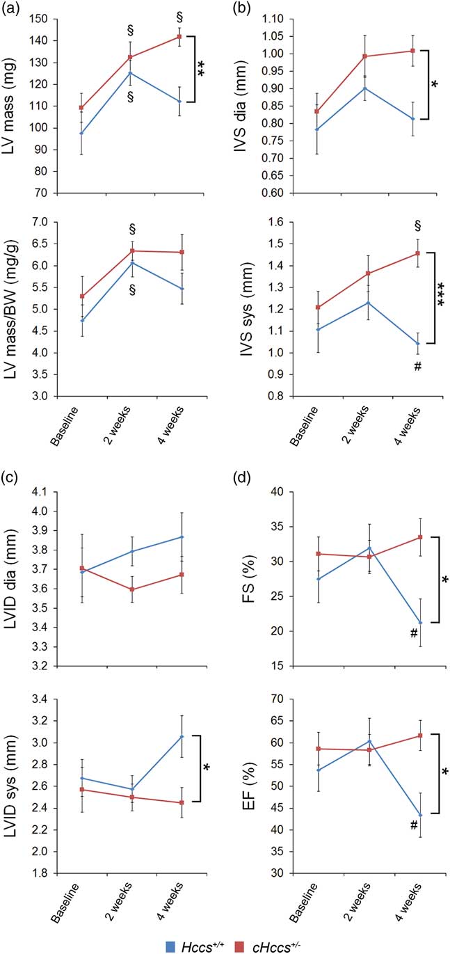
To more dynamically investigate the response of control and cHccs +/− hearts to pressure overload over time, we performed longitudinal analyses of echocardiography data before as well as 2 and 4 weeks after AAC on the same group of animals. These data revealed that hypertrophic growth of both control and cHccs +/− hearts is similar over the first 2 weeks, resulting in significantly increased LV mass and LV mass/BW ratios compared with baseline levels (Fig. 3a and Supplementary Table S2). However, LV mass as well as wall thickness decline in controls between 2 and 4 weeks, such that wall thickness returns to baseline values (Fig. 3b). The latter might indicate wall thinning at the onset of LV dilation as a result of persistent pressure overload. In contrast, hypertrophy parameters are well maintained or even further increase in cHccs +/− females during the same period, resulting in significant differences between genotypes 4 weeks after AAC (Fig. 3a and 3b). Similarly, LVID as well as contractility do not differ between genotypes over the first 2 weeks of pressure overload, but both FS and EF significantly deteriorate in controls between 2 and 4 weeks. In contrast, LVID and LV contractility is constant in cHccs +/− females over the full 4-week period (Fig. 3c and 3d and Supplementary Table S3). In summary, whereas control hearts show mild but significant LV dysfunction after 4 weeks AAC, cHccs +/− females can fully compensate cardiac stress by pressure overload resulting in significantly better outcome.
Longitudinal echocardiography measurements of left ventricular (LV) dimensions and function over the 4 weeks abdominal aortic constriction (AAC) period. (a) Absolute LV mass and LV mass normalized to body weight similarly increase in both genotypes during the first 2 weeks after AAC compared with baseline levels. Between 2 and 4 weeks LV mass declines in controls but not heterozygous cardiac Holocytochrome c synthase knockout (cHccs +/− ) females. (b) In parallel to LV mass, interventricular septum (IVS) thickness increases in both genotypes over the first 2 weeks after AAC, but declines thereafter in controls only. (c) Whereas LV internal diameter (LVID) in diastole (dia) is not different between genotypes or compared with baseline levels over the full 4-week period, LVID in systole (sys) increases in controls but not cHccs +/− females between 2 and 4 weeks of AAC. (d) LV contractility is fully preserved in cHccs +/− hearts during the 4-week period of pressure overload. In contrast, LV function is maintained in controls over the first 2 weeks but significantly decreases thereafter (n=6 per genotype, *P<0.05, **P<0.01, ***P<0.001, § P<0.05 compared with baseline, # P<0.05 compared with 2 weeks).

Cardiomyocyte hypertrophy is attenuated in cHccs +/− females in response to LV pressure overload
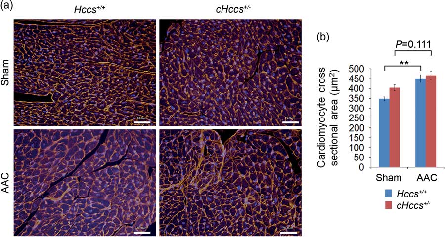
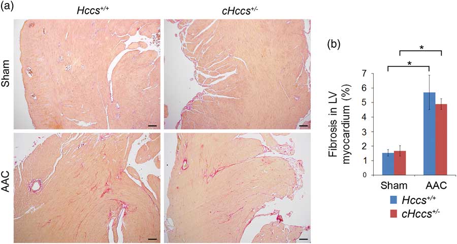
To investigate whether the better functional outcome of cHccs +/− females compared with controls 4 weeks after AAC is reflected by differences in myocardial tissue composition or remodeling, we analysed cardiomyocyte size in LV tissue sections by determining their CSA. In sham operated mice, cell size is increased in cHccs +/− hearts compared with controls, as previously reported for untreated animals.Reference Drenckhahn, Strasen and Heinecke 21 Surprisingly, this difference is no longer observed after AAC, such that CSA is similar between the genotypes (Fig. 4a and 4b). This is due to an attenuated hypertrophic response in cHccs +/− hearts, which show a slight but non-significant increase in cell size when comparing AAC and sham operated animals. In contrast, CSA significantly increases in control hearts after pressure overload (Fig. 4a and 4b). To further evaluate pathological myocardial remodeling in both genotypes after AAC, we visualized extracellular matrix deposition and quantified interstitial fibrosis within the LV myocardium. Sham operated animals did not show differences between genotypes and AAC induces LV fibrosis to a similar extent and to comparable levels in both cHccs +/− and control hearts (Fig. 5a and 5b). In conclusion, reduced LV function in controls after AAC coincides with a more pronounced cardiomyocyte hypertrophy compared with cHccs +/− hearts, whereas fibrosis as a parameter of pathological myocardial tissue remodeling is not different between genotypes.
Cardiomyocyte hypertrophy is attenuated in heterozygous cardiac Holocytochrome c synthase knockout (cHccs +/− ) hearts compared with controls 4 weeks after abdominal aortic constriction (AAC). (a) Fluorescence microscopy images of cross-sectioned cardiomyocytes within the left ventricular (LV) myocardium 4 weeks after AAC or sham operation, respectively. Cell membranes are stained in red (using wheat germ agglutinin) and nuclei are stained in blue (scale bar=50 μm). (b) Cardiomyocyte cross-sectional area is significantly increased in control hearts after AAC when compared with the respective sham group. In contrast, cell size slightly increases in cHccs +/− females in response to pressure overload, but this does not reach statistical significance when compared with the sham group (n=3 for sham groups and n=5 for AAC groups, **P<0.01).

Induction of myocardial fibrosis in response to pressure overload is not different between heterozygous cardiac Holocytochrome c synthase knockout (cHccs +/− ) and control hearts. (a) 4 weeks after abdominal aortic constriction (AAC) or sham operation fibrosis within the left ventricular (LV) myocardium is detected using Sirius Red staining, which marks extracellular matrix and collagen deposition in pink while the remaining myocardium is stained in yellow (scale bar=100 μm). (b) Quantification of interstitial fibrosis in the LV myocardium revealed that it is significantly induced in both cHccs +/− and control hearts in response to pressure overload (when comparing AAC and sham groups), but no difference was observed between genotypes (n=3 for sham groups and n=6 for AAC groups, *P<0.05).

Programmed cell death and proliferation are unaltered in cHccs +/− hearts upon pressure overload
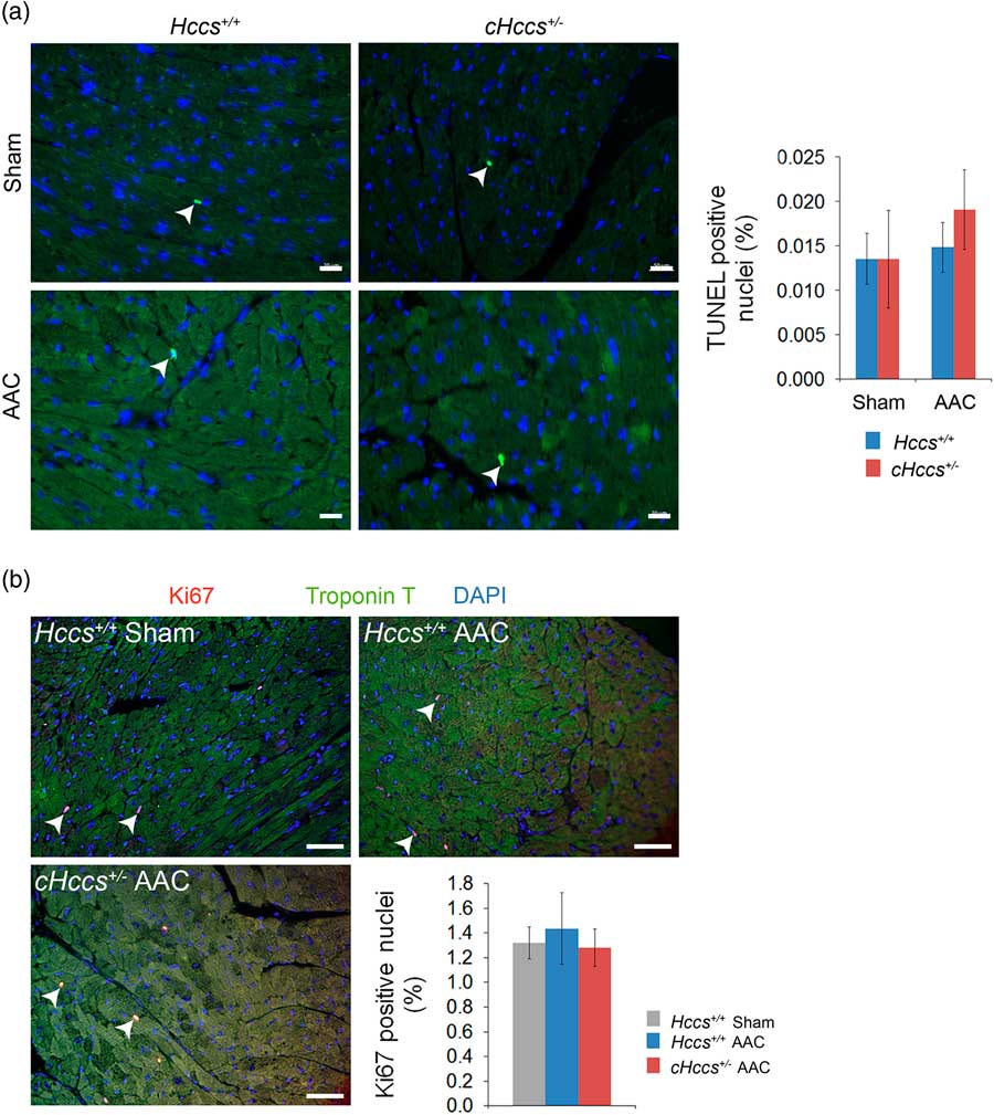
The seemingly contradictory findings of increased LV mass and wall thickness (Fig. 1) but similar cardiomyocyte size in cHccs +/− hearts compared with controls after AAC (Fig. 4) could indicate a progressive cardiomyocyte loss in controls but not cHccs +/− females over the 4-week period. This might also explain a regression in LV mass in AAC controls between 2 and 4 weeks to the level of sham controls despite a significantly increased cell size after 4 weeks pressure overload. To address this question, we determined programmed cell death (apoptosis) by TUNEL staining in the LV myocardium. No differences in apoptosis rates were detected between genotypes or treatment groups (Fig. 6a). In addition, we have previously shown that cardiac stress imposed by chronic Ang II infusion increases proliferation of the non-myocyte cell population in cHccs +/− hearts compared with controls.Reference Drenckhahn, Strasen and Heinecke 21 Here we show that such proliferative response is not observed upon pressure overload, as revealed by unaltered numbers of cycling (determined by immunofluorescence for Ki67) interstitial cells in both cHccs +/− and control hearts after AAC compared with sham operated controls (Fig. 6b). These data furthermore suggest that AAC induced differences in LV mass between controls and cHccs +/− mice are unlikely to be caused by excessive proliferation of a non-myocyte cell population.
Apoptosis and cell proliferation are not differentially regulated by pressure overload in heterozygous cardiac Holocytochrome c synthase knockout (cHccs +/− ) compared with control hearts. (a) Apoptosis in the left ventricular (LV) myocardium was detected by TUNEL staining 4 weeks after abdominal aortic constriction (AAC) or sham operation. Nuclei are stained in blue using 4',6-Diamidino-2-Phenylindole (DAPI). Terminal deoxynucleotidyl transferase dUTP nick end labeling (TUNEL) positive nuclei were identified by green fluorescence co-localized with DAPI staining (see arrowheads). Apoptosis rates were not different between genotypes or treatment groups (n=3 for sham groups and n=5 for AAC groups, scale bar=20 µm). (b) Immunofluorescence staining for the cell cycle marker Ki67 (in red) and the cardiomyocyte marker troponin T (in green) detected proliferating cells within the LV myocardium 4 weeks after AAC or sham operation. Cycling cells are located between troponin T positive cardiomyocytes, indicating their non-myocyte identity (see arrowheads, nuclei are stained in blue, scale bar=50 μm). Quantification of Ki67 positive cells revealed no difference between AAC or sham operated controls or between cHccs +/− and control hearts 4 weeks after AAC (n=3 for sham controls and n=6 for AAC groups).

Activation of p38 MAP-kinase in cHccs +/− hearts after AAC
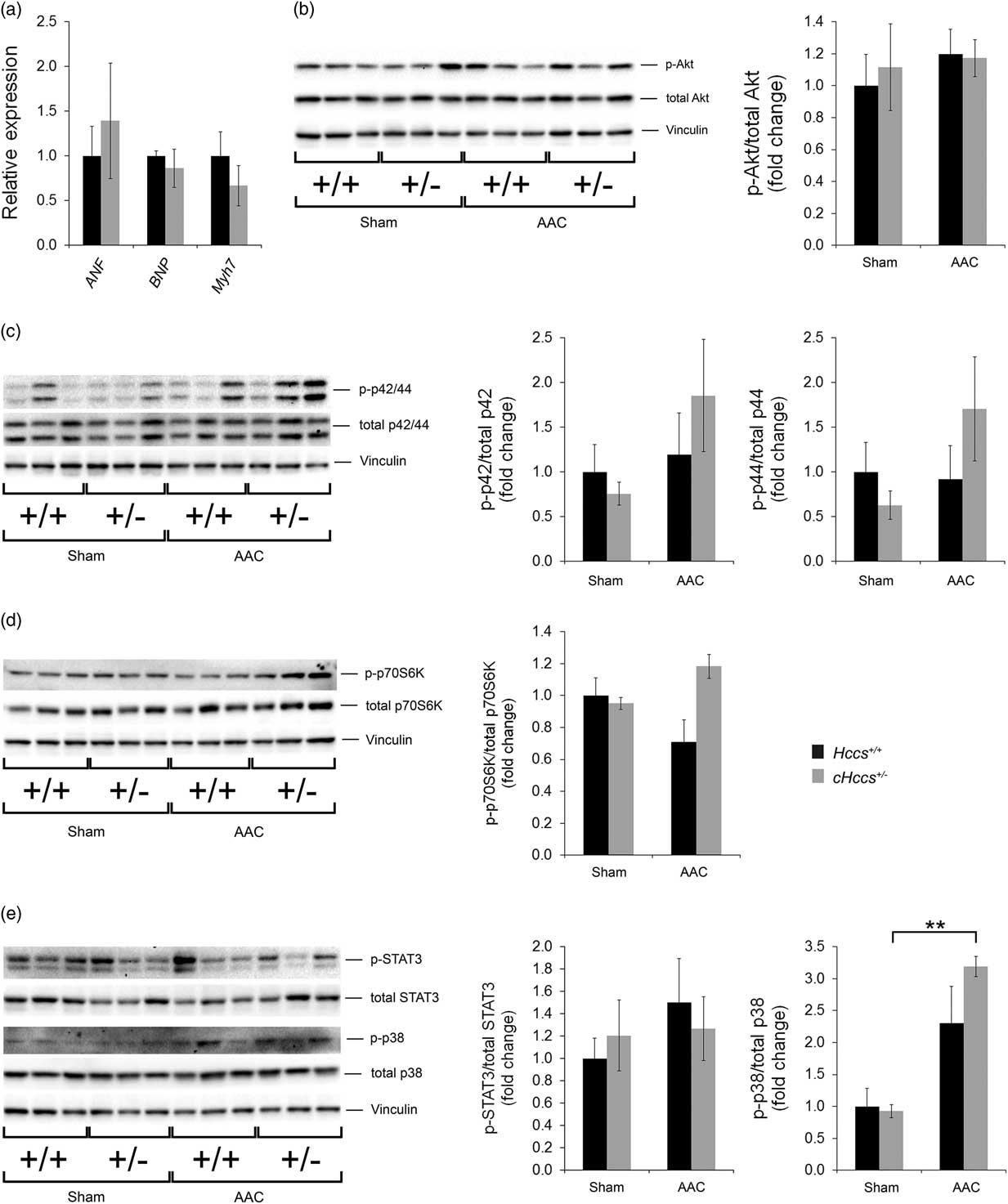
Altered tissue remodeling in response to pressure overload might be accompanied by differential expression of the fetal gene program that is reactivated by pathological conditions in the heart. We therefore analysed RNA expression of atrial natriuretic factor (Nppa), brain natriuretic peptide (Nppb) and β-myosin heavy chain (Myh7) in hearts 4 weeks after AAC, but did not find differences between cHccs +/− and control females (Fig. 7a). In combination with a similar induction of myocardial fibrosis by AAC in both genotypes (Fig. 5), certain aspects of the response of cHccs +/− hearts to pressure overload appear to be comparable to controls, but differences occur in the regulation of cardiomyocyte hypertrophy (Fig. 4).
Investigation of signaling pathways involved in cardiomyocyte growth in heterozygous cardiac Holocytochrome c synthase knockout (cHccs +/− ) compared with control hearts after abdominal aortic constriction (AAC). (a) RNA expression of the fetal genes atrial natriuretic factor (ANF, Nppa), brain natriuretic peptide (BNP, Nppb) and β-myosin heavy chain (Myh7) is not different in cHccs +/− compared with control hearts after AAC (quantitative real-time PCR data, n=6/genotype). (b) Phosphorylation of Akt Ser473 (n=4–6/group), (c) p42/44 MAP-kinase Thr202/Tyr204 (n=3–4) and (d) the mTORC1 downstream target p70 S6 kinase Thr389 (n=3) is not significantly different between genotypes or treatment groups. (e) Whereas phosphorylation of STAT3 Tyr705 (n=4–6/group) is not different between groups, phosphorylation of p38 MAP-kinase Thr180/Tyr182 (n=3) is significantly increased in cHccs +/− hearts 4 weeks after AAC compared to sham operated animals (**P<0.01).

Differences in the cardiomyocyte growth response upon pressure overload could be due to alterations in intracellular signaling associated with cardiac hypertrophy. Consequently, we analysed a subset of the underlying pathways with a special focus on those that have previously been shown to be differentially regulated in cHccs +/− v. control hearts.Reference Drenckhahn, Strasen and Heinecke 21 Whereas phosphorylation of Akt (Fig. 7b), p42/p44 MAP-kinases (Erk1/2) (Fig. 7c) and the mTORC1 downstream target p70S6K (Fig. 7d) was not different between genotypes 4 weeks after AAC, phosphorylation of p38 MAP-kinase was increased in cHccs +/− hearts compared with the respective sham operated group (Fig. 7e). We have previously shown that exaggerated cardiomyoyte growth in cHccs +/− hearts upon Ang II infusion depends on JAK/STAT3 signaling.Reference Drenckhahn, Strasen and Heinecke 21 Here we show that STAT3 phosphorylation in the heart is not different between genotypes 4 weeks after AAC (Fig. 7e, see Supplementary Discussion). In conclusion, activation of p38 MAP-kinase might be involved in restricting cardiomyocyte growth in cHccs +/− hearts in response to pressure overload.
Discussion
The intrauterine environment determines susceptibility toward cardiovascular disease in adulthood, which is proposed to involve a reduced cardiomyocyte complement in growth restricted neonates. Using heart conditional Hccs KO mice, which exhibit a 25% reduction in cardiomyocyte number at birth,Reference Drenckhahn, Strasen and Heinecke 21 we show here that such hypoplastic hearts have a better functional outcome after pressure overload in adulthood compared with controls. We certainly do not claim the Hccs KO model to mimic or replace other IUGR models. cHccs +/− mice are not generally growth restricted and exhibit normal birth weight,Reference Drenckhahn, Strasen and Heinecke 21 thereby lacking a hallmark of IUGR. Nevertheless, the impairment of heart development results in a phenotype very similar to IUGR, that is cardiac hypoplasia at birth. Given the various organism-wide alterations induced by IUGR and the resulting complexity of adult outcomes, animal models of organ-specific growth impairment could be useful to dissect the mechanisms of developmental programming. We therefore believe that the heart conditional Hccs KO model could help to generate new hypotheses regarding fetal programming in the heart which should subsequently be validated in classical IUGR animal studies.
Our previous study has shown that in adult cHccs +/− hearts the consequences of neonatal cardiac hypoplasia can be functionally compensated, such that upon Ang II stress LV contractility is not different from controls.Reference Drenckhahn, Strasen and Heinecke 21 Using chronic LV pressure overload we show here, that control mice exhibit reduced LV contractility after 4 weeks, whereas LV function was unaltered in cHccs +/− hearts over the full AAC period. This unexpected finding raises the interesting hypothesis, that the cHccs +/− heart might be preconditioned and better equipped to response to pressure overload than the normally developed heart. Alterations in stress response of the hypoplastic cHccs +/− heart in adulthood appear to converge on cardiomyocyte growth control but not pathological tissue remodeling, given that no differences in fibrosis or fetal gene expression were observed compared with controls after AAC (this study) or Ang II.Reference Drenckhahn, Strasen and Heinecke 21 Therefore, in cHccs +/− mice aberrant prenatal cardiac development potentially preserves or programs a beneficial growth plasticity which allows a better outcome after pressure overload. Certainly, it will have to be established whether such ‘fetal preconditioning’ is also observed in classical IUGR models or whether it is specific to cHccs +/− hearts. Preconditioning has been well established in the context of myocardial ischemia, where short exposures of the heart to hypoxic conditions reduce myocardial damage after a subsequent prolonged ischemic period.Reference Hausenloy and Yellon 23 IUGR does not seem to provide ischemic protection, given that a multitude of studies have shown increased susceptibility of restricted offspring to I/R injury in adulthood.Reference Xu, Williams, O’Brien and Davidge 14 – Reference Xue and Zhang 17 But this might be different for challenges not primarily associated with acute and large scale tissue damage and cell loss. Programming of cardiomyocyte growth plasticity could allow for better adaptation to challenges requiring a hypertrophic response. In this regard, disturbing cell growth by certain postnatal risk factors or drugs might be fatal in the developmentally impaired heart. Given that fetal programming has been proposed to permanently alter cellular metabolism after birth,Reference Martínez, Pentinat and Ribó 24 , Reference Keating and El-Osta 25 it is also possible that IUGR induces a metabolic profile in the adult heart that is beneficial under certain stress conditions. In this regard, the postnatal heart switches from fatty acid and glucose oxidation to glycolysis upon stress, thereby returning to a fetal metabolic program that is thought to be protective.Reference Kolwicz and Tian 26 In summary, alterations of intrauterine development and growth could precondition the heart for certain pathological stimuli during postnatal life, thereby mediating a beneficial effect. Such an idea would certainly contradict the basic principles of fetal programming, which assume that disease susceptibility is increased by intrauterine insults. Nevertheless, our data urge for future studies investigating this hypothesis in classical animal models of IUGR.
A reduced cardiomyocyte complement in the postnatal heart is believed to increase disease susceptibility by decreasing the ability to respond and adapt to pathological conditions.Reference Botting, Wang and Padhee 11 In support of this notion, heart conditional inactivation of the mitochondrial apoptosis regulator survivin (BIRC5) results in a reduction in cardiomyocyte number and survivin KO mice develop spontaneous heart failure in adulthood even without a specific challenge.Reference Levkau, Schäfers and Wohlschlaeger 27 Similarly, mice heterozygous for a KO of the cardiac transcription factor GATA4 show a cardiomyocyte deficit in the fetal heart and postnatally develop mild LV dysfunction under physiological conditions. Importantly, upon pressure overload these mice exhibit more severe contractile dysfunction compared with controls.Reference Bisping, Ikeda and Kong 28 It is important to consider, however, that in KO models of autosomal genes described above the functional consequences of the genetic alteration persists in cardiomyocytes throughout life. Even in heterozygous KO animals permanently reduced gene dosage (e.g. haploinsufficiency) or dominant-negative effects could disturb the adaptive capacity of the heart upon pathological conditions. In contrast, transcriptional silencing of one X chromosome occurs physiologically in mammalian female cells in order to compensate gene dosage compared with males.Reference Payer 20 One active copy of the Hccs gene is the normal state, such that cardiomyocytes in cHccs +/− hearts, which inactivated the X chromosome harboring the mutated Hccs allele, are uncompromised. Therefore, the vast majority (⩾90%) of cardiomyocytes in cHccs +/− females are per se healthy and able to utilize the complete program of adaptive growth or stress response. This resembles the situation in most IUGR models, in which the prenatal insult is released after birth such that programming of a more flexible metabolism and growth plasticity could be beneficial under certain conditions.
We acknowledge certain limitations of the data reported here. Due to the X chromosomal localization of the Hccs gene, it is not possible to include Hccs KO males in postnatal studies. The latter are hemizygous for the mutated Hccs allele, resulting in the complete lack of HCCS in all cardiomyocytes and embryonic lethality at mid-gestation.Reference Drenckhahn, Schwarz and Gray 18 Given that gender has been shown to have an influence on developmental programmingReference Alexander, Dasinger and Intapad 2 , Reference Blackmore and Ozanne 3 with IUGR males often being more susceptible to cardiac stress in adulthood,Reference Elmes, Gardner and Langley-Evans 15 , Reference Xue and Zhang 17 it is possible that hypoplastic male hearts respond differently to pressure overload as compared with cHccs +/− females. Furthermore, despite a clear hypertrophic response in control mice 2 weeks after AAC surgery, LV mass partially regresses thereafter and wall thickness returns to normal levels after 4 weeks. It is unclear whether this represents an adaptation to pressure overload or a beginning wall thinning due to LV dilation. It has previously been reported, that AAC in wild-type mice might not significantly alter LV mass if determined by echocardiography.Reference Wu, Hagaman, Kim, Reddick and Maeda 29 AAC is a relatively mild challenge compared with the more widely used thoracic aortic constriction. A variety of different outcomes has been reported for both models, ranging from mild hypertrophy with fully sustained contractilityReference Hu, Zhang and Swenson 30 to LV dilation and heart failure.Reference Barrick, Rojas, Schoonhoven, Smyth and Threadgill 31 Such differences are likely caused by the species used (rats v. mice), different strains and genetic background,Reference Barrick, Rojas, Schoonhoven, Smyth and Threadgill 31 gender,Reference Douglas, Katz and Weinberg 32 the duration of pressure overload (ranging from 1 to 20 weeks), differences in the surgical procedure and the localization and degree of the aortic constriction. Given the reduced LV contractility and increased LVID (even though statistically not significant in diastole) in control mice 4 weeks after AAC we would favor an interpretation of the partial normalization of LV mass and wall thickness as the onset of LV dilation rather than adaptation.
Along these lines, cardiomyocyte CSA and myocardial fibrosis significantly increase in control mice 4 weeks after AAC compared with the sham group, whereas LV mass is only marginally (and non-significantly) increased. This could potentially be explained by a progressive cardiomyocyte loss upon pressure overload, such that a compensatory increase in cell size is insufficient to elevate LV mass due to a parallel increase in cell death. However, we did not detect differences in apoptosis between groups 4 weeks after AAC, similar to previously published data.Reference Uozumi, Hiroi and Zou 33 Therefore, a potential apoptotic cardiomyocyte loss might occur earlier during the AAC period but is no longer observed after 4 weeks. Alternatively, other cell death mechanisms not detectable by TUNEL staining might be involved (as e.g. necrosis). A peak in cardiomyocyte apoptosis has been reported a few days after the onset of pressure overload, and cell death returns to baseline levels thereafter.Reference Teiger, Than and Richard 34 , Reference Hang, Huang and Jiang 35 Others, however, show a continuous induction or even increasing apoptosis after aortic banding with time.Reference Li, Ma and Yang 36 Therefore, potential differences in cell death between AAC controls, sham controls and AAC cHccs +/− hearts, respectively, will have to be determined at different time points after surgery. Interestingly, p38 has been proposed to be primarily required for cardiomyocyte survival rather than hypertrophy upon pressure overload.Reference Nishida, Yamaguchi and Hirotani 37 Given that p38 is activated in cHccs +/− hearts upon AAC, it is tempting to speculate that it might be involved in an altered cell death regulation compared with controls. At the same time, cardiomyocyte CSA significantly increases in control hearts after AAC compared with the sham operated group, whereas this response is markedly attenuated in cHccs +/− mice. Given the proposed anti-hypertrophic effects of p38,Reference Yokota and Wang 38 its activation could alternatively be required to restrict cardiomyocyte growth upon pressure overload in the latter. Thereby, cHccs +/− hearts potentially prevent excessive pathological hypertrophy and concomitant maladaptive cellular alterations to maintain LV contractility. Another possible explanation for a significantly increased LV mass in cHccs +/− hearts compared with controls after AAC despite similar cardiomyocyte CSA are differences in cell length. Interestingly, in heterozygous GATA4 KO mice, which exhibit a cardiomyocyte deficit in the heart (as described above), pressure overload induces an over-proportional increase in cardiomyocyte length rather than width compared with controls.Reference Bisping, Ikeda and Kong 28 Similarly, cardiomyocyte volume might be increased in cHccs +/− hearts compared with controls after AAC due to differences in cell length but not CSA.
The data presented in our current and previousReference Drenckhahn, Strasen and Heinecke 21 study indicate, that the stress response of the hypoplastic heart in adulthood differs depending on the type of the pathological stimulus. This suggests that various cardiovascular risk factors can have very different impacts on the developmentally programmed heart in humans. Whereas an increased disease susceptibility of the adult heart after IUGR is primarily studied in regard of ischemia and myocardial infarction, the impact of other conditions such as pressure overload, Ang II or β-adrenergic stimulation is largely unknown. Most investigators choose to study fetal programming by applying I/R injury to ex vivo perfused IUGR hearts, thereby showing an increased susceptibility of the adult heart after prenatal dietary protein restriction, placental insufficiency or fetal hypoxia.Reference Xu, Williams, O’Brien and Davidge 14 – Reference Xue and Zhang 17 Although this approach has led to important breakthroughs in the field, its general impact is clearly limited, given that it only allows studying the acute and short-term consequences of a single insult in an artificial system (i.e. the explanted, retrogradely perfused heart). In addition, myocardial ischemia only covers a fraction of the possible conditions that can impact on the developmentally programmed heart in humans. Challenges imposed by arterial hypertension, neurohumoral stimulation, drug induced cardiotoxicity or metabolic syndrome appear to be neglected instead. Consequently, long-term in vivo studies using different pathological insults in different animal models of IUGR would greatly enhance our understanding how altered intrauterine development causes heart disease in adulthood.
Acknowledgments
The authors thank Martin Taube and Stefanie Schelenz for performing echocardiography, Friederike Skole, Saskia Fiedler and Simon Pyschny for technical assistance and Christina Eichhorn for advice regarding statistical analyses.
Financial Support
This work was partially supported by the German Research Foundation (Deutsche Forschungsgemeinschaft DFG, grant number DR 446/3-1)
Conflicts of Interest
None.
Ethical Standards
The authors assert that all procedures contributing to this work comply with the ethical standards of the relevant national guides on the care and use of laboratory animals (GV-SOLAS) and has been approved by the responsible authorities [Landesamt für Gesundheit und Soziales (LaGeSo), Berlin, approval number G 0191/06].
Supplementary Material
To view supplementary material for this article, please visit https://doi.org/10.1017/S2040174417000514