Introduction
The genus Menegazzia A. Massal. is an important, species-rich component of the lichen biota of cool temperate regions in the Southern Hemisphere. Whilst revising the 30 known Tasmanian species (Kantvilas Reference Kantvilas2012), three additional, undescribed, non-Tasmanian species were discovered; these are described below. For a general introduction to the genus, outlining previous research, diagnostic features, ecology and biogeography, see Kantvilas (Reference Kantvilas2012).
Materials and Methods
This study is based chiefly on collections in the Tasmanian Herbarium (HO), supplemented by reference to critical holdings in other herbaria, especially the Natural History Museum (BM), the Allan Herbarium, New Zealand (CHR), and the herbarium of the late Henry Imshaug (MSC). Routine chemical analysis of all specimens followed standard methods of thin-layer chromatography (Orange et al. Reference Orange, James and White2001), with solvent A being the preferred medium. All fertile material was sectioned and dimensions of ascospores given in the descriptions are based on at least 100 observations each and presented in the format: 5th percentile–mean–95th percentile, with outlying measurements given in parentheses. Hand-cut apothecial and thallus sections were routinely mounted in water for observation and measurement, but subsequently eluted with 15% KOH or ammoniacal erythrosin for further examination.
The Species
Menegazzia bjerkeana Kantvilas sp. nov.
MycoBank No: MB800687
Menegazziae eperforatae similis et item thallo potius olivaceo-viridi, acidum sticticum continenti et dense isidiato sed perforatis numerosis et lobis parum latioribus, 1–2 mm latis differt.
Typus: Australia, New South Wales, Wiangaree Forest Drive, Tweed Range, near “The Pinnacle”, on Nothofagus moorei, 3 August 1988, G. Kantvilas 639/88 (HO—holotypus; NSW, TROM—isotypi).
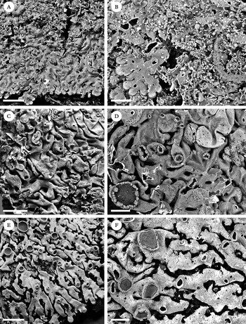
New species of Menegazzia. A, M. bjerkeana habit; B, M. bjerkeana detail, showing the maculate, perforate lobes with isidia; C, M. brattii habit; D, M. brattii detail, showing perforate lobes and apothecia with a crenulate, non-inflated margin; E, M. gallowayi, habit; F, M. gallowayi detail, showing maculate lobes, perforations with upturned margins, and apothecia with a pruinose disc. Scales: A, C, E=5 mm; B, D, F=2 mm.

Thallus relatively tightly adnate, very brittle and fragile, typically forming rather neat rosettes to c. 10 cm wide, isidiate. Lobes 1·0–1·5(–2·0) mm wide, rather flattened and undulate, densely imbricate and congested at the centre of the thallus, at the margins dichotomously branched and palmately radiating; apices discrete, adnate, decumbent or ± ascending. Upper surface perforate, pale olive-green to greyish green, brownish bronze at the apices, glossy, epruinose, with conspicuous, whitish, effigurate maculae, especially in younger, marginal parts. Perforations scattered, abundant, roundish to ellipsoid, rather variable in size and shape, 0·1–0·5(–0·8) mm wide, with margins flush with the thallus surface or turned slightly inward, occasionally rather ragged. Medullary cavity byssoid, persistently white on the upper surface, black with a few white, cobweb-like hyphae on the lower surface. Isidia laminal, very numerous, covering most of the thallus except for the lobe apices, particularly crowded in the centre of the thallus, erect or decumbent, at first rather knob-like, occasionally swelling and resembling minute lobes but more commonly elongate, cylindrical, to 1 mm long and 0·10–0·25 mm wide, simple or coralloid branched with the axils constricted.
Apothecia and conidia not seen.
Chemistry. Atranorin, stictic acid, constictic acid, menegazziaic acid (trace), cryptostictic acid (±trace), peristictic acid (±trace), 3-O-methylconsalazinic acid (trace), plus additional traces of related compounds; medulla K+ yellow, P+ orange, C−, KC−, UV−.
Etymology. This species is named in honour of my colleague Dr Jarle Bjerke of Tromsø, Norway, who has made significant contributions to the study of Menegazzia and has given me much assistance and encouragement in my study of the genus in Australasia.
Distribution and ecology. Menegazzia bjerkeana is known only from the type locality, in the Nothofagus-dominated rainforests of the coastal ranges along the New South Wales-Queensland border. It grows in the shaded forest interior on rough, rather dry, flaky bark of a mature Nothofagus moorei tree. Although locally very common, it was not found elsewhere, despite the author's detailed lichen studies in numerous stands of Nothofagus-dominated forests throughout New South Wales. Menegazzia bjerkeana is a further addition to a small group of Menegazzia species confined to this region, viz.: M. conica P. James, M. enteroxantha (Müll. Arg) R. Sant. and M. grandis P. James.
Remarks. Menegazzia bjerkeana is closely related to M. eperforata and M. nothofagi. These three species are superficially similar, sharing an olive-green thallus, relatively narrow, fragile and brittle, conspicuously maculate lobes and the stictic acid chemosyndrome (see Kantvilas Reference Kantvilas2012). It is most similar to M. eperforata in that both species have isidia, an uncommon feature in Menegazzia. However, it is clearly different in having very obvious and numerous perforations. Like M. bjerkeana, M. nothofagi is also perforate, but this species differs in having soredia that develop from inflated, subglobose to coralloid, isidia-like vesicles that rupture or become abraded (Kantvilas Reference Kantvilas2012). Menegazzia bjerkeana differs further from both of these related species by having slightly broader lobes.
Menegazzia brattii Kantvilas sp. nov.
MycoBank No: MB800688
Menegazziae pertransitae similis et item ascis octosporis sed atranorinum et acidum lecanoricum, plerumque cum pigmento aurantiaco ignoto continenti differt.
Typus: Îles Kerguelen, Bras de la Fonderie, summit of coastal cliffs W of Col Demi-Lune, near sea level, 8 March 1971, G. C. Bratt 71/535A (HO—holotypus; MSC—isotypus).
Thallus tightly adnate, forming irregular rosettes to c. 8 cm wide (herbarium material), lacking soredia or isidia. Lobes 1–3 mm wide, ±cylindrical to a little flattened, at the centre of the thallus irregular and densely imbricate, at the margins more neatly radiating but nevertheless rather disorganized and contiguous; apices inflated. Upper surface perforate, presumably a shade of pale greenish grey but discolouring pale yellowish brown in storage, here and there streaked with black along the margins, abundantly speckled with black, scattered pycnidia, rather glossy, smooth to a little puckered in older parts, epruinose, mostly emaculate. Perforations occasional, round to irregularly oval, 0·5–2·0 mm wide, with margins somewhat elevated when young, soon becoming flush with the thallus surface and at length turned inward, sometimes markedly so. Medullary cavity byssoid, white to suffused pale yellowish, K− to K± brownish, also with patches of an orange, K+ purple pigment on the upper and lower surfaces, especially towards the lobe apices, in the oldest parts of the thallus becoming blackened, with sparse, white cobweb-like hyphae.
Apothecia scattered, occasional, to 3 mm wide, sessile to subpedicellate; thalline margin 0·1–0·2 mm thick, not inflated, smooth and entire to crenulate; disc matt, orange-brown to brown, epruinose, persistently concave. Hymenium 95–120 µm thick, orange-brown in the upper part, not inspersed; asci 8-spored, occasionally with some spores aborted during development; paraphyses with apices not capitate, unpigmented, to 2–3 µm thick. Ascospores broadly ellipsoid, (26–)27–32·1–36(–38)×16–20·1–24(–25) µm.
Pycnidia abundant, scattered, especially towards the lobe apices, visible as black or black-brown specks 0·05–0·08 mm wide. Conidia filiform, 6–8×0·5 µm.
Chemistry. Atranorin (trace), lecanoric acid plus unidentified pigments; medulla K−, C− or ± very weak pink, KC−, P−, UV−; orange-pigmented areas K+ purple. As with several other species [e.g. M. myriotrema (Müll. Arg.) R. Sant. and M. tarkinea Kantvilas], the concentration of lecanoric acid is too weak to yield a reliable spot test; however, it can be discerned by eluting squash preparations with bleach solution. The suffused yellowish, K± brownish pigment is probably related to secalonic acid. The K+ purple pigment is unknown and does not show up on TLC plates, possibly due to its low concentration; it may also occur in the subhypothecial medulla where it reacts K+ orange-red in squash preparations.
Etymology. This new species is named after the Tasmanian lichenologist, Geoff Bratt (1931–1977), who undertook pioneering lichen collection in Tasmania in the 1960s and 1970s, and collected the type specimen while visiting Kerguelen with Henry Imshaug's expedition in 1971 (Bratt Reference Bratt1971; Imshaug Reference Imshaug1971).
Remarks. Menegazzia brattii is characterized by the combination of 8-spored asci, and the presence of lecanoric acid and yellow and orange pigments. Lecanoric acid is found in some South American eight-spored taxa [e.g. M. cincinnata (Ach.) Bitter and M. valdiviensis (Räsänen) R. Sant.; Bjerke Reference Bjerke2005] but these are yellowish, usnic acid-containing lichens. In contrast, M. brattii is greyish, contains atranorin and closely resembles the Australasian species, M pertransita (Stirt.) R. Sant. The latter differs from M. brattii chiefly in its chemistry, which comprises protolichesterinic and/or lichesterinic acids, as well as suffused yellowish secalonic acid-type pigments. Another related, superficially similar species is M. jamesii Louwhoff & Kantvilas, which also contains protolichesterinic acid plus an additional yellow, K+ purple pigment, skyrin. All the compounds in M. brattii occur in relatively low concentrations. I have tried repeatedly to establish whether protolichesterinic acid is present but cannot be certain. A more detailed discussion of the pigments of the M. pertransita group is provided by Kantvilas & Louwhoff (Reference Kantvilas and Louwhoff2004).
Distribution and ecology. Menegazzia brattii is known only from a rich collection, made from the bark of an unidentifed tree or shrub at the type locality. The specimen labels offer no further ecological information, nor could any further details be gleaned from the collector's notebooks, held in the archives of the Tasmanian Herbarium (HO). Associated with the new species are scraps of a sorediate Parmelia (probably P. protosulcata Hale) and Mycoblastus dissimulans (Nyl.) Zahlbr. Of greater interest is that it was collected together with a specimen of Menegazzia sanguinascens (Räsänen) R. Sant. (G. C. Bratt 71/542B, MSC), a species previously unrecorded for the island. The only other species of Menegazzia recorded for Kerguelen is M. castanea P. James & D. J. Galloway (Øvstedal & Gremmen Reference Øvstedal and Gremmen2009).
Additional specimen examined. Îles Kerguelen: Bras de la Fonderie, summit of coastal cliffs W of Col Demi-Lune, near sea level, 1971, G. C. Bratt 71/542A (MSC).
Menegazzia gallowayi Kantvilas sp. nov.
MycoBank No: MB800689
Thallo perforato, griseo-albido, esorediato, lobis 1·2–2·5 mm latis, acidum fumarprotocetraricum continenti, discis apotheciorum laete albido-pruinosis, asci octospori et ascosporis 28–40 µm longis et 12–24 µm latis distinguibilis.
Typus: New Zealand, South Island, Canterbury, Craigieburn Range, Craigieburn Stream, 43°07′15″S 171°42′48″E, 1050 m alt., on young Nothofagus solandri var. cliffortioides, 27 November 2010, G. Kantvilas 311/10 (HO—holotypus; CHR—isotypus).
Thallus loosely to tightly adnate, mostly forming rather neat rosettes to c. 10 cm wide, lacking soredia or isidia. Lobes 1·2–2·0(–2·5) mm wide, mostly rather flattened, usually cylindrical only in central parts of the thallus, sparsely dichotomously branched, imbricate, congested and convoluted centrally, at the margins palmately radiating, discrete or contiguous; apices flattened. Upper surface perforate, grey-white, pale greenish grey in more shaded situations, usually black to blackish brown along the lobe margins and at the apices, matt to glossy, epruinose, with scattered blotch-like maculae, smooth when young but increasingly wrinkled, dimpled or somewhat ridged in older lobes. Perforations scattered, numerous, roundish, 0·3–1·0 mm wide, at first with margins flush with the thallus surface or slightly turned inward, soon becoming slightly to conspicuously elevated and with the margins turned upwards. Medullary cavity byssoid and white throughout.
Apothecia scattered and numerous, to 3·5(–5·0) mm wide, subpedicellate ± persistently; thalline margin 0·2–0·5 mm thick, not inflated, smooth and entire, or crenulate and with occasional radial cracks; disc matt or glossy, pinkish or yellowish brown to brown, with a patchy to continuous whitish pruina at least when young, becoming dark red-brown and mostly epruinose when older. Hymenium 120–170 µm thick, orange-brown in the upper part, not inspersed (although overlying pruina granules fluoresce in polarized light and dissolve in K); asci 8-spored; paraphyses with apices mostly not capitate and unpigmented, to 2–4 µm thick. Ascospores narrowly to broadly ellipsoid, sometimes ovate, (28–)30–33·8–38(–40)×12–16·1–22(–24) µm.
Pycnidia black, speck-like, scattered on the upper surface, mainly towards the lobe tips, not common. Conidia filiform, 6–9×0·5 µm.
Chemistry. Atranorin (trace), fumarprotocetraric acid plus additional traces of related compounds; medulla K−, P+ red, C−, KC−, UV−.
Etymology. This species is named after the New Zealand lichenologist, David Galloway, in appreciation of his monumental Flora of New Zealand Lichens (1985, 2007) which has greatly advanced knowledge of the lichens of the Southern Hemisphere.
Distribution and ecology. Menegazzia gallowayi is known only from the type locality where it was abundant on the trunks and upper limbs of young trees of Nothofagus solandri var. cliffortioides in low, closed forest fringing a subalpine grassland-heathland mosaic. It grew within a rich assemblage of macrolichens that included species of Usnea, Pseudocyphellaria and several other Menegazzia species [M. abscondita Kantvilas, M. caliginosa P. James & D. J. Galloway, M. globulifera R. Sant., M. pertransita (Stirt.) R. Sant. and M. lucens P. James & D. J. Galloway]. The genus Menegazzia is now relatively well studied and collected in New Zealand, and thus the author was highly surprised to encounter this undescribed species growing so abundantly, albeit intermixed with several other, superficially similar members of the same genus.
Remarks. Menegazzia gallowayi is characterized by the unique combination of 8-spored asci and fumarprotocetraric acid, a metabolite that is relatively uncommon in the genus. In New Zealand, it is known only in M. hypernota Bjerke, which is sorediate and 2-spored (Bjerke Reference Bjerke2004). In Australia, it occurs in the saxicolous M. petraea Kantvilas and the epiphytic M. conica P. James, both of which are esorediate but have two-spored asci (James & Galloway Reference James and Galloway1992; Kantvilas Reference Kantvilas2012). In South America, there is the two-spored, sorediate species M. fumarprotocetrarica Calvelo & Adler (Adler & Calvelo Reference Adler and Calvelo1996), whereas in New Guinea, there is M. menyamensis Elix (syn. M. fumarprotocetrica Elix nom. illegit.) (Elix Reference Elix2007, Reference Elix2008). Eight-spored species are not common in the genus, although there are six others in New Zealand (Galloway Reference Galloway2007), all of which have a different chemical composition. Pruinose apothecia are also uncommon and notably occur in the New Zealand endemic M. dielsii (Hillman) R. Sant., which has eight-spored asci but differs in containing psoromic and echinocarpic acids (Galloway Reference Galloway2007). In the field, M. gallowayi is best recognized by the combination of the very pale grey, rather maculate thallus, the elevated perforations and pruinose, semi-pedicellate apothecia (at least when young).
Additional specimens examined. New Zealand:South Island: Canterbury, Craigieburn Range, Craigieburn Stream, 43°07′15″S 171°42′48″E, 1050m altitude, 2010, G. Kantvilas 350/10, 351/10 (HO).
I thank Dr Jean Jarman who photographed all of the species and prepared the plate that illustrates this paper. For the speedy loan of specimens from MSC I thank Dr Alan Fryday. Fieldwork in New Zealand was greatly assisted by Dr David Glenny.