Introduction
Case presentation
A 34-year-old woman with Turner syndrome, diagnosed at age 15, was referred due to aortic coarctation. Two years prior, she presented to the emergency room with a sudden-onset headache and right-sided body weakness. Her blood pressure in the left arm was severely elevated (192/110 mmHg), and a brain CT scan revealed findings consistent with a haemorrhagic stroke. The patient was hospitalised and received medical therapy for blood pressure management in a hospital in her city of origin. Later, it was decided to perform an echocardiogram to rule out a cardiac abnormality.
On physical examination, a patient with Turner’s phenotype had right hemiparesis with asymmetry on the left side of the face. During a subsequent cardiology evaluation, a systolic murmur was detected. An echocardiogram confirmed aortic coarctation. She was started on antihypertensive therapy (valsartan and amlodipine) and referred to cardiothoracic surgery for further management.
The diagnostic workup revealed no evidence of myocardial hypertrophy or cardiac enlargement. Coronary angiography further excluded the presence of coronary artery disease.
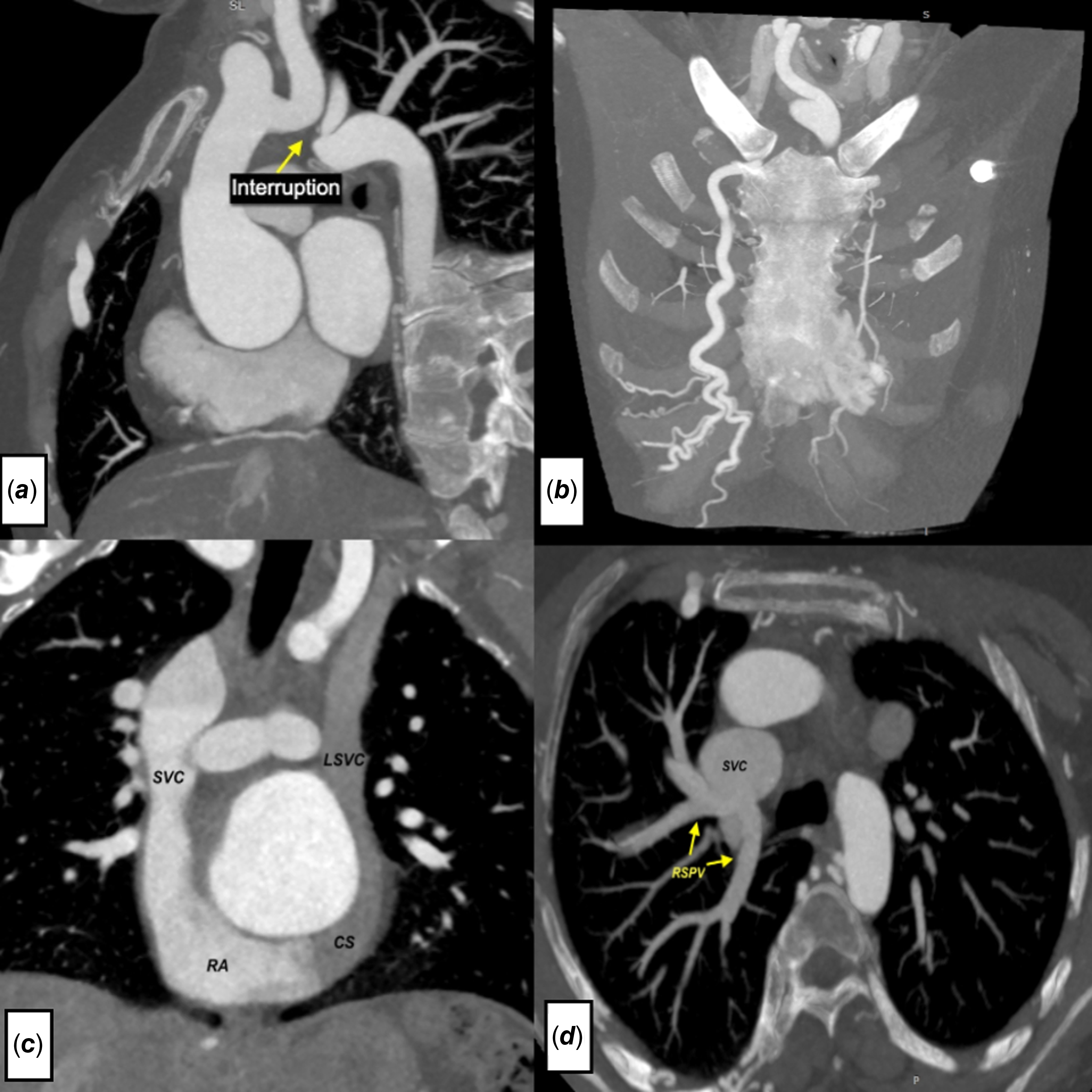
As an initial management, a transthoracic ultrasound was performed, where the suprasternal view found continuity between the left carotid and left subclavian arteries. A contrast-enhanced CT angiography showed interruption of type B aortic arch with a significant proliferation of collateral circulation observed surrounding the descending aorta, notably featuring dilated right internal mammary artery and hypertrophied intercostal vessels, partial anomalous pulmonary venous connection, and persistent left superior vena cava. Diameters of ascending aorta and aortic root were 42.5 × 40 mm (Indexed 36 mm/m2) and 35 × 30 mm (Indexed 28.4 mm/m2), respectively (Figure 1). Transesophageal echocardiographic evaluation detected a bicuspid aortic valve configuration accompanied by mild stenosis. The valve morphology was Sievers type 0 with a latero-lateral variant. Discrepant findings between modalities were noted, with echocardiography showing a non-significant gradient while CT demonstrated the interrupted aortic arch morphology.
( a ) CT angiography shows interrupted aortic arch between the left carotid and left subclavian artery. ( b ) Large bilateral collateral vessels connect the subclavian arteries with the descending segment of the thoracic aorta and enlarged internal mammary arteries. ( c ) Persistent left superior vena connected to the coronary sinus. ( d ) Anomalous right superior pulmonary vein connected to the superior vena cava.

The case was presented to the heart team, and surgical management was chosen. It was agreed to perform Tyrone David surgery and aortic arch restoration. Given the patient’s young age, the David procedure was selected to eliminate the need for lifelong anticoagulation. Furthermore, modern assisted reproductive technologies offer viable options for future childbearing. The approach was by median sternotomy. The arterial cannulation was done in the femoral artery and brachiocephalic trunk, and bicaval cannulation was performed for venous drainage.
Once inside cardiopulmonary bypass with crystalloid cardioplegic solution, the first part of the operation began, interposition of a 30 mm straight vascular Dacron tube was anastomosed to reconnect the interrupted aorta between the ascending aorta and the descending portions of the aorta. Subsequently, the dilated root was replaced by a 23 mm diameter Dacron graft with aortic valve sparing. The correction of the anomalous venous drainage consisted of disconnecting the right superior lobular pulmonary venous drainage from the superior vena cava and redirecting it to the right superior pulmonary vein by anastomosing an 8 mm polytetrafluoroethylene graft.
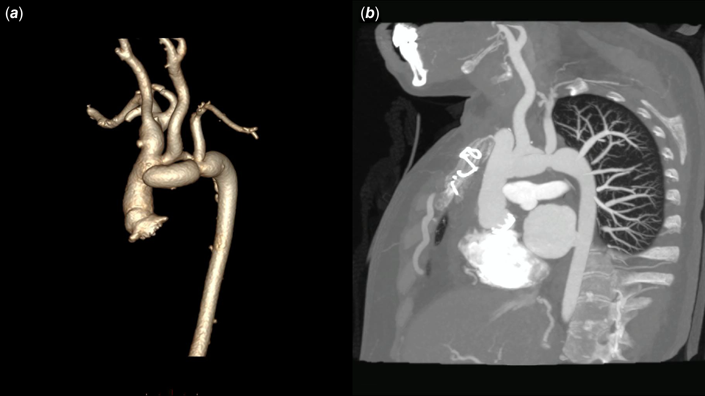
The procedure involved valve-sparing root replacement employing a 26 mm corrugated Dacron graft (later trimmed to 23 mm) (Figure 2). Post-reimplantation TEE demonstrated significant regurgitation, requiring CPB reinstitution. Surgical inspection identified a torn right coronary leaflet, ultimately requiring replacement with a 19 mm mechanical prosthesis.
( a ) Post-operative CT angiography reveals a patent graft, with the narrowest diameter (10 mm) located at the distal anastomosis. ( b ) Around the descending aorta, there were multiple collateral branches, and the graft located in the aortic arch distal to the left subclavian artery.

Due to the prolonged time of extracorporeal circulation, haemostasis was difficult, and bleeding was controlled with three gauze pads that were removed after 48 hours. Post-procedural recovery was uneventful with a good haemodynamic response. The patient presented with atelectasis that resolved after 7 days. Tracheostomy was not required. Likewise, the patient presented dysphagia in the control before discharge; a laryngoscopy was performed, which showed evidence of a left recurrent laryngeal nerve palsy. At 6-month follow-up, the patient reported marked clinical improvement: dysphagia resolved to occasional solid-food difficulty (Grade 2 on the FOIS scale), dyspnoea decreased by ≥1 NYHA class, and blood pressure remained controlled on single-agent amlodipine. Cardiac rehabilitation was completed uneventfully, enabling full return to employment.
Discussion
Interrupted aortic arch occurs in one in every 3 million live births. Type B, the most common form, has a complete loss of continuity between the left carotid artery and the left subclavian artery. Reference Kumanan, Guruparan, Gnanakanthan and Ratnayake1 Severe congestive heart failure is the major presentation of interrupted aortic arch in infants, and its mortality rate is 90% in the first year. Survival into adulthood without treatment relies on developing an extensive collateral network, essential for the maintenance of distal flow. Reference Sharma, Neupane, Dhakal, Sharma, Sitaula and Rajlawot2,Reference Jonas3
Recent Italian cohort data identified CHD in 22.9% of Turner syndrome patients (136/594 cases). While aortic root dilation occurs in approximately 2% of this population, the specific incidence of interrupted aortic arch remains unreported. Notably, interrupted aortic arch presentation in adulthood constitutes an exceptional clinical scenario across all populations. Reference Ribé, Shihadeh, Afifi, Estrera and Prakash4
The presentation of interrupted aortic arch in adults differs significantly from that in neonates. It can range from no symptoms to resistant hypertension, headache, claudication, malaise, differential blood pressure in the arms and legs, and congestive heart failure. Undiagnosed aortic coarctation may progress to complete blockage in the position of the obstruction beyond the left subclavian artery. This could explain the development of widespread collateral vessels supplying the distal descending aorta and the delayed symptoms. Reference Vaideeswar, Marathe, Singaravel and Anderson5 Asymmetric blood pressure, a classic feature of interrupted aortic arch, was evident in this case with significantly lower readings on the left side compared to the right.
Turner syndrome has a generalised arteriopathy and is an independent risk factor for thoracic aortic dilation. According to small body size, aortic dimension must be normalised to the body surface area. Elective surgery for aneurysms of the aortic root should be considered for women who are >16 years of age, have an ascending aortic size index >25 mm/m2. Reference Michelena, Della Corte and Evangelista6
Surgery is indicated during the neonatal period when the metabolic state is appropriate and consists of direct aortic arch anastomosis. The first week of life is the ideal time for surgical intervention. In adulthood, surgical correction remains the preferred treatment, although there have been some cases that have received anti-hypertensive therapy, and close follow-up is necessary. Reference Jiang, Wang, Jiang and Chen7 The majority of documented interrupted aortic arch cases undergoing surgical reconstruction show an improvement in patients’ symptoms following surgery. A review performed by Jiang et al shows that patients who receive anti-hypertensive therapy did not have problems, even mentioning that medical treatment is comparable with surgical correction. Reference Jiang, Wang, Jiang and Chen7 In the presented case, the patient presented with an interrupted aortic arch, and dilated aortic root, and anomalous pulmonary veins connection, because the heart team decided on surgical treatment.
Exclusion of an interrupted aortic arch should be warranted in the presence of severe systemic hypertension. Anti-hypertensive triage may have comparable results to surgery on symptoms and quality of life of patients if the functional signs of arterial hypertension are considered the main presenting symptoms.
Patients with Turner syndrome may be at an increased risk for post-operative complications after aortic surgery. One study showed that respiratory complications were the most common, with pleural effusions being the most common post-surgery complication. Reference Gravholt8
Conclusion
Complex heart anomalies in Turner syndrome can be successfully treated with surgical intervention. This case represents a successful management of a rare congenital malformation. In developing countries, the management of these heart malformations continues to be challenging, and their management must be multidisciplinary with a comprehensive approach by cardiologists, cardiovascular surgeons, and rehabilitators is essential to ensure success in these patients.
Given the high prevalence of heart abnormalities in Turner syndrome, every patient should be seen by a cardiologist with a lifelong and regular multidisciplinary follow-up at an expert centre.
Acknowledgements
We thank the Cardiovascular Surgery Department of the National Cardiovascular Institute and the Cardiac Imaging Service for their contribution to the development of this work. We also thank Thatiane Ramirez for her contribution in the language correction in this work.
Financial support
This work was self-funded.
Competing interests
The authors do not present any type of conflict of interest.
Ethical standards
Informed consent was obtained from the patient and was approved by the institutional committee.