Patients with a right aortic arch can be classified into several categories according to the nature of the origin of the branches of the arch.
Isolation of the left innominate artery is very rare, because its occurrence requires partial regression of the aortic sac and the left fourth aortic arch in early foetal life, which is very uncommon.
Only six cases have been reported thus far. Isolation of the left common carotid artery – that is, the left common carotid artery arising from the pulmonary artery – is even rarer, with only two cases having been reported.Reference Shiu-Feng and Mei-Hwan 1
We report a case of isolation of the left common carotid from the main pulmonary artery in a neonate with aortic valve atresia and hypoplastic right aortic arch.
To the best of our knowledge, this association has not been reported before.
The clinical implications are discussed.
Case report
A 20-day-old girl was referred to our clinic for systolic murmur. She had poor weight gain, cyanosis, and dyspnoea with feeding.
Her physical examination revealed mild cyanosis, micrognathia, normal pulses, single S1, loud S2, and a systolic murmur grade II/VI in the left sternal border. Oxygen saturation in her right and left hands were 80 and 70%, respectively.
She was born via normal vaginal delivery at term with a weight of 2100 g. She had mild respiratory distress and difficulty in breast feeding, and was, therefore, admitted to the neonatal intensive care unit for 10 days.
Family history of CHD was negative.
Electrocardiography showed normal sinus rhythm, right axis deviation, and right ventricular hypertrophy.
In the chest roentgenography, the cardiothoracic ratio and pulmonary vascular markings were increased.
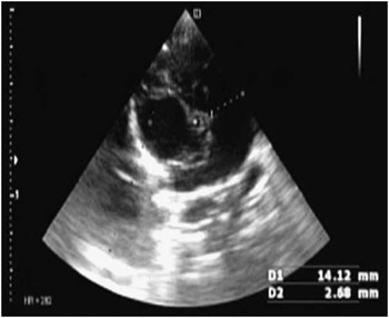
Echocardiography revealed an atretic aortic valve and hypoplasia of the ascending aorta and aortic arch without interruption or coarctation of the aorta, a moderate-sized perimembranous ventricular septal defect, very dilated pulmonary artery, a moderate-sized patent ductus arteriosus with right-to-left shunt, and reversal flow in the aortic arch (Fig 1).
The five-chamber echo view shows the aortic valve atresia.

To evaluate the hypoplastic aortic arch and its branches, we performed CT angiography.
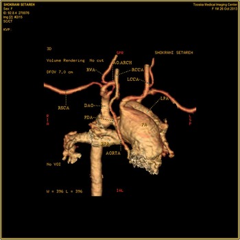
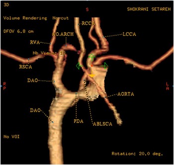
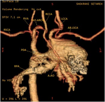
The CT angiography revealed hypoplastic ascending aorta (with diameter 1.5–2mm) and severe hypoplastic right sided aortic arch (diameter 1mm), where the right common carotid, right vertebral and right subclavian arteries were originated respectively. Then the diameter of aorta increased (3.7 mm) which may be due to steal phenomenon from the brain circulation. The diameter of descending aorta after the patent ductus arteriosus junction was normal (5.8 mm). The dilated pulmonary artery originated from the right ventricle, and the left common carotid artery originated from the distal main pulmonary artery ( an incidental finding). In addition, there was a retro-oesophageal aberrant left subclavian artery that originated from the descending aorta after the patent ductus arteriosus junction. The patent ductus arteriosus, with diameter of 3.5 mm and length of 6.3 mm, attached transversely to the main pulmonary artery between the origins of the right and left pulmonary arteries (Figs 2–4).
CT angiography shows the left common carotid originating directly from the dilated pulmonary artery (PA). AO arch=aortic arch; DAO=descending aorta; PDA=patent ductus arteriosus; LCCA=left common carotid artery; RCCA=right common carotid artery; RSCA=right subclavian artery; RVA=right vertebral artery; PA=pulmonary artery; RPA=right pulmonary artery; LPA=left pulmonary artery; SPR=superior-posterior-right; RIA=right-inferior-anterior; IAL=inferior-anterior-left; LSP=left-superior-posterior

CT angiography shows the aberrant left subclavian from the descending aorta (DAO) and the patent ductus arteriosus (PDA), as well as the hypoplastic ascending aorta (AAO). AO arch=aortic arch; DA=descending aorta; PDA=patent ductus arteriosus; LCCA= left common carotid artery; RCCA=right common carotid artery; RSCA=right subclavian artery; RVA=right vertebral artery; AB.LSCA=aberrant left subclavian artery; RP=right posterior; LA=left anterior; S=superior; I=inferior.

CT angiography in posterior view shows severe aortic arch hypoplasia. AO arch=aortic arch; PDA=patent ductus arteriosus; LCCA=left common carotid artery; RCCA=right common carotid artery; RSCA=right subclavian artery; RVA=right vertebral artery; AB.LSCA=aberrant left subclavian artery; RPA=right pulmonary artery; RP=right posterior; LA=left anterior; S= superior; I=inferior.

The patient was checked for signs of DiGeorge syndrome because of her abnormal facial structure and probability for associated anomalies, and it was ruled out with respect to normal serum calcium levels and the presence of a thymic shadow in her chest roentgenogram.
Her problems were discussed in the Joint Cardiology and Cardiac Surgeon Conference for decision-making, and the plan was patent ductus arteriosus stenting and bilateral pulmonary artery banding at first stage and then follow-up for any possible future intervention.
Discussion
The Edwards’ hypothetical double aortic arch explains aortic arch abnormalities by selective regression of various parts of either arch. In cases of isolation of the left innominate artery, the regression occurs at two sites: in the left posterior arch distal to the ductus arteriosus and in the left anterior arch proximal to the left common carotid artery, and also accompanied by concurrent migration of the left subclavian artery distal to the left ductus arteriosus. This resulted in an aberrant left subclavian artery as well as isolation of the left common carotid artery.Reference Shiu-Feng and Mei-Hwan 1
As the origin of aortic root and its branches was not delineated well by echocardiography, we performed CT angiography to determine whether there was any association with aortic arch anomalies.
Absence or hypoplasia of the thymus is commonly associated with aortic arch anomalies or tetralogy of Fallot;Reference Shiu-Feng and Mei-Hwan 1 therefore, we checked our patient for possible association with DiGeorge syndrome.
To the best of our knowledge, ours is the first case of isolation of the common carotid artery connecting to the main pulmonary artery associated with aortic valve atresia and hypoplasia of the ascending aorta, but without association with the Shone complex.
Therefore, although such cases are very rare, in similar cases such as aortic atresia, it seems that the evaluation of the aortic arch and its branches is necessary with non-invasive methods such as CT angiography or MRI, especially in cases where the brain supply will be impaired with the closure of the ductus arteriosus.
Acknowledgements
We are very grateful to Dr. Sofia Saburi for reconstructing these CT angio imagings. This study was approved by our local Ethic committee.
Financial Support
This research received no specific grant from any funding agency, commercial, or not-for-profit sectors.
Conflicts of Interest
None.