


Introduction
Novel agents displaying antimicrobial activity are urgently needed to tackle the public health hazards posed by the global spread of antimicrobial-resistant bacterial pathogens (Miethke et al., Reference Miethke, Pieroni, Weber, Brönstrup, Hammann, Halby, Arimondo, Glaser, Aigle, Bode and Moreira2021). Bioprospecting studies of endophytic microbes for antimicrobial activity are fundamental for discovering novel human antimicrobial agents (Strobel and Long, Reference Strobel and Long1998; Strobel, Reference Strobel2002; Strobel and Daisy, Reference Strobel and Daisy2003). Around the world, investigations are being carried out to explore endophytic fungi of various plants to discover new, potentially valuable secondary metabolites (Tiwari and Bae, Reference Tiwari and Bae2022). Among different plants, mangrove plants are attractive biodiversity hotspots for prospecting novel bioactive compounds, including those having potential medicinal applications (Cadamuro et al., Reference Cadamuro, da Silveira Bastos, Silva, da Cruz, Robl, Sandjo, Alves, Lorenzo, Rodríguez-Lázaro, Treichel and Steindel2021). As endophytic fungi of plants can produce similar biologically active constituents similar to their hosts (Khan et al., Reference Khan, Gilani, Waqas, Al-Hosni, Al-Khiziri, Kim, Ali, Kang, Asaf, Shahzad and Hussain2017), mangrove-associated fungi have received great attention in the past two decades. The mangrove ecosystem is a significant source of novel fungal strains, constituting the second-largest ecological category of marine fungi (Li et al., Reference Li, Li, Li, Dang, Proksch and Wang2008). Accordingly, worldwide, several mangrove species, such as Avicennia officinalis, Avicennia marina, Acanthus ilicifolius, Aegiceras corniculatum, Arthrocnemum indicum, Bruguiera gymnorrhiza, Ceriops decandra, Excoecaria agallocha, Kandelia candel, Lumnitzera racemosa, Rhizophora mucronata, Rhizophora apiculata, Sesuvium portulacastrum, Suaeda fruticosa, Suaeda maritima, and Sonneratia caseolaris have been investigated for their endophytic fungus relationships (Kumaresan and Suryanarayanan, Reference Kumaresan and Suryanarayanan2001; Ananda and Sridhar, Reference Ananda and Sridhar2002; Chi et al., Reference Chi, Pang, Chen, Wang and Lee2019; Yanti and Anwarrudin, Reference Yanti and Anwarrudin2021). These studies revealed that each mangrove plant's dominant endophytic fungi species were specific, although many common endophytes existed in several hosts (Kumaresan and Suryanarayanan, Reference Kumaresan and Suryanarayanan2001). Furthermore, the fungi in mangrove plants are shown as a mixture of soil, marine, and freshwater fungi (Ananda and Sridhar, Reference Ananda and Sridhar2002). Chi et al. (Reference Chi, Pang, Chen, Wang and Lee2019) and Yanthi and Anwarrudin (Reference Yanti and Anwarrudin2021) investigated the potential antimicrobial and anti-inflammatory activities of six fungal species isolated from A. ilicifolius, namely, Corynespora cassiicola, Phellinus noxius, Xylaria sp., Geotrichum sp., Humicola sp. and Aspergillus sp. The findings suggested that endophytic fungi associated with A. ilicifolius leaves and stems could be a source of several novel active substances.
India's mangroves are well recognized for their remarkable biodiversity and spatial spread. India's mangroves comprise a total area of 4740 km2, representing ~3% of the world's vegetated area (FSI, 2019). On the west coast of India, the mangroves of Kerala constitute one of the most critical ecosystems (Sreelekshmi et al., Reference Sreelekshmi, Veettil, Nandan and Harikrishnan2021). A. ilicifolius, belonging to Acanthaceae, is one of the most common mangrove species in Kerala (Sreelekshmi et al., Reference Sreelekshmi, Veettil, Nandan and Harikrishnan2021) and is widely utilized in traditional medicine for a variety of medicinal uses (Ragavan et al., Reference Ragavan, Saxena, Mohan, Jayaraj and Ravichandran2015; Saranya et al., Reference Saranya, Ramanathan, Kesavanarayanan and Adam2015). It has been established that this plant is an abundant source of alkaloids, long-chain alcohols, flavonoid glycosides, benzoxazinoid glucosides, megastigmane glucosides, triterpenes, triterpenoid saponins, and steroids, especially stigmasterol (Bandaranayake, Reference Bandaranayake2002; Bai et al., Reference Bai, Seasotiya, Malik, Bharti and Dalal2014). The plant showed several medicinal properties, viz., antioxidant, hepatoprotective, anticarcinogenic, cytotoxic, antimicrobial, anti-inflammatory, anti-ulcer, and antibacterial activities (Zhang et al., Reference Zhang, Vareed and Nair2005), pointing out the high opportunity for finding endophytic fungi with high bioactivities. Phomopsis sp. and Colletotrichum sp. are reported as the dominant fungal endophytes of A. ilicifolius (Suryanarayanan and Kumaresan, Reference Suryanarayanan and Kumaresan2000; Chaeprasert et al., Reference Chaeprasert, Piapukiew, Whalley and Sihanonth2010). Further, three independent studies in Indonesia, Thailand, and Taiwan demonstrated that endophytic fungi affiliated with A. ilicifolius possess strong antimicrobial actions (Chaeprasert et al., Reference Chaeprasert, Piapukiew, Whalley and Sihanonth2010; Chi et al., Reference Chi, Pang, Chen, Wang and Lee2019; Yanti and Anwarrudin, Reference Yanti and Anwarrudin2021). The endophytic fungus of A. ilicifolius from the genera Geotricum, Humicola, and Aspergillus exhibited a high in vitro antagonistic activity against harmful bacteria such as Escherichia coli and Staphylococcus aureus (Yanti and Anwarrudin, Reference Yanti and Anwarrudin2021). Chi et al. (Reference Chi, Pang, Chen, Wang and Lee2019) revealed that endophytic fungi from C. cassiicola and Xylaria sp. have potent antimicrobial activity against Bacillus subtilis, S. aureus, and E. coli. According to Chaeprasert et al. (Reference Chaeprasert, Piapukiew, Whalley and Sihanonth2010), Xylaria sp. found in the leaves of A. ilicifolius from Thailand possesses extensive antibacterial activities. These results imply that a range of biologically active endophytic fungi may exist in A. ilicifolius. However, only a few studies have identified biologically active endophytic fungi associated with the Indian mangrove trees (Ananda and Sridhar, Reference Ananda and Sridhar2002; Maria and Sridhar, Reference Maria and Sridhar2003; Priyadharshini et al., Reference Priyadharshini, Ambikapathy and Panneerselvam2015). In this context, the present study analyses the cultural diversity of the fungal endophyte assemblage on the healthy leaves of A. ilicifolius established on the mangrove habitat of the west coast of India, with an ultimate aim to elucidate their potential antimicrobial applications.
Materials and methods
Sample collection
The leaves of two apparently healthy mature trees of A. ilicifolius (Figure 1A) were collected from Thevara (9.924348705°N, 76.28840469°E) on the south coast of Kerala, India, during the pre-monsoon season (April and May 2021). The physicochemical parameters at the sampling site were recorded as follows: salinity: 25 ppt, temperature: 31°C, pH: 7.8, and dissolved oxygen: 2.8 mg l−1. The samples were placed in sterile sampling bags, placed in clean thermocol boxes with ice packs, brought to the lab (Cha et al., Reference Cha, Chiang, Guo, Lin and Pang2021), and analysed within 24 h of collection.
Mangrove leaves in the study and results of endophytic fungi enumeration: (A) leaves of A. ilicifolius used in the study and (B) results of endophytic fungi enumeration. Observed means of log CFU ± SD per g of leaves are given. *Indicates significant difference at P < 0.05.

Isolation and enumeration of endophytic fungi
The trituration procedure was used to prepare the samples to isolate endophytic fungus (Kloepper et al., Reference Kloepper, Hallmann and Rodroa1999). The leaves were sequentially washed in 70% ethanol for 1 min and 4% sodium hypochlorite solution for 1 min to sanitize the surface. The leaves were then rinsed thrice in distilled sterile water. To test the efficacy of the surface disinfection step, the disinfected samples were cultured on Martin's Rose Bengal streptomycin agar plates, followed by incubation for 2 weeks at 28°C to confirm the absence of epiphytes. The surface-disinfected leaf samples were then triturated using a mortar and pestle with sterile potassium phosphate buffer. Serial ten-fold dilutions of the triturate were prepared, and each dilution was spread plated in duplicates onto Martin Rose Bengal streptomycin agar medium. All plates were incubated at 28°C for 48 h. The total viable count was calculated by multiplying with the dilution factor and expressed as the number of colony-forming units (CFU) per g. Using potato dextrose agar (PDA) media, the cultivated fungal colonies were subcultured and purified for subsequent identification and activity testing. Further, the PDA plates with pure isolates were photographed. The glycerol stocks were prepared in a 30% glycerol solution and preserved at the Microbial Culture Collection, Fish Microbiology Lab, ICAR-Central Marine Fisheries Research Institute, Kochi.
Morphological characterization
Endophytic fungal isolates were grown in pure culture on PDA media, and the colony characters such as colour, texture, etc., were studied. Morphological characters were studied by observing the slide culture (Harris, Reference Harris1986) of fungi under a microscope. The septation of hyphae, conidiophore, and conidia characters were observed, and photomicrographs were recorded. Established taxonomic keys (Kohlmeyer and Kohlmeyer, Reference Kohlmeyer and Kohlmeyer1979; Kohlmeyer, Reference Kohlmeyer1984; Kohlmeyer and Volkmann, Reference Kohlmeyer and Volkmann-Kohlmeyer1991; Jones et al., Reference Jones, Sakayaroj, Suetrong, Somrithipol and Pang2009) were used to identify the isolates.
Molecular characterization
Fresh axenic cultures were cultivated on PDA media in the dark for 7 days at 28°C to extract genomic DNA. Fungal hyphae (100 mg) were scraped from the agar plate surface and ground in liquid nitrogen. Following that, fungal DNA was collected using the NucleoSpin® Plant II Kit according to the manufacturer's protocol (Macherey-Nagel GmbH & Co., Düren, Germany). The internal transcribed spacer (ITS) sequences were then amplified by polymerase chain reaction using the primers ITS1 and ITS4 (White et al., Reference White, Bruns, Lee, Taylor, Innis, Gelfand, Sninsky and White1990) (ITS1: 5′-TCCGTAGGTGAACCTGCGG-3′; ITS4: 5′-TCCTCCGCTTATTGATATGC-3′). The initial denaturation was conducted at 98°C for 30 s, followed by 40 reaction cycles (denaturation at 98°C for 30 s, annealing at 58°C for 10 s, extension at 72°C for 15 s), and final extension at 72°C for 60 s The amplicons were then sequenced in both directions at the Regional Facility for DNA Fingerprinting, Rajiv Gandhi Centre for Biotechnology, Thiruvananthapuram. The sequence data from forward and reverse sequencing reactions were edited and compiled using Editseq (DNASTAR, Lasergene, Madison, WI, USA). Next, we BLAST-ed (with default settings) the nucleotide sequence to check for similar sequences in the GenBank database at the National Center for Biotechnology Information (NCBI). The results of molecular characterization were then compared with those of morphological characterization for the final identification. The representative gene sequences of each distinct endophyte species were then deposited in GenBank and allocated accession numbers.
In vitro antimicrobial assay
The fungal isolate representing the most dominant species in the leaves of A. ilicifolius was only used for the downstream experiments. The antibacterial activity was tested against a panel of six indicator human pathogens (Table 1) by the well diffusion method (Taye et al., Reference Taye, Giday, Animut and Seid2011) and disc diffusion method (Mabrouk et al., Reference Mabrouk, Effat, Sadek, Tawfiek, Hassan and Magdoub2014). Briefly, 18 h old cultures of the indicator pathogens were centrifuged, and the pellet was resuspended by adjusting the optical density of the suspension test culture to 0.1 at 600 nM. Each indicator strain's resuspension was applied separately on Mueller–Hinton agar (HiMedia, Mumbai, India) to create a lawn culture. Subsequently, for the well diffusion method, the wells were prepared on each plate, and 100 μl of supernatant of 7 day old fungal culture was transferred to the wells. Simultaneously, the disc diffusion method was also applied, for which 50 μl of supernatant of fungal culture was added to 10 mm sterile disc (HiMedia, Mumbai, India) and allowed to dry. Discs containing the supernatant were placed on the surface of the medium containing the lawn culture of each pathogen. The plates (both well and disc diffusion) were incubated for 2 days at 37°C. For positive cases, the inhibitory zone developed after the incubation period was registered and measured. To validate the antimicrobial activity, the test was repeated thrice for positive instances. The mean zone diameter was then measured for the study. Further, to confirm the results of the disc and well diffusion methods the antibacterial efficacy in terms of minimum inhibitory concentration (MIC) and minimum bactericidal concentration (MBC) against the indicator pathogens was determined by the microtitre plate method (Marshall et al., Reference Marshall, Jones, Wanger, Washington, Doern, Leber and Haugen1996; Sharma et al., Reference Sharma, Sumithra, Suja, Azhar, Sanil, Ashok Kumar and Patil2021). In brief, the fungal culture supernatant mentioned above for the disc and well diffusion methods was used as the stock. Then, two-fold serial dilutions of the stock were prepared in Mueller–Hinton broth, and 100 μl of each dilution was added to polystyrene sterile flat-bottom 96-well plates in duplicates. All the wells were then inoculated with 100 μl of bacterial suspension (106 CFU ml−1) followed by incubation at 37°C for 24 h. The wells containing bacterial inoculum without fungal supernatant served as the control. At the end of incubation, the lowest concentration of supernatant that did not show visible bacterial growth was noted as MIC. Subsequently, 50 μl of suspension from each well was cultured onto Mueller–Hinton agar plates and incubated at 37°C for 24 h. The plates were inspected for bacterial growth after incubation, and the lowest concentration showing no bacterial growth on agar plates was noted as MBC.
Details of human pathogenic bacteria used in the study

Phylogenetic analysis
The evolutionary relationship of the most dominant endophytic fungal isolate was inferred through phylogenetic analysis using the sequences representing 18S rRNA gene partial sequence, 5.8S rRNA gene, 28S rRNA gene partial sequence, ITS1, and ITS2. Initially, a 551 bp-sized sequence representing the partial sequences of the 18S rRNA gene, ITS1, 5.8S rRNA gene, ITS2, and 28S rRNA gene of the fungus was obtained by editing and compiling the sequences obtained from forward and reverse sequencing reactions using Editseq (DNASTAR, Lasergene, Madison, WI, USA). Then, using CLUSTALW, multiple sequence alignment of this sequence, corresponding sequences of other fungi of the same family (Periconiaceae), and two representatives from the nearest family, viz., Massarinaceae was then performed (Altschul, Reference Altschul1997). Molecular Evolutionary Genetics Analysis, version 10 (MEGA 10), was then employed to perform a phylogenetic analysis on the aligned data (Kumar et al., Reference Kumar, Stecher, Li, Knyaz and Tamura2018). The Kimura two-parameter model (Nei and Kumar, Reference Nei and Kumar2000), which had the lowest Bayesian information criterion score was used to calculate the evolutionary distances. Following that, the phylogenetic tree was built using the neighbour-joining method with 1000 bootstrap replications (Saitou and Nei, Reference Saitou and Nei1987), and the gamma distribution with five discrete categories was employed to model the variations in evolutionary rates among sites. For the purpose of rooting, the corresponding sequence from an uncultivated Agaricomycetes was used (GenBank accession number: GQ268657.1).
Data analysis
Data on the enumeration of endophytic fungi were recorded as the log colony-forming units per g of leaf tissue (log CFU ± SD). The inhibition zone diameter (the difference between the total zone diameter and the bacterial colony/disc diameter) of the three-triplicate testing (mean ± SD) was used to express the antimicrobial assay results. The colony counts and the inhibition zone diameters (antibacterial tests) were compared using one-way analysis of variance (ANOVA), with a P-value of <0.05 set to indicate a significant difference. Tukey's test was employed for post-hoc analysis following one-way ANOVA (SPSS software program, ver. 16). The relative frequency (R F) of each distinct fungal isolate was then estimated as the ratio of the number of isolates of a particular taxon (n) to the total number of isolates (N) [(n/N) × 100] (Du et al., Reference Du, Yao, Li, Sun, Xia, Wang, Shi and Ren2020). The zone of inhibition diameter was used to score each isolate against each pathogen (Sumithra et al., Reference Sumithra, Reshma, Christo, Anusree, Drisya, Kishor, Revathi and Sanil2019). High (>10 mm), moderate (5–10 mm), low (1–4 mm), and no inhibition activities were given scores of 4, 3, 2, 1, and 0, respectively.
Results
Isolation and enumeration of endophytic fungi
Three different endophytic fungi, designated AFE-1, AFE-2, and AFE-3, were successfully isolated from the leaf segments of A. ilicifolius. The results of the enumeration of these fungi are depicted in Figure 1B. A statistical method was used to test the overall significant difference (P < 0.05) between the log10 CFU values of different fungal isolates, with significantly higher counts of AFE-3 compared to the other two isolates. In brief, the results showed that the most predominant fungus was AFE-3, with an average log10 CFU g−1 of leaf tissue of 7.70 ± 0.06.
Identification of the isolates
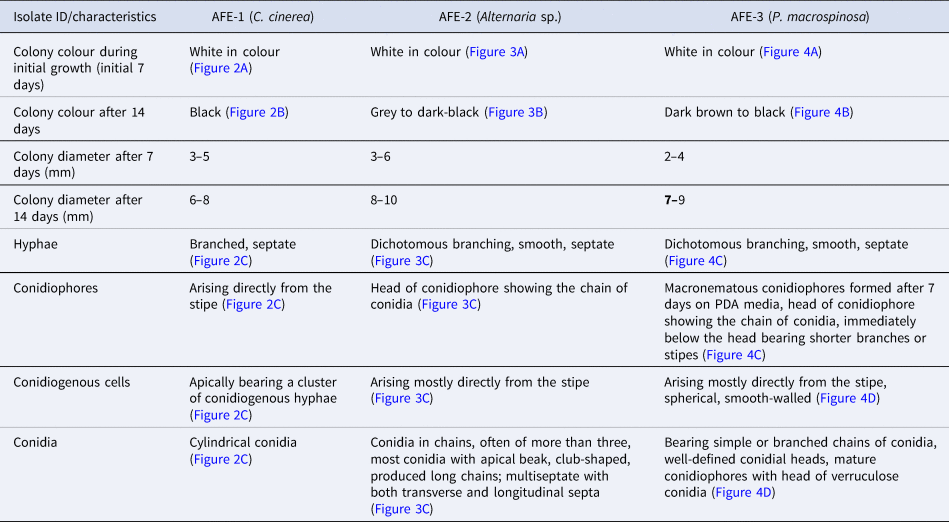
The morphological characteristics of the three fungal isolates on PDA media are depicted in Table 2 and Figures 2–4. During molecular characterization, an amplicon of 484, 439, and 546 bp was obtained from AFE-1, AFE-2, and AFE-3 isolates, respectively. GenBank accession numbers ON897769, ON898017, and OM085665 were assigned to the submitted sequencing data derived from the three fungal isolates. In total, three different fungus species belonging to the genera Alternaria sp., Coprinopsis sp., and Periconia sp. were identified (Table 3). A combined analysis of the morphological and molecular characteristics classified the first two fungi as Periconia macrospinosa (strain: CMFRI/fPM-01) and Coprinopsis cinerea (strain: CMFRI/fCC-01). The third fungus (strain: CMFRI/fAl-01) could not be classified to the species level. The R F of each unique fungal isolate was calculated as 35.22, 32.64, and 32.13% for CMFRI/fPM-01, CMFRI/fCC-01, and CMFRI/fAl-01, respectively.
Morphological characteristics of C. cinerea: (A) colony morphology during initial growth, (B) colony morphology after 14 days of growth, and (C) microscopic morphology after lactophenol cotton blue staining.

Morphological characteristics of Alternaria sp.: (A) colony morphology during initial growth, (B) colony morphology after 14 days of growth, and (C) microscopic morphology after lactophenol cotton blue staining.

Morphological characteristics of P. macrospinosa: (A) colony morphology during initial growth, (B) colony morphology after 14 days of growth and (C) microscopic morphology after lactophenol cotton blue staining.

Morphological characteristics of the isolates
Composition of endophytic fungi from the leaves of A. ilicifolius

In vitro antimicrobial assay
The antibacterial activity of the fungal isolate of the most prevalent species in A. ilicifolius leaves was investigated. The results are shown in Figure 5. In essence, the P. macrospinosa strain CMFRI/fPM-01 demonstrated a broad-spectrum antibiotic efficacy against all indicator pathogens tested. There was no significant difference in the antibacterial activity against the tested pathogens (P > 0.05). More importantly, the fungus scored 4, indicating high inhibitory activity against various pathogens. MIC and MBC values of the culture supernatant against the tested pathogens were 1:8 and 1:2 dilutions, respectively
Antibacterial activity of P. macrospinosa (CMFRI/fPM-01). VC, V. cholerae; VP, V. parahaemolyticus; VV, V. vulnificus; KP, K. pneumoniae; SA, S. aureus; EC, E. coli.

Phylogenetic analysis
The outcome of the phylogenetic analysis of the promising fungal isolate AFE-3 with potential antibacterial action is shown in Figure 6. Based on the derived phylogenetic tree, the Periconiaceae family of the Pleosporales order is distinguished from the Massarineae as a sister taxon with a distinct lineage. There were two well-separated subclades in the family ‘Periconiaceae’ clade (order: Pleosporales). The first subclade contained P. macrospinosa, Periconia igniaria, Periconia epilithographicola, Periconia variicolor, Periconia homothallica, Periconia digitata, Periconia elaeidis, Periconia cyperacearum, and Periconia psuedodigitata. The strains belonging to Periconia echinochloae, Periconia pseudobyssoides, and Periconia byssoides formed a different subclade (II). The isolate CMFRI/fPM-01 was clustered in the first subclade with close similarity to P. macrospinosa strains.
Phylogenetic analysis of P. macrospinosa strain CMFRI/fPM-01 based on ITS sequence.

Discussion
Among different plants, mangrove plants are the most attractive biodiversity hotspots for prospecting novel bioactive compounds (Cadamuro et al., Reference Cadamuro, da Silveira Bastos, Silva, da Cruz, Robl, Sandjo, Alves, Lorenzo, Rodríguez-Lázaro, Treichel and Steindel2021). A. ilicifolius is a widely used mangrove species for various medicinal purposes, especially in the field of conventional medicines (Ragavan et al., Reference Ragavan, Saxena, Mohan, Jayaraj and Ravichandran2015; Saranya et al., Reference Saranya, Ramanathan, Kesavanarayanan and Adam2015). The previous limited studies on A. ilicifolius (Chaeprasert et al., Reference Chaeprasert, Piapukiew, Whalley and Sihanonth2010; Chi et al., Reference Chi, Pang, Chen, Wang and Lee2019; Yanti and Anwarrudin, Reference Yanti and Anwarrudin2021) suggested that A. ilicifolius may possess a wide variety of biologically active endophytic fungi. In this context, the cultural diversity of the endophytic fungal assemblage on the healthy leaves of A. ilicifolius established on the mangrove habitat of the west coast of India was explored to elucidate their possible antimicrobial applications.
Only three taxonomically distinct endophytic fungi could be identified from the leaves of A. ilicifolius in the current investigation. The diversity of endophytic fungi in the plants relates to the host species and their environment. The number of distinct endophytic fungal species identified from the mangrove plants varies among the studies. According to Petrini (Reference Petrini, Fokkema and van den Huevel1986) and Rajagopal et al. (Reference Rajagopal, Meenashree, Binika, Joshila, Tulsi, Arulmathi, Kathiravan and Tuwar2018), only one or a small number of endophytic fungi dominate within a single host species due to inter-competition. The same might be a reason for the low diversity of the fungal endophytes obtained in the present research. In accordance with our results, Rodrigues and Samuels (Reference Rodrigues and Samuels1999) and Rajagopal et al. (Reference Rajagopal, Sasiprabha, Sabarish, Arulmathi, Meenashree, Kathiravan and Kathireshan2020) recovered only 13 and six endophytic fungal species from Spondias mombin and Eichhornia crassipes, respectively. Similarly, Mao et al. (Reference Mao, Zhang, Wu, Feng, Peng, Shahid, Cui, Ding and Shan2021) could isolate only 15 different endophytic fungi from the branches and fruits of Eucalyptus exserta, using PDA media. Wibowo et al. (Reference Wibowo, Prachyawarakorn, Aree, Mahidol, Ruchirawat and Kittakoop2016) isolated 20 compounds from the mangrove-associated fungus Pseudolagarobasidium acaciicola, originally isolated from the B. gymnorrhiza tree. The work underscored the importance of mangrove fungi as a rich source of novel bioactive compounds. Because most endophytic fungi cannot be cultured on artificial media and PDA media is not always an optimal medium, the three species in this study may represent only a subset of the total endophytic fungi found in A. ilicifolius. Arnold and Lutzoni (Reference Arnold and Lutzoni2007) recommended using 2% malt extract agar to encourage the growth of the most diversity of fungi, which has to be explored in future studies. Bosshard (Reference Bosshard2011) recommended an incubation period of 2–4 weeks to recover the maximum possible diverse fungi from the samples; accordingly, a shorter incubation period might be another reason for the observed low diversity in the present study. Further, Yu et al. (Reference Yu, Ding, Shen, Bu, Newcombe and Liu2021) showed that the diversity of foliar endophytes in plants varies significantly with age and is shown to be spread out and insignificant on young leaves. Accordingly, using older leaves, diverse isolation media, prolonged incubation, trying other isolation methods, such as surface sterilization method and application of high-throughput sequencing, are recommended to isolate more diverse endophytic fungi from A. ilicifolius or to confirm the low diversity; these are the future perspectives of the present study.
The three fungal isolates of the present study were identified as C. cinerea, Alternaria sp., and P. macrospinosa belonging to the phylum Basidiomycota, Ascomycota, and Ascomycota, respectively. The results support the hypothesis of Du et al. (Reference Du, Yao, Li, Sun, Xia, Wang, Shi and Ren2020) that all endophytic fungi belong to either Ascomycetes or Basidiomycetes. Also, Mishra et al. (Reference Mishra, Singh, Passari, Yadav, Gupta and Singh2016) and Pecoraro et al. (Reference Pecoraro, Caruso, Cai, Gupta and Liu2018) reported that Ascomycetes are the most prevalent members of endophytic fungal communities isolated using traditional separation techniques, as in agreement with our findings. Nevertheless, the diversity analysis of endophytes based on culture-dependent methodologies underestimates the actual diversity, and many isolates could still be identified by the culture-independent methods (Wang et al., Reference Wang, García-Fernández, Mas and Esteve-Zarzoso2015). Our study identified the dominant endophytes on the healthy leaves of A. ilicifolius in the mangrove ecosystem of the west coast of India, which will be helpful in screening and setting the groundwork for their application. It is important to mention that none of the species identified in the present study has been previously reported as the endophyte of A. ilicifolius leaves. Among the three species identified in the current research, Alternaria sp. is already described as a dominant endophytic fungal species of different plants (Rashmi et al., Reference Rashmi, Kushveer and Sarma2019) and has been isolated from other mangrove species such as A. marina, Sonneratia alba, Myoporum bontioides, and Rhizophora annamalayana (Kjer, Reference Kjer2009; Elavarasi et al., Reference Elavarasi, Peninal, Rathna and Kalaiselvam2014; Wang et al., Reference Wang, Cox, Ding, Huang, Lin and Li2014; Liu et al., Reference Liu, Chen, Chen, Liu, Lu, Chen, Lin, Huang and She2016).
Similarly, C. cinerea has been identified in Eugenia jambolana leaf tissue (Rashmi et al., Reference Rashmi, Kushveer and Sarma2019) but not from any mangrove species. However, other fungal species of the same genus Coprinopsis, have been reported in certain mangrove plants (Li et al., Reference Li, Sun, Chen and Guo2016; Devadatha et al., Reference Devadatha, Jones, Pang, Abdel-Wahab, Hyde, Sakayaroj, Bahkali, Calabon, Sarma, Sutreong and Zhang2021). Similarly, P. macrospinosa and other Periconia spp. are reported as the endophytes of the leaves of Acer truncatum and different plants, respectively (Rashmi et al., Reference Rashmi, Kushveer and Sarma2019). Further, other Periconia spp. have been reported as the endophytes of certain mangrove plants (Costa et al., Reference Costa, Maia and Cavalcanti2012; Hamzah et al., Reference Hamzah, Lee, Hidayat, Terhem, Faridah-Hanum and Mohamed2018). However, the study forms the first report of P. macrospinosa and C. cinerea as endophytes in any mangrove plant species. It was also interesting to note that the common endophytes of mangrove plants such as Aspergillus and Penicillium could not be obtained in this study (Costa et al., Reference Costa, Maia and Cavalcanti2012; Salini, Reference Salini2014).
The fungal isolate representing the most dominant species in the leaves of A. ilicifolius, P. macrospinosa (strain: CMFRI/fPM-01), was then evaluated for possible antimicrobial activity. Surprisingly, P. macrospinosa showed antibacterial properties against all the tested indicator pathogens, displaying a broad-spectrum antibacterial efficacy. More importantly, the fungus scored 4, indicating the solid inhibitory action against several pathogens. These findings concur with the report of Azhari and Supratman (Reference Azhari and Supratman2021), identifying Periconia spp. as an exciting resource for natural product research. The secondary metabolites of this genus have been found to have strong antimicrobial, cytotoxic, anti-HIV, and anti-inflammatory properties, including terpenoids, polyketides, cytochalasin, meroterpene, macrolides, macropshelides, aromatic compounds, and carbohydrate derivatives (Azhari and Supratman, Reference Azhari and Supratman2021). The results are exciting as the spectrum of the antimicrobial action includes three ESKAPE pathogens (S. aureus, Klebsiella pneumoniae, and E. coli), which cause the vast majority of nosocomial infections and have been shown to exhibit rising rates of antibiotic resistance and pathogenicity (Mulani et al., Reference Mulani, Kamble, Kumkar, Tawre and Pardesi2019). It is worth mentioning that the usage of E. coli ATCC 35218 in the study, which is the quality control organism for β-lactam–β-lactamase inhibitor compounds, further signifies the antimicrobial action of P. macrospinosa.
Conclusion
The present study demonstrated the cultural diversity of endophytic fungal assemblages on the healthy leaves of A. ilicifolius established in the mangrove habitat of the west coast of India. The study has identified the lesser-known genera Alternaria, Coprinopsis, and Periconia as endophytic members of the A. ilicifolius leaf fungal community. More importantly, this study is the first report of P. macrospinosa and C. cinerea as endophytes in mangrove plant species. The results also demonstrated the high and wide spectrum of antimicrobial action of the isolated P. macrospinosa against human pathogens, including ESKAPE pathogens. The isolate could serve as a potential resource for developing novel antimicrobial drugs, which could aid in the fight against antibiotic-resistant bacteria. Future studies shall be carried out using different media and high-throughput sequencing to isolate more endophytic fungi from the same plant or to confirm the low diversity found in this study. Also, further efforts are warranted to adequately characterize the isolated organism's biotechnological potential.
Acknowledgements
The authors are grateful for the facilities provided by the Director, ICAR-CMFRI, and Vice-Chancellor, KUFOS. The authors also thank the Vice Chancellor, KAU, for the support extended during the study.
Data availability statement
The authors confirm that the data supporting the findings of this study are available within the article. All the sequences generated were submitted to NCBI under unique accession numbers (ON897769, ON898017, and OM085665).
Author's contribution
All authors contributed to the study's conception and design. Conceptualization was done by KR Sreenath and TG Sumithra. Material preparation, data collection, and analysis were performed by P. Sainamole Kurian, VN Anusree, and VP Sruthi. The first draft of the manuscript was written by VP Sruthi and all authors contributed to the previous versions of the manuscript. All authors read and approved the final manuscript.
Financial support
The lead author received Kerala e-Grantz as a research fellowship during the study.
Competing interests
None
Ethical Approval
No ethical approval of research ethics committee was required to accomplish the goals of this study because no live animals were used for the experiments.



