Case report
A 39-year-old female presented for evaluation of shortness of breath on exertion and lower extremity oedema. She had no angina-type chest pain, orthopnea, and paroxysmal nocturnal dyspnoea. She reported a lifelong history of exercise intolerance, to which she attributed her fluctuating weight and chronic pain. Her medical history includes obesity (body mass index of 43.3), fibromyalgia, chronic pain syndrome, degenerative disc disease, Addison’s disease, suspected Ehlers-Danlos syndrome, hyperlipidaemia, and recurrent vein thrombosis. She smokes four cigarettes a day.
She was referred to our centre for further evaluation by our adult congenital team. An electrocardiogram showed sinus rhythm with normal axis and intervals. Her initial investigations included a transthoracic echocardiogram that showed normal left ventricle wall motion, normal left ventricular diastolic function left ventricle ejection fraction of 60%, and normal right ventricular systolic pressure estimated at 19 mmHg.
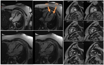
Chest CT angiography showed cardiac enlargement with focal outpouching at the left ventricular apex, which was potentially aneurysmal. Cardiac MRI then demonstrated double-chambered left ventricle (DCLV) (Fig 1), normal right ventricle size, and no obstruction or thrombus in either left ventricular chamber. The two left ventricle chambers were separated by a muscle band that extended from the apex to the mid-ventricle with both chambers joining together at the mid-region. Left ventricular systolic function was normal with a calculated ejection fraction at 70%. Both left ventricle chambers contracted normally with normal wall motion. Right ventricular systolic function was normal with a calculated right ventricular ejection fraction 57%. There was no delayed contrast enhancement seen in either ventricle.
Cardiac MRI: 4 chamber view and HLA stack showing the two LV chambers separated by muscle band that extends from the LV apex to the mid LV with both LV chambers joining together at the mid LV.

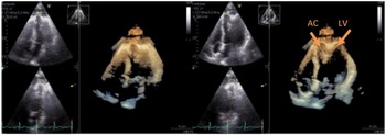
A congenital echocardiogram with 3D imaging confirmed the diagnosis (Fig 2). The study showed prominent muscular shelf from apex to mid-cavity that divided the left ventricle into two chambers with two distinct contracting apices that appeared to protrude into the apical pericardial fat tissue. Left ventricle EF was again normal at 60–65%. No other significant abnormalities were noted.
3D echocardiogram: showing prominent muscular shelf from apex to mid cavity that divided the left ventricle into two chambers with two distinct contracting apices that appeared to protrude into the apical pericardial fat tissue.

Her case was discussed at our weekly adult CHD conference, and after review of available limited literature on DCLV, we felt that surgical intervention was not warranted as this would be indicated in the setting of left ventricle pseudoaneurysm and not in DCLV. She was recommended to have additional baseline work up including brain natriuretic peptide (4.6 pg/mL, normal 0–100 pg/mL) and a 30-day loop recorder (no noted arrhythmias). A stress echocardiogram was done as part of her ischaemic work up that showed the medial left ventricle cavity became smaller and slit-like during stress. The apical portion of the medial cavity dilated mildly during stress. There was no obvious mid-cavitary gradient of either the medial or lateral left ventricle chambers. There were no wall motion abnormalities consistent with ischaemia at peak heart rate.
Her symptoms were medically treated with diuretics. In addition, after the stress echocardiography, a beta-blocker was added to allow for better diastolic filling, which corresponded with subjective improved exercise capacity.
Discussion
DCLV is a very rare, isolated, and sporadic CHD. In a 12-patient series with double-chambered left ventricle, Zhang et al found five of their patients were relatives, all of which had a MYH7 gene mutation known to be associated with myopathies including hypertrophic and dilated cardiomyopathy. Reference Zhang, Cao and Yuan1
Double-chambered left ventricle is often an incidental finding Reference Masci, Pucci, Fontanive and Coceani2–Reference Destounis, Tountas and Theodosis‐Georgilas4 . However, a few case reports had patients present with ventricular arrhythmia as noted by Sharma et al, Köz et al, and Kato et al. Reference Sharma, Dinwoodey and Chaudhry5–Reference Koz, Yokusoglu and Uzun7 It is unclear that the arrhythmia was associated with DCLV as the cardiac MRI findings reported in two of these case reports did not show findings suggestive of scar or fibrosis. However, Bravo-Jaimes et al reported a case of DCLV with inducible ventricular tachycardia (VT)Reference Bravo-Jaimes, Marah and Raghunathan 8 where there were cardiac MRI finding of late Gadolinium enhancment (LGE). Another interesting finding by Zhang et al reported reduced left ventricle ejection fraction in five patients and left ventricular dilation in eight of their patients; however, this is not typically reported elsewhere in the literature.
Considering its rarity and the uncertainty of its clinical course, the most appropriate management plan is likely serial clinical and imaging surveillance with consideration for implantable cardioverter-defibrillator (ICD) placement in patients who present with ventricular arrhythmia or unexplained syncope. In addition, LGE might be useful in assisting with decision and guiding management. Heart failure symptoms should be addressed medically as there is lack of evidence that surgical obliteration of the so-called accessory chamber is necessary. In our patient, given the unexplained symptoms, the interesting changes in the shape of the DCLV were noted on stress echo, which was thought to contribute to her symptoms as the decrease in the chamber size during exercise resulted in a relatively reduced cardiac output compared to resting state. She had good response to the beta-blocker with improved exercise capacity. This highlights the benefit of dynamic imaging when exam limitations are not explained by further resting imaging.
Conclusions
DCLV is a rare, usually asymptomatic condition but could present with heart failure or ventricular arrhythmias. Congenital echocardiography and cardiac MRI are important tools in the diagnosis of congenital DCLV and differentiation from acquired disease, such as post-infarction pseudoaneurysmReference Nacif, Mello, Lacerda and Sibley 9 , Reference Mordi, Carrick and Tzemos 10 .
Acknowledgements
None.
Financial support
This research received no specific grant from any funding agency, commercial, or not-for-profit sectors.
Conflicts of interest
None.
Ethical standards
The authors assert that all procedures contributing to this work comply with the ethical standards of the relevant national guidelines on human experimentation and with the Helsinki Declaration of 1975, as revised in 2008. All authors made a significant contribution to this study. All authors have read and approved the final version of the manuscript.