




No CrossRef data available.
Article contents
Serpin expression in the parasitic stages of Trichostrongylus vitrinus, an ovine intestinal nematode
Published online by Cambridge University Press: 27 August 2004
Abstract
Members of the genus Trichostrongylus, such as T. vitrinus, being endemic in Northern Europe, are among the principal causative nematodes which contribute to parasitic gastro-enteritis in sheep world-wide, inhabiting the proximal small intestine and causing damage to the mucosa. This results in impaired nutrient absorption as well as a pronounced inflammatory response with cellular infiltration of the mucosa, including a pronounced mast cell response. These mast cells release serine proteinases that enhance the passage of effector cells and macromolecules across epithelial boundaries and into direct contact with the invading parasite. The adult and larval stages of T. vitrinus release a number of serine proteinases in vitro that may contribute to tissue invasion and nutrient acquisition in vivo. This study describes the molecular cloning and characterization of a serine proteinase inhibitor (serpin)Sequence reported here is available with GenBank Accession number Y12233. that is present in extracts of all the parasitic stages, becoming more abundant as the life-cycle progresses. The serpin is present in the in vitro excretory/secretory products (ES) of 4th-stage larval and adult parasites, being more abundant in the former. The serpin was expressed in E. coli and the recombinant protein was a potent inhibitor of several host serine proteinases including mast cell proteinases. The serpin may regulate the activity of the parasite serine proteinases or it may modulate the host immune response to the parasite by inhibiting the activity of serine proteinases released from host inflammatory cells.
Information
- Type
- Research Article
- Information
- Copyright
- 2004 Cambridge University Press
INTRODUCTION
Trichostrongylus vitrinus is one of the principal causative nematodes of ovine parasitic gastroenteritis in Northern Europe and is the predominant nematode in the autumn months. It is closely related to Trichostrongylus colubriformis which is endemic in other temperate regions of the world including Southern Britain, Europe and Australasia (Urquhart, et al. 1997). It inhabits the proximal small intestine where the mucosal surface is damaged by the larval stages emerging from subepithelial tunnels, distortion and flattening of the villi. Disease results from the resultant reduction in the surface area available for nutrient absorption and from the inflammatory response to infection (Urquhart et al. 1987).
There continues to be extensive interest in the role of parasite and host proteinases at the host–parasite interface. Adult and 4th-stage larval T. vitrinus release a number of serine proteinases, as judged by molecular size during in vitro maintenance and these proteinases can degrade a variety of biological macromolecules (MacLennan, Gallagher & Knox, 1997). These proteinases may facilitate parasite survival in the host by mediating, for example, tissue penetration, feeding and immunoevasion. Proteinases also play an important role in the host immune response, exemplified by the release of serine proteinases from mast cells in response to gastrointestinal nematode infection (Miller, 1984). These proteinases may loosen intracellular junctions to facilitate the passage of effector cells and macromolecules across epithelial boundaries and, hence, into direct contact with the invading nematode (Emery, McClure & Wagland, 1993). The proteinases may also have direct effects on the surface of nematodes (McKean & Prichard, 1989) but the authors did point out that the target proteins, namely collagens, would, in normal circumstances, be unlikely to be exposed to proteinase digestion due to masking by other molecules present on the nematode surface.
There has been sustained interest in parasite-derived proteinase inhibitors since they may regulate endogenous proteinases or those involved in host physiological and immunological effector mechanisms (Leid, 1987; Modha & Doenoff, 1994; Morris & Sakanari, 1994; Rhoads, Fetterer & Hill, 2000). In the mammalian host, a range of proteinase-mediated physiological processes are regulated by serpins, including blood coagulation (Carrell, Evans & Stein, 1991), complement activation and inflammation (Travis & Salvesen, 1983) while parasite-derived serpins may simply regulate endogenous proteinases or modulate host immune responses, the latter possibility being pioneered in studies of a proteinase inhibitor from Taenia taeniaformis (e.g. Suquet, Green-Edwards & Leid, 1984). Recently, Zang et al. (1999) described the properties of a serpin expressed by Brugia malayi microfilariae that included the ability to inhibit human neutrophil serine proteinases. In the intestinal environment, it can be anticipated that invading nematodes will encounter host digestive proteinases and proteinases released from immune effector cells that accumulate as part of the immune response to the parasite.
A number of different immunopositives were isolated by screening a cDNA library prepared from adult T. vitrinus with antiserum to adult excretory/secretory products (ES), one of which had close sequence homology to a serine protease inhibitor (serpin). The molecular cloning and characterization of this serpin are described here.
MATERIALS AND METHODS
Parasites, extracts and excretory-secretory products (ES)
Adult and 4th-stage larvae (L4) were harvested from the small intestine of previously worm-free donor lambs as described previously (Knox & Jones, 1990). After extensive washing in phosphate-buffered saline, pH 7·4 (PBS), aliquots (200 mg) were blotted dry and snap-frozen in liquid nitrogen for DNA procedures or stored at −70 °C for the preparation of worm extracts. The remaining parasites (500 adults or 1000 L4/ml) were maintained in vitro for 16 h in sterile RPMI-1640 containing antibiotics (MacLennan et al. 1997). The resultant ES were then centrifuged (10000 g for 10 min) and concentrated as described previously (MacLennan et al. 1997). Extracts were prepared on ice in PBS containing 2% Triton X-100 using a glass/glass homogenizer and the protein concentration adjusted by dilution in PBS to 1 mg/ml.
Antisera for library screening and immunoblots
Rabbit antisera to adult ES were raised by subcutaneous injection at 3-week intervals of antigen (50 μg protein) in Freund's Complete (priming injection) and Incomplete adjuvant (2 boosts) using standard methods. Serum was harvested by ear-bleed 2 weeks after each boost to monitor the antibody response and by cardiac puncture following euthanasia 2 weeks after the final boost.
Sera and lymph harvested from lambs rendered immune to T. vitrinus infection by repeated challenge were kindly provided by Dr F. Jackson using methods described previously (Smith et al. 1983).
Lambda gt11 library preparation and immunoscreening
A cDNA library was prepared in the bacteriophage lambda gt11 from 500 mg adult (21 days) T. vitrinus using the procedures described in the manufacturer's instructions (‘cDNA cloning system – λgt11’; Amersham, UK). The resultant primary library had a titre of 5·2×105 pfu/ml. Among 34 immunopositives selected from 105 plaques by screening with the rabbit anti-ES serum, 1 plaque contained a 398 bp insert, the sequence of which showed significant similarity to a number of serpins. This was selected for further analysis.
Cloning and expression of the full-length cDNA
The full-length coding sequence was isolated from an adult T. vitrinus cDNA preparation using 5′- and 3′-RACE PCR primed using the nematode spliced leader sequence, SL1 [5′-GGTTTAATTACCCAAGTTTGAG-3′] and an antisense gene specific primer to aas 111 to 120 (5′-AACTTTTCCACCCATTCAGCAGTG-3′). The full-length cDNA was amplified using a sense primer directed to the 5′ extremity of the coding region and an antisense primer to the 3′ polyA+ region of the target transcript. The product of this reaction was subcloned and sequenced and the data used to design primers to the 5′ [5′-GTCGGATCCATGTTTTTAACGGCGGAA-3′] and 3′ [5′-GTCCCGGGTCACCGGAACTGCCAA-3′] extremities of the coding region. The primers contained BamH1 and Sma1 restriction enzyme recognition sequences to enable directional subcloning into the expression vector pGEX 4-T. The resultant plasmid was transformed into E. coli JM109 cells which were then grown in L-broth containing ampicillin (50 μg/ml) at 37 °C for 4 h and for a further 3 h after the addition of isopropyl-1-β-D-galactopyranoside (IPTG, 0·1 M, Sigma, Poole, UK) to induce recombinant protein expression. Aliquots (1 ml) of induced cultures were taken and soluble and insoluble bacterial extracts prepared (Sambrook, Fritsch & Maniatis, 1989). The insoluble extract was resuspended in 1 ml of phosphate-buffered saline, pH 7·4 (PBS) and thrombin (Sigma, Poole, UK) was added to a concentration of 50 U/ml after which the solution was incubated for 14 h at 25 °C. After centrifugation for 10 min at 11000 g, the pellet was resuspended in 0·5% Sarkosyl in PBS, centrifuged again, and the supernatant retained for further analyses.
Sequence analysis
Sequences were determined using ABI rhodamine dye terminators and an ABI 377 automated sequencer. Sequence analysis was conducted using DNAstar (DNAstar.com) and compared to those in the public databases using the BLAST alogorithm with subsequent alignments performed using CLUSTAL X. Unrooted phylogenetic trees were constructed by the Neighbour-joining method using Phylip and visualized using TreeView (Page, 1996).
Northern and Southern blotting
These procedures were conducted using standard methods (Sambrook et al. 1989) with 1 μg mRNA or 2 μg genomic DNA, the latter digested with HindIII, EcoR1 or BamH1, being blot-transferred onto nylon membranes after fractionation in agarose gels. Blots were probed using the 398bp insert labelled with [32P]-α-dCTP and washed at high stringency.
Electroelution
The putative serpin released by thrombin digestion was purified from 12% SDS–PAGE gels using the method described by Schagger, Cramer & von Jagow (1994), an elution procedure which removes SDS and Coomassie blue stain. Rabbit antisera were prepared to the eluted protein using the same immunization schedule outlined above. The N-terminal amino acid sequence of the eluted protein was determined.
Immunoblots
Parasite extracts and ES (~10 μg protein/lane) were fractionated using 10% SDS–PAGE, blot-transferred to nylon membranes (Amersham) and probed with the rabbit anti-serpin serum. Antibody binding was visualized using horseradish peroxidase (HRP)-conjugated goat secondary antibody and diaminobenzidine staining. In addition, serpin recognition during natural infection was sought by probing immunoblots of the bacterially expressed recombinant with sera and lymphs from lambs rendered immune to T. vitrinus, binding being visualized with HRP-conjugated anti-sheep IgG (Scottish Antibody Production Unit). Equivalent blots were also probed with serum from the same rabbit, pre-immunization, to act as a negative control.
Proteinase inhibition assays
Enzyme (10 μl of a 10 units/ml stock in PBS where 1 unit hydrolyses 1 μM substrate per min at 25 °C) was mixed with varying amounts of the serpin or control in 10 μl total volume. Then 100 μl of assay buffer (0·1 M Tris–HCl, pH 7·5) were added and the mixture incubated at 25 °C for 15 min. After addition of substrate (5 μl of a 100 μM stock solution in 0·1 M Tris–HCl, pH 7·5), the increase in absorbance with time was measured at 405 nm. Assays were conducted with porcine pancreatic elastase, human leucocyte elastase and human cathepsin G using benzoyloxycarbonyl-L-alanine-4-nitrophenol ester as substrate. Sheep mast cell protease II (SMCP; prepared from ovine intestinal mast cells (Huntley et al. 1986)) and bovine chymotrypsin and bovine pancreatic trypsin, using benzoyl-tyrosine-4-nitrolanilide and benzoyl-arginine-4-nitroanilide, respectively, as substrates. Enzymes and substrates were all purchased from Sigma.
RESULTS
Isolation and characterization of T. vitrinus serpin cDNA
Immunoscreening the adult λgt11 cDNA library yielded, amongst others, a positive clone containing a 398 bp insert, the predicted amino acid sequence of which showed homology with a number of serpins and which contained a putative methionine start codon preceded by the nematode SL1 spliced-leader sequence. The amplification from SL-1 to a gene-specific antisense primer yielded 3 different-sized products of 728, 584 and 528 bp, each containing a putative ATG start codon at nucleotides 223, 79 and 24 respectively, after which the sequences were identical. Northern blot analysis showed that the original cDNA hybridized to a region from ~1·2 kb to 1·5 kb while Southern blotting indicated that the serpin was encoded by a single copy gene (data not shown). The 3′ untranslated region contained a putative polyadenylation signal (AATAAA) 10 bases upstream from the poly (A)22 tail.
The predicted amino acid sequence is shown in Fig. 1. The T. vitrinus serpin showed between 24 and 33% identity with other serpin sequences in the databases, including those of nematode origin (Fig. 2), containing 31 of 51 residues that are characteristic of the serpin superfamily (Huber & Carrell, 1989; Zang et al. 1999). On the basis of this similarity, Fig. 1 shows predicted features relating to the secondary protein structure. Four out of five residues that comprise the serpin motif or hinge region (Fig. 1) correspond to the consensus (Fig. 2) but the predicted serpin signature (residues 341 to 352, Trichostrongylus numbering) shows little homology with the other serpins in the alignment containing additional residues to the other sequences. A dipeptide in helix F, Ile-Lys (aas 127-128), differs from the highly conserved consensus of Ile-Asn. The predicted protein sequence does not contain a signal peptide (Signal P server). The sequence contains 4 predicted N-linked glycosylation sites at asparagine 20, 68, 176 and 289. A phylogenetic comparison of the sequence with other serpins of vertebrate and invertebrate origin (Fig. 3) showed that the protein was most closely related to a serpin derived from C. elegans and the serpins identified from Brugia malayi, but was in a distinct group to the serpin from T. spiralis and those of schistosome origin. In general terms, 3 distinct groups were indicated in this analysis with the nematode serpins derived from a common ancestor. There was a suggestion, with limited bootstrap confidence, that the nematode group could be divided into 2 branches. When the same analysis was conducted with the peptide sequences containing the motif, reactive site loop and signature of each serpin shown in Fig. 3, the tree branches changed considerably, the T. vitrinus serpin then being closest to the Bombyx AT and Manduca 1 although the relationships had limited boot strap confidence (not shown).

Fig. 1. Predicted amino acid sequence of Tv Serp showing the position of conserved alpha helices and beta strands based on the structure of alpha-1-antitrypsin (Huber & Carrell, 1989).

Fig. 2. Multiple sequence alignment of the predicted amino acid sequences of serpins from human (human leucocyte elastase inhibitor, lei; alpha-1-antitrypsin, Alpha1AT), Trichinella spiralis (Tspiralis), Trichostrongylus vitrinus (Tvitrinus) and Brugia malayi (BmSPN1, BmSPN2). [squf ]=serpin motif; =serpin signature.

Fig. 3. Phylogenetic analysis of serpin sequences from invertebrate and vertebrate sources. The analysis was conducted using Clustal X and visualized with TreeView. The numbers at the nodes indicate the degree of bootstrap support for each branch. GenBank and SWISS-PROT Accession numbers for the sequences were: alpha-1-antitrypsin (alpha1AT), P01009; S. japonicum, Q967L9; S. mansoni, Q26606; Bombyx mori (Silkworm) anti-chymotrypsin (BombyxAC), P80034; Bombyx mori antitrypsin (BombyxAT), D00738; C. elegans (AAS13530); Brugia malayi SPN1, O18656; Brugia malayi SPN2, O18656; Tachypleus tridentatus (horseshoe crab; Ttrident), D14483; Manduca Sexta (tobacco hornworm, Manduca) 1, U58361; Manduca Sexta 2, U79184; T. spiralis, Q9NH65; T. vitrinus, Y12233.
Parasite-derived protein characterization by Western blot
This was investigated using rabbit antiserum to the approximately 40 kDa band released by thrombin digestion of the fusion protein expressed in pGEX (Fig. 4, Lanes 2 and 3 respectively compared to the undigested control, Lane 1). N-terminal sequence (FLTAETD) confirmed that this peptide was, as expected, the serpin. Immunoblot analysis of the products with anti-GST antibody indicated that the 60 kDa and 28 kDa bands were the undigested fusion protein and liberated GST (not shown).

Fig. 4. Expression of TvSERP in E. coli as a glutathione-S-transferase fusion protein. Lane 1, the insoluble bacterial pellet solubilized with 0·1% Triton X-100; Lane 2, the solubilized pellet digested with thrombin; Lane 3, the supernatant of the resultant digest. The arrows indicate the remaining undigested fusion protein (upper), the liberated serpin (middle) and the free GST (lower). Molecular weight markers indicated in kDa.
The rabbit antiserum recognized a number of proteins in adult and L4 extracts ranging in size from ~40 kDa to 100 kDa, a band at ~55 kDa being particularly prominent (Fig. 5). The antiserum reacted with a doublet between ~42 and 45 kDa, consistent with the predicted molecular weight of the serpin (41793 Da), this reactivity being particularly strong in adult extracts. Only weak, high molecular weight banding was evident in L3 extracts (not shown). The rabbit antiserum recognized, weakly, proteins at ~45 and 55 kDa in adult ES and 45 and 66 kDa proteins in L4 ES, the L4 ES, recognition being much stronger than the adult despite equal protein loading on the blot. Pre-immunization serum from the same rabbit did not react with any of the protein bands (not shown).

Fig. 5. Immunoblot of adult and L4 extracts and ES probed with rabbit antiserum to the bacterially expressed TvSERP. The arrow indicates the putative free serpin. Molecular weight markers indicated in kDa.
Serpin recognition by sera and lymph from lambs infected with T. vitrinus
The electro-eluted serpin was probed with sera and lymph harvested up to 1 week post-infection of immune lambs (Fig. 6). The serpin was recognized faintly as early as 3 days post-infection, with this being particularly strong at 8 days post-infection.

Fig. 6. Antibody binding to TvSERP by serum and lymph (indicated by the arrows) harvested from a lamb immune to T. vitrinus infection. The positive control is the soluble thrombin digest product of the bacterially expressed recombinant protein. pi=pre-infection; 3 & 8=days after a single larval challenge infection.
Protease inhibition assays
TvSERP inhibited all the serine proteinases tested in a dose-dependent manner (Fig. 7) being a particularly potent inhibitor of leucocyte and pancreatic elastases, inhibiting trypsin, the sheep mast cell proteinase (SMCP) and cathepsin G, and showing weak activity against chymotrypsin.

Fig. 7. Inhibition of a panel of serine proteases by TvSERP. Chymo=bovine chymotrypsin; Try=bovine trypsin; Lelast=human leukocyte elastase; Pelast=pancreatic elastase; SMCP=sheep mast cell protease; CathG=human cathepsin G.
DISCUSSION
The present study has demonstrated the presence of a serpin (TvSERP) in extracts and ES of 4th-stage larval and adult Trichostrongylus vitrinus. Immunoblot analysis suggested that the serpin is complexed to other proteins in parasite extracts, indicating a role in regulation of parasite proteinases, while it also exists in its native form in adult and L4 ES, being more prevalent in the latter. The serpin is recognized by serum and lymph from immune lambs, suggesting in vivo release. The recombinant protein inhibited a panel of serine proteinases including those of parasite origin and several proteinases of potential importance in the host anti-parasite immune response. This, and evidence for in vivo release, suggests a posssible role in immunoevasion.
Multiple sequence alignment showed that the TvSERP shared many features characteristic of the serpin superfamily, an analysis that suggests that the TvSERP has a similar secondary structure to the other members of the superfamily (Huber & Carrell, 1989). However, similarity to other serpins was limited in the serpin signature and in the reactive site loop. The latter shows extensive variation in the sequences aligned here and in the serpin superfamily (Jiang & Kanost, 1997). The dipeptide in helix F, Ile-Lys (aas 127-128), differed from the consensus of Ile-Asn in the serpin superfamily but it is notable that the lysine residue is also not conserved in Bm-SPN-2 (Zang et al. 1999). In addition, the T. vitrinus serpin showed only limited overall similarity to other serpins that have been characterized from parasitic nematodes such as Brugia malayi (Yentbur & Scott, 1995; Zang et al. 1999) and Trichinella spiralis (Rhoads, Fetterer & Hill, 2000). Phylogenetic analysis of a variety of invertebrate serpin sequences indicated that TvSERP was most closely related to serpins from C. elegans and B. malayi (BmSPN1, Yentbur & Scott, 1995; BmSPN2, Zang et al. 1999) with the serpins of nematode origin tending to group as a separate branch, although the bootstrap support was weak. It was interesting to note that this grouping was lost when the analysis was conducted with only the predicted reactive-site components alone suggesting that the serpins might have quite diverse functional specificity. This may indicate that serpins have evolved some nematode-specific characteristics, a trend which will be clarified as more serpin sequences from helminths become available for analysis.
Immunoblot analysis of parasite extracts and ES with antiserum raised to the ~40 kDa electro-eluted protein revealed a number of protein bands in extracts and ES with only a single band in adult and L4 extracts coinciding with the molecular weight (41793 kDa) predicted from the cDNA sequence. Sequence analysis predicted 4 potential N-linked glycosylation sites and this type of post-translational modification could account for some of the heterogeneity in the sizes of the proteins recognized, although we have no direct evidence that these sites are actually used here. The prominent higher molecular weight (>55 kDa) bands probably result from recognition of the serpin bound to endogenous proteinases, serpin–proteinase complexes being stable during SDS-PAGE (Patston et al. 1991). Fourth-stage larvae and adult T. vitrinus both contain, and release in vitro, several serine proteinases with different apparent molecular weights as judged by gelatin-substrate gel analysis (MacClennan et al. 1997) The blot results indicate that the serpin is present in adult and L4 ES, being more prominent in the latter. T. vitrinus L4 are intimately associated with the intestinal mucosa and are likely to be constantly exposed to host digestive proteinases as well as to proteinases released from host immune effector cells such as mast cell proteinases.
The GST–TvSERP fusion protein could not be eluted from the affinity column using the standard procedures. We have found this to be the case with other proteins expressed in the authors' laboratory and a possible explanation is that the protein is binding to the column via an interaction quite separate from the specific glutathione binding.
Antibody present in serum and lymph from a lamb rendered immune to T. vitrinus challenge by repeated infection, bound to recombinant TvSERP as early as 3 days post-challenge infection, an observation which supports the view that the serpin is released in vivo. A serpin (Bm-SPN-2) secreted exclusively by B. malayi microfilariae elicited strong, short-lived serpin-specific Th1 responses in microfilariae-infected mice while human patients displayed potent humoral responses to Bm-SPN-2 in both IgG1 and IgG4 subclasses (Zang et al. 2000).
Sequence analysis did not predict a signal peptide indicative of secretion. Adult and L4 T. vitrinus release several serine proteinases in vitro and it is possible that the serpin is found in ES as a result of being bound to a released proteinase. Alternatively, the serpin could be secreted using a non-classical protein secretion pathway not involving signal peptide cleavage (Nickel, 2003). A recent report identified a Drosophila serpin gene that had 4 distinct reactive centre-coding exons. The mutually exclusive use of these exons along with alternatively selectable 5′ and 3′ exons allowed the generation of 10 different transcripts, all of which were expressed in Drosophila embryos. The authors (Kruger et al. 2002) noted that these transcripts could code for 8 different forms of the protein with differing biological functions and they could either be secreted, be in the endoplasmic reticulum or be in the cytoplasm of the cell. Serpin genes encoding 2 or 3 alternative reactive centre exon cassettes were also identified in C. elegans (Kruger et al. 2002). In attempting to amplify the full-length coding sequence for Tv-SERP with coding sequence-specific primers, 3 different-sized transcripts were amplified, all of which contained identical core coding sequences but with distinct sequences in other regions (not shown) suggesting the same phenomenon may occur with some nematode serpin genes.
Serpins form stable 1[ratio ]1 complexes with the target proteinase, the serpin being cleaved in the reactive centre before being covalently linked to the target proteinase (Gettings, Patston & Schapira, 1993). Also, serpins can become substrates for the target serine proteinases (Yentbur & Scott, 1995; Gettings et al. 1993), again being cleaved in the reactive site with the carboxy-terminus released without complex formation (Patston et al. 1991). Therefore, the different-sized bands observed in the immunoblots may result from different elements of the serpin–proteinase interaction. Immunolocalization experiments were not conclusive (data not shown) but suggested that the serpin is present in the intestinal cells and pharynx of adult parasites.
The recombinant protein inhibited a variety of proteinases that may be encountered by the parasite within the intestinal mucosa. These data in themselves do not prove a specific function but they do demonstrate that the serpin has the capacity to inhibit a variety of serine proteinases, including the immunologically important leucocyte elastase, SMCP and cathepsin G. SMCP, for example, is found in the duodenal contents of immune sheep challenged with T. colubriformis (Emery et al. 1993) and mast cell proteinases are associated with changes in the permeability of the intestinal mucosa, mucus secretion and epithelial shedding (Huntley, 1992) all of which contribute to worm expulsion from the host. Cathepsin G and leucocyte elastase are released from stimulated polymorphonuclear neutrophils and these proteases have a variety of important functional capabilities including degradation of extracellular matrix, vasoregulation and IL-8 processing (Padrines et al. 1994). Serpins have already been implicated as factors that help infectious organisms to evade host immune defences (Zang et al. 1999), and it is worth noting that the same authors described preliminary results where a specific immune response against BmSPN2 resulted in clearance of microfilariae. Subsequent work has shown that Bm-SPN-2 is recognized by both murine and human T and B cells, with the responses being under stringent temporal control (Zang et al. 2000).
A recent report cast doubts on the enzyme-inhibitory role for BmSPN2, stating that a recombinant version of the inhibitor had no effect on cathepsin G and did not form SDS-stable complexes with the protease typical of serpin–proteinase interactions (Stanley & Stein, 2003). The TvSERP used for the enzyme assays here was eluted from SDS-PAGE gels. Despite the fact that the procedure removed SDS, there is a presumption that the serpin refolded correctly and, hence, that the enzyme inhibition arises from a specific interaction between the serpin and the proteinase active site. This view is supported to an extent by the differing degrees of inhibition observed with chymotrypsin only being partially inhibited by relatively high concentrations of TvSERP. Whatever the reality of this interaction, given its potential function, TvSERP may be critical for worm survival in the intestine and, hence, a good target for immunologically based control strategies.
This work was funded by the Scottish Executive Environment and Rural Affairs Department and by a grant from the Moredun Foundation. The authors are very grateful to Frank and Liz Jackson and colleagues for provision of parasite material.
References
REFERENCES
CARRELL, R. W., EVANS, D. L. &
STEIN, P. E. (
1991).
Mobile reactive centre of serpins and the control of thrombosis.
Nature, London 353, 576–578.CrossRefGoogle Scholar
EMERY, D., McCLURE, S. &
WAGLAND, B. (
1993).
Production of vaccines against gastrointestinal nematodes of livestock.
Immunology and Cell Biology 71, 463–472.CrossRefGoogle Scholar
GETTINS, P., PATSTON, P. A. &
SCHAPIRA, M. (
1993).
The role of conformational change in serpin structure and function.
Bioessays 15, 461–467.CrossRefGoogle Scholar
HUBER, R. &
CARRELL, R. W. (
1989).
Implications of the three-dimensional structure of alpha 1-antitrypsin for structure and function of serpins.
Biochemistry 28, 8951–8966.CrossRefGoogle Scholar
HUNTLEY, J. F. (
1992).
Mast cells and basophils: a review of their heterogeneity and function.
Journal for Comparative Pathology 107, 349–372.CrossRefGoogle Scholar
HUNTLEY, J. F., GIBSON, S., KNOX, D. &
MILLER, H. R. P. (
1986).
The isolation and purification of a proteinase with chymotrypsin-like properties from ovine mucosal mast cells.
International Journal for Biochemistry 18, 673–682.CrossRefGoogle Scholar
JIANG, H. &
KANOST, M. R. (
1997).
Characterization and functional analysis of 12 naturally occurring reactive site variants of serpin-1 from Manduca sexta.
Journal of Biological Chemistry 272, 1082–1087.CrossRefGoogle Scholar
KNOX, D. P. &
JONES, D. G. (
1990).
Studies on the presence and release of proteolytic enzymes (proteinases) in gastro-intestinal nematodes of ruminants.
International Journal for Parasitology 20, 243–349.CrossRefGoogle Scholar
KRUGER, O., LADEWIG, J., KOSTER, K. &
RAGG, H. (
2002).
Widespread occurrence of serpin genes with multiple reactive center-containing exon cassettes in insects and nematodes.
Gene 293, 97–105.Google Scholar
LEID, R. (
1987).
Parasite defense mechanisms for evasion of host attack; a review.
Veterinary Parasitology 25, 147–162.CrossRefGoogle Scholar
MacLENNAN, K., GALLAGHER, M. P. &
KNOX, D. P. (
1997).
Stage-specific serine and metallo-proteinase release by adult and larval Trichostrongylus vitrinus.
International Journal for Parasitology 27, 1031–1036.CrossRefGoogle Scholar
McKEAN, P. G. &
PRITCHARD, D. I. (
1989).
The action of a mast cell protease on the cuticular collagens of Necator americanus.
Parasite Immunology 11, 293–297.CrossRefGoogle Scholar
MILLER, H. R. P. (
1984).
The protective mucosal response against gastrointestinal nematodes in ruminants and laboratory animals.
Veterinary Immunology and Immunopathology 6, 167–259.CrossRefGoogle Scholar
MODHA, J. &
DOENHOFF, M. J. (
1994).
Schistosoma mansoni host–parasite relationship: interaction of contrapsin with adult worms.
Parasitology 109, 487–495.CrossRefGoogle Scholar
MORRIS, S. R. &
SAKANARI, J. A. (
1994).
Characterization of the serine protease and serine protease inhibitor from the tissue-penetrating nematode Anisakis simplex.
Journal of Biological Chemistry 269, 27650–27656.Google Scholar
NICKEL, W. (
2003).
The mystery of nonclassical protein secretion – A current view on cargo proteins and potential export routes.
European Journal of Biochemistry 270, 2109–2112.CrossRefGoogle Scholar
PADRINES, M., WOLF, M., WALZ, A. &
BAGGIOLINI, M. (
1994).
Interleukin-8 processing by neutrophil elastase, cathepsin G and proteinase-3.
FEBS Letters 352, 231–235.CrossRefGoogle Scholar
PAGE, R. D. M. (
1996).
TREEVIEW: An application to display phylogenetic trees on personal computers.
Computational and Applied Bioscience 12, 357–358.Google Scholar
PATSTON, P. A., GETTINS, P., BEECHEM, J. &
SCHAPIRA, M. (
1991).
Mechanism of serpin action: evidence that C1 inhibitor functions as a suicide substrate.
Biochemistry 30, 8876–8882.CrossRefGoogle Scholar
RHOADS, M. L., FETTERER, R. H. &
HILL, D. E. (
2000).
Trichuris suis: A secretory serine protease inhibitor.
Experimental Parasitology 94, 1–7.CrossRefGoogle Scholar
SAMBROOK, J., FRITSCH, E. F. &
MANIATIS, T. (
1989).
Molecular Cloning: A Laboratory Manual. 2nd Edn.
Cold Spring Harbor Laboratory Press, Cold Spring Harbor, USA.
SMITH, W. D., JACKSON, F., JACKSON, E. &
WILLIAMS, J. (
1983).
Local immunity and Ostertagia circumcincta: changes in the gastric lymph of immune sheep after a challenge infection.
Journal for Comparative Pathology 93, 479–488.CrossRefGoogle Scholar
SCHAGGER, H., CRAMER, W. A. &
VON JAGOW, G. (
1994).
Analysis of molecular masses and oligomeric states of protein complexes by blue native electrophoresis and isolation of membrane protein complexes by two-dimensional native electrophoresis.
Analytical Biochemistry 217, 220–230.CrossRefGoogle Scholar
STANLEY, P. &
STEIN, P. E. (
2003).
BmSPN2, a serpin secreted by the filarial nematode Brugia does not inhibit human neutrophil proteinases but plays a noninhibitory role.
Biochemistry 42, 6141–6248.Google Scholar
SUQUET, C., GREEN-EDWARDS, C. &
LEID, R. W. (
1984).
Isolation and partial characterization of a Taenia taeniaeformis metacestode proteinase inhibitor.
International Journal for Parasitology 14, 165–172.CrossRefGoogle Scholar
TRAVIS, J. &
SALVESEN, G. S. (
1983).
Human plasma proteinase inhibitors.
Annual Reviews of Biochemistry 52, 655–709.CrossRefGoogle Scholar
URQUHART, G. M., ARMOUR, J., DUNCAN, J. L., DUNN, A. M. &
JENNINGS, F. W. (
1987).
Veterinary Parasitology.
Longman Scientific and Technical, UK.
YENBUTR, P. &
SCOTT, A. L. (
1995).
Molecular cloning of a serine proteinase inhibitor from Brugia malayi.
Infection and Immunity 63, 1745–1753.Google Scholar
ZANG, X., YAZDANBAKHSH, M., JIANG, H., KANOST, M. R. &
MAIZELS, R. M. (
1999).
A novel serpin expressed by blood-borne microfilariae of the parasitic nematode Brugia malayi inhibits human neutrophil serine proteinases.
Blood 94, 1418–1428.Google Scholar
ZANG, X., ATMADJA, A. K., GRAY, P., ALLEN, J. E., GRAY, C. A., LAWRENCE, R. A., YAZDANBAKHSH, M. &
MAIZELS, R. M. (
2000).
The serpin secreted by Brugia malayi microfilariae, Bm-SPN-2, elicits strong, but short-lived, immune responses in mice and humans.
Journal for Immunology 165, 5161–5169.CrossRefGoogle Scholar
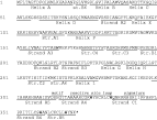
Fig. 1. Predicted amino acid sequence of Tv Serp showing the position of conserved alpha helices and beta strands based on the structure of alpha-1-antitrypsin (Huber & Carrell, 1989).
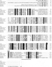
Fig. 2. Multiple sequence alignment of the predicted amino acid sequences of serpins from human (human leucocyte elastase inhibitor, lei; alpha-1-antitrypsin, Alpha1AT), Trichinella spiralis (Tspiralis), Trichostrongylus vitrinus (Tvitrinus) and Brugia malayi (BmSPN1, BmSPN2). [squf ]=serpin motif; =serpin signature.
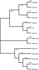
Fig. 3. Phylogenetic analysis of serpin sequences from invertebrate and vertebrate sources. The analysis was conducted using Clustal X and visualized with TreeView. The numbers at the nodes indicate the degree of bootstrap support for each branch. GenBank and SWISS-PROT Accession numbers for the sequences were: alpha-1-antitrypsin (alpha1AT), P01009; S. japonicum, Q967L9; S. mansoni, Q26606; Bombyx mori (Silkworm) anti-chymotrypsin (BombyxAC), P80034; Bombyx mori antitrypsin (BombyxAT), D00738; C. elegans (AAS13530); Brugia malayi SPN1, O18656; Brugia malayi SPN2, O18656; Tachypleus tridentatus (horseshoe crab; Ttrident), D14483; Manduca Sexta (tobacco hornworm, Manduca) 1, U58361; Manduca Sexta 2, U79184; T. spiralis, Q9NH65; T. vitrinus, Y12233.
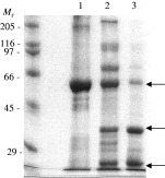
Fig. 4. Expression of TvSERP in E. coli as a glutathione-S-transferase fusion protein. Lane 1, the insoluble bacterial pellet solubilized with 0·1% Triton X-100; Lane 2, the solubilized pellet digested with thrombin; Lane 3, the supernatant of the resultant digest. The arrows indicate the remaining undigested fusion protein (upper), the liberated serpin (middle) and the free GST (lower). Molecular weight markers indicated in kDa.
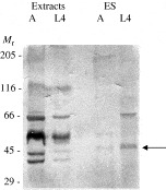
Fig. 5. Immunoblot of adult and L4 extracts and ES probed with rabbit antiserum to the bacterially expressed TvSERP. The arrow indicates the putative free serpin. Molecular weight markers indicated in kDa.
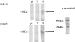
Fig. 6. Antibody binding to TvSERP by serum and lymph (indicated by the arrows) harvested from a lamb immune to T. vitrinus infection. The positive control is the soluble thrombin digest product of the bacterially expressed recombinant protein. pi=pre-infection; 3 & 8=days after a single larval challenge infection.
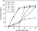
Fig. 7. Inhibition of a panel of serine proteases by TvSERP. Chymo=bovine chymotrypsin; Try=bovine trypsin; Lelast=human leukocyte elastase; Pelast=pancreatic elastase; SMCP=sheep mast cell protease; CathG=human cathepsin G.
 You have
Access
- 12
- Cited by